



Abstract
Few human tissues have functions as closely linked to the composition of their extracellular matrices as the intervertebral disc. In fact, the hallmark of intervertebral disc degeneration, commonly accompanying low back and neck pain, is the progressive loss of extracellular matrix molecules - specifically the GAG-substituted proteoglycans. While this loss is often associated with increased extracellular catabolism via metalloproteinases and pro-inflammatory cytokines, there is strong evidence that disc degeneration is related to dysregulation of the enzymes involved in GAG biosynthesis. In this review, we discuss those environmental factors, unique to the disc, that control expression and function of XT-1, GlcAT-I, and ChSy/ChPF in the healthy and degenerative state. Additionally, we address the pathophysiology of aberrant GAG biosynthesis and highlight therapeutic strategies designed to augment the loss of extracellular matrix molecules that afflict the degenerative state.
Keywords: Intervertebral disc, extracellular matrix, intervertebral disc degeneration, GAG synthesis, proteoglycans
1. Background
The human spinal column contains 23 intervertebral discs, each flanked by sequential pairs of vertebrae forming a basic spinal motion segment. Each segment represents a polyaxial diarthrodial joint, where the intervertebral disc is specifically tasked with permitting motion and transmitting applied biomechanical loads [1].
The intervertebral disc comprises an outer fibrocartilaginous annulus fibrosus (AF), encompassing a hydrophilic nucleus pulposus (NP), bordered superiorly and inferiorly by hyaline cartilaginous endplates (CEP). Although cellularity is low in both the NP and AF compartments, resident cells produce and secrete the complex ECM molecules that accommodate the compressive and tensile spinal loads, respectively. Underscoring the importance of the ECM, intervertebral disc degeneration is intricately associated with decreased hydrophilic extracellular matrix (ECM) molecules and alterations in disc cell phenotype, leading to structural changes and instability of the spine [2]. The loss of disc ECM is the combined result of decreased synthesis and increased ECM catabolism through pro-inflammatory cytokines induced metalloproteases [3–5]. Disc degeneration is the main cause of herniation and is linked to low back pain, the leading cause of disability in the United States [2,6]. Moreover, greater than 96% of autopsy specimens from donors over the age of 60 exhibit signs of disc degeneration making it the predominant feature of spinal ageing [7].
The molecular structure of the healthy disc ECM, especially the NP tissue compartment, contributes to the unique hypoxic and hyperosmolar characteristics of the niche itself. It is known that blood vessels reach the superficial regions of the CEP and AF, but do not infiltrate the NP [8]. As a consequence of this avascularity, to survive in this effectively hypoxic environment NP cells robustly and constitutively express transcription factors HIF-1/2α [9–11]. In addition, due to high negative charge density of constituent proteoglycans (PG), the disc ECM is hyperosmolar; osmolarity ranges between 430 to 496 mOsm/Kg [12,13]. To prevent potentially damaging effects of the hypertonic environment, the transcription factor TonEBP drives the expression of a panoply of genes that facilitate the cytosolic accumulation of non-ionic osmolytes in the NP [14,15].
There is a reciprocal relationship between specific niche conditions of the disc and the ECM composition; on one hand, the ECM structure contributes to the generation of the hypoxic and hyperosmolar niche of the disc. On the other hand, downstream signaling pathways controlled by HIF-1α and TonEBP are responsible for regulating PG synthesis, especially through enzymes involved in the GAG and core protein synthesis. In this review, we will detail the current knowledge of factors regulating enzymes in the GAG biosynthetic pathway in the disc and show how they affect ECM composition during health and disease.
2. Extracellular matrix composition of the disc
The healthy disc ECM contains PGs covalently substituted with glycosaminoglycans (GAG) chains, collagens and other glycoproteins [16–20]. The three disc compartments are composed of unique patterns of these ECM molecules, based on their specific mechanical and cellular function. Importantly, the most abundant PG in the NP is aggrecan [21]. The aggrecan core protein is bound by both chondroitin sulfate (CS) and keratan sulfate (KS) GAG chains, providing aggrecan with its osmotic properties including swelling potential and compressive resistance - hallmarks of healthy disc function [22]. The hexose, hexuronic acid, and hexosamine residues on these chains are sulfated, providing a net negative charge to the aggrecan molecule [23]. Furthermore, aggrecan, so named because of its tendency to aggregate with hyaluronan, is a nonsulfated GAG composed of alternating N-acetylglucosamine and glucuronic acid residues [24]. The high negative charge density of charged aggrecan molecules electrostatically attracts and binds water thereby maintaining the hydration status of the healthy disc. In addition to aggrecan, versican, another well-studied large aggregating CSPG, confers tissue hydration and viscoelasticity to the disc. Versican is diffusely distributed throughout the NP compartment, in lower concentrations than aggrecan, yet in greater abundance in the disc than in articular cartilage[25]. In fact, a shift from a versican-rich ECM to one rich in collagen is a hallmark of tissue fibrosis and degeneration [26]. Swelling of the NP is partially balanced by tensile forces imposed by collagen II fibrils within the tissue compartment. However, given that the PG to collagen ratio in the NP is approximately 27:1, swelling resistance is mainly provided by collagen fibrils in the AF which bear large circumferential tensile stress [27,28].
The AF is composed of concentric lamellae of collagen fibers arranged circumferentially at ±30 degrees alternating between consecutive lamellae. The major collagens within the AF are fibril-forming type I and type II collagen [16]. Within the AF there are regional variations in ECM composition depending on mechanical function. Similar to the NP, the inner region of the AF bears compressive load and is mostly composed of PG and collagen II fibers. Since the tensile strength of the outer AF is of the major importance this region is rich in organized collagen I lamellae [29].
While aggrecan, versican, and the fibrillar collagens maintain the mechanical function and/or hydration in the disc, PGs are also concerned with signal transduction pathways as they can bind and interact with ECM cytokines/growth factors. In addition, NP cells expresses perlecan, syndecans, glypicans, and various small leucine-rich repeat proteoglycans (SLRPs) including decorin, biglycan, fibromodulin, chondroadherin and aspirin, molecules that are pivotal for mediating signaling pathways necessary for cell survival (Fig. 1) [18,21,30]. Furthermore, all of the PGs of the disc contain CS/DS or HS chains; the exceptions are fibromodulin, which has a KS chain, while CHAD and asporin have no GAG chains [31,32].
Figure 1.

Major proteoglycans in the intervertebral disc.
Schematic of the GAG-substituted proteoglycans in the intervertebral disc showing their subcellular localization, core protein structure, and GAG-chains. Abbreviations: heparan sulfate (HS), chondroitin sulfate/dermatan sulfate (CS/DS), keratin sulfate (KS), and hyaluronic acid (HA).
3. Regulation of GAG biosynthesis in the disc
The GAGs are divided into four major groups depending on their core disaccharide structures: chondroitin sulfate/dermatan sulfate (CS/DS), heparan sulfate (HS), keratan sulfate (KS), and hyaluronic acid (HA) [33]. Both CS/DS and HS are synthesized in the Golgi apparatus, where protein cores are post-translationally modified at specific serine residues with a common linkage tetrasaccharide: GlcAβ1- 3Galβ1- 3Galβ1- 4Xylβ1-O- Ser. Biosynthesis of the linkage tetrasaccharide starts with transfer of xylose to the serine residue on the core protein by xylosyltransferases (XT-1/2), followed by the sequential addition of two galactose residues by galactosyltransferases (GalT-I/II), and a final glucuronic acid residue by β-1,3-glucuronosyltransferase 3 (GlcAT-I). All monosaccharides are transfered from their corresponding UDP-sugars [23]. After the linker region is synthesized, addition of GalNAc to the tetrasaccharide commits the molecule to CS synthesis through alternating addition of GlcA and GalNAc by chondroitin synthase (ChSy) and chondroitin polymerization factor (ChPF) (Fig 2). Alternatively, if GlcNAc is added to the linkage tetrasaccharide, it results in HS synthesis though the addition of alternating GlcA and GlcNAc residues by HS copolymerases (EXT-1/2). Following formation and elongation of the CS/DS and HS GAG chains, coordinated actions of sulfotransferases and sulfatases promote rapid changes in the sulfation pattern [34]. Furthermore, KS can modify proteins through N- or O-linked glycosylation of PGs in the Golgi, meanwhile HA is synthesized by integral membrane proteins that immediately secrete the nascent chain [33].
Figure 2.

Regulation of enzymes involved in GAG biosynthesis in the intervertebral disc.
Schematic of the hexoses and hexotransferases involved in CS/DS and HS GAG chain biosynthesis. The common CS/DS and HS linkage chain is thought to be synthesized in either the pre-golgi or endoplasmic reticulum exit sites. The GAG repeating disaccharide region shown is specific for CS chains and occurs in the Golgi. Transcription factors and environmental cues regulating enzyme expression in the disc are shown for enzymes highlighted in green. Enzymes with regulation not yet known in the disc are shown in black. Abbreviations:
It has been well documented that while the rate of GAG synthesis, is directly controlled by the transfer of monosaccharides from their corresponding UDP-sugars to the intermediate GAG chain, in the disc it can also be affected by environmental factors. Such factors include inflammatory cytokine and growth factor signaling, calcium flux, oxygen tension, age and degenerative state. Given that CSPG and HSPGs are the most abundant in the disc, research has focused on elucidating the regulation of enzymes specifically involved in their biosynthesis; these include XT-1 and GlcAT-I which synthesize the initial and final steps of the common CS and HS linker region, and ChSy1 and ChPF which synthesize the CS-specific repeating disaccharide chain.
A. Regulation of xylosyltransferase-1 (XT-1)
Biosynthesis of CS and HS chains is initiated by the formation of a common linker tetrasaccharide (D-GlcA- β 1, 3-Gal- β 1, 3-Gal- β 1, 4-Xyl-Ser) formed by the sequential transfer of individual sugar residues from their corresponding UDP-sugars. The first step in the synthesis of the chain involves attaching xylose to a specific serine residue on the core protein; this step is catalyzed by xylosyltransferase-1 (XT-1) and, thus, serves as an important rate-limiting step in GAG biosynthesis [35,36]. Noteworthy, it has been shown that changes in XT-1 expression levels and, thus, changes in the biosynthesis of CS and HS chains occur in diseases such as osteoarthritis (OA), dilated cardiomyopathy, and skin fibrosis [37–39]. In chondrocytes, it is known that XT-1 expression is suppressed by inflammatory cytokine IL-1β, and signaling through Sp1 and Sp3 transcription factors exert opposing effects on XT-1 transcription such that the ratio of the two transcription factors determines XT-1 expression levels [40,41]. Interestingly, unlike chondrocytes, in the NP, XT-1 expression was refractory to two inflammatory cytokines associated with degenerative disc disease: TNF-α and IL-1β [42].
Meanwhile, signaling through the Jun/AP-1 axis and complementary actions of Sp1 and Sp3 transcription factors, all positively controlled XYLT1 (XT-1) at the transcript level through cognate binding sites on the XT-1 promoter [42]. Since XT-1 represents the first rate-limiting step in GAG biosynthesis, these researchers hypothesized that its expression was modulated during the disease process. Again, in contrast to osteoarthritis, pro-inflammatory cytokines did not influence XT-1 expression. Instead, expression levels of XT-1 in degeneration correlated with levels of Jun, Fos, and AP1 expression highlighting their role in maintaining XT-1 expression under physiologic and pathophysiologic conditions in NP cells [42].
B. Regulation of glucuronosyltransferase- I (GlcAT-I)
The GlcAT-I enzyme catalyzes the synthesis of the common linker region of CS and HS GAGs by transferring the final glucuronic acid (D-GlcA-β1) to the core tetrasaccharide chain. It has been shown that GlcAT-I is highly expressed in neonatal and mature rat NP and AF cells [43]. This latter observation is not unexpected as the inner AF is rich in PGs and bears compressive loads. GlcAT-I expression in the disc is regulated by a host of factors including intracellular calcium concentration, canonical and non-canonical growth factor signaling, and hypoxia [43–46].
It has been documented that shifts in intracellular calcium flux can affect aggrecan and PG synthesis in multiple cell types including chondrocytes and Sertoli cells [47–49]. Recent findings demonstrated that the expression of GlcAT-I in the disc is indeed calcium-dependent, a critical transducer of mechanical load [43]. Further studies showed that TonEBP (NFAT5), a transcription factor also known to regulate aggrecan synthesis, is a positive regulator of B3GAT3 (GlcAT-I) [43,50]. On the other hand, it was observed that the calcium-dependent calcineurin-NFAT transcription factor, a member of the same family as TonEBP, is a negative regulator of GlcAT-I [43]. From this perspective, by controlling GAG as well as aggrecan synthesis, TonEBP permits NP cells to autoregulate the osmotic environment of the disc.
Other studies focused on the effect of the cytokine TGF-β, and its downstream signaling pathways on GlcAT-I regulation in the disc [44,45]. TGF-β functions by binding to transmembrane serine/threonine protein kinase receptors to phosphorylate downstream targets. While the Smad transcription factor is a canonical target, ligand-bound TGF-β receptors are also able to activate ERK, p38, JNK MAPK and PI3K/AKT pathways [51]. The role of TGF-β in the NP has been previously studied, for example, TGF-β has been shown to regulate PG synthesis and stimulate chondroitin sulfate biosynthesis [52]. In the NP, expression of GlcAT-1 was regulated by TGF-β and BMP-2. When treated with these growth factors, there was an induction in GlcAT-1 promoter activity and expression. A second major observation was that TGF-β and BMP-2 both regulate GlcAT-1 at the transcriptional level by signaling through specific MAPKs (including ERK1), thereby directly promoting AP1, Sp1, and TonEBP transcriptional activity [44]. Thus, from a functional perspective, regulation of expression is mediated by members of the TGF-β family of proteins as well as environmental stimuli - including osmotic pressure and biomechanical loading (calcium flux)- factors that transduce TonEBP. In this way, TGF-β growth factor signaling and TonEBP work in tandem to maintain GAG and PG biosynthesis in NP cells.
Although regulation of GlcAT-I by TGF-β via a non-canonical signaling pathway was discovered first in NP cells, it was quickly followed by a study demonstrating that this enzyme is also regulated by Smad3-dependent canonical TGF-β signaling pathway [45]. Smad3 regulated GlcAT-1 transcription by directly controlling the activity of GlcAT-1 promoter. Furthermore, the in vitro findings were supported by the observation that there was a decrease in GlcAT-1 expression in discs of Smad3-null mice. Lastly, studies showed that ALK-5 signaling in particular controlled basal expression of GlcAT-1. Taken together, both the canonical and non-canonical TGF-β signaling pathways promote GAG and PG biosynthesis in NP cells of the disc.
It is important to note that although levels of TGF-β are elevated during disc degeneration, resulting from inflammatory or degenerative stimuli, there is a disconnect between TGF-β and GlcAT-1 levels in later stages of disc degeneration [45,53,54]. The lack of GlcAT-1 induction following TGF-β treatment in cultured cells from degenerate human disc samples, suggested that the disease process may have altered cellular responsiveness to this growth factor. This finding may explain the inability of cells to mount a reparative response to enhance GAG synthesis, despite elevated TGF-β levels seen during disc degeneration.
Hypoxia is another key environmental factor shown to regulate GlcAT-I expression in NP cells [46]. Earlier studies indicated while that there is a stimulatory effect of hypoxia on aggrecan synthesis in NP cells, it was not known if this was through increased GlcAT-I expression,. Subsequently, Gogate et al. demonstrated that while hypoxia increases expression of GlcAT-I in NP cells and chondrocytes, HIF-1 and HIF-2 in fact serve as negative regulators of GlcAT-I expression in NP cells. These studies suggest that hypoxia and HIF proteins form a regulatory loop that maintains the expression of GlcAT-I. Furthermore, while ERK1 was involved in TGF-β mediated GlcAT-I regulation, in hypoxia, the ERK2 isoform contributed to regulation of GlcAT-I promoter activity [44]. This finding indicated that these MAPK isoforms are not redundant, but are modulated by, and responsive to, separate physiological stimuli.
C. Regulation of chondroitin sulfate synthase 1 (ChSy1) and chondroitin polymerizing factor (ChPF)
While GlcAT-1 catalyzes the transfer of GlcA to the common linkage tetrasaccharide, the ChSy1 enzyme possesses dual glucuronyl-transferase and galactosaminyl-transferase activity critical for the biosynthesis of CS [33]. Therefore, unlike GlcAT-1, ChSy1 can transfer both GlcA and N-acetylgalactosamine (GalNAc) from their UDP-donors to the non-reducing end of the elongating CS chain [33]. Since ChSy1 alone lacks polymerase activity, an enzyme complex of ChSy1 and ChPF synthesizes the repeating disaccharide region of the CS backbone. Importantly, cartilage from ChPF-deficient mice contain a similar number of truncated CS chains, implying that ChPF is necessary for extension of the CS backbone [55]. Due to the importance of inflammatory cytokines in regulating XT-1 expression and TGF-β signaling in GlcAT-I expression, Hu et al. elucidated their respective roles in CS-specific synthesis via ChSy1 and ChPF [56–58]. It was confirmed that TGF-β treatment in NP cells resulted in increased expression of ChSy1 at both protein and mRNA levels [56]. Knockdown of ChSy1 led to reduced level of GAGs and decreased sensitivity to TGF-β stimulation. Importantly, the effects of TGF-β on ChSy1 expression in NP cells was partially mediated through the canonical Smad signaling pathway, such that when endogenous Smad2 was depleted, the induction of ChSy1 by TGF-β was attenuated. In addition, it was confirmed that transcription factors Ap1 and Sp1 directly controlled ChSy1 promoter activity in response to TGF-β in a MAPK-dependent manner. Taken together, the results indicated that activation of both Smad2 and MAPK pathways are required for the full induction of ChSy1 and subsequent synthesis of CS chains. As the same regulatory mechanisms are involved in GlcAT-I, it is likely that both canonical and non-canonical TGF-β signaling pathways work synergistically in the disc.
Another study by the same group demonstrated that treatment of NP cells with TGF-β upregulated ChPF expression [57]. However, in this case, Smad3, but not Smad2 induced ChPF expression and sGAG synthesis induced by TGF-β. RhoA/ROCK1 signaling also induced expression of ChPF in a c-Jun N-terminal kinase (JNK), but not the p38 or ERK1/2, dependent manner. Meanwhile, chemical inhibitors of the JNK, p38, and ERK1/2 signaling pathways block the increase in ChPF expression and sGAG synthesis induced by TGF-β in a Smad3-independent manner. In a separate study, Hu et al. revealed that the gene expressions of CHSY-1, -2, -3 are affected by inflammatory cytokines [58]. They showed that IL-1β- and TNF-α- stimulated expression of microRNA-194/-515 which significantly reduced ChSy expression blocking CS biosynthesis. This study provided novel information about the involvement of ChSy in the pathogenesis of disc degeneration and established a cytokine-microRNA regulatory network that modulated CS synthesis in NP cells.
4. The consequences of impaired GAG synthesis in the disc
Aging and degeneration of the disc is principally characterized by a loss of ECM molecules resulting in compromised mechanical function [2]. It is well documented that degradation of the ECM is induced by catabolic inflammatory cytokines, including IL1-β and TNF-α, which activate ECM degrading enzymes such as metallopeptidases and disintegrin and metalloproteinase with thrombospondin motif (ADAMTS)[4]. During degeneration, loss of ECM integrity can also be due to decreased ECM anabolism, ranging from decreased de novo PG and collagen synthesis to aberrant changes in GAG chain substitutions. For optimal disc function, it is essential that the PG concentration and charge remain as high as possible, thus, functional PGs should possess maximal substitution of its sulfated GAG chains, and with respect to aggrecan, form large aggregates with HA. However, the importance of truncated aggrecan molecules in the disc with fewer and shortened CS chains has been studied in the disc field for over 30 years [22,59]. Accumulation of truncated forms of aggrecan can alter tissue distribution, impair ability to aggregate with HA, and cause further fragmentation of the PG, all of which may contribute to the pathogenesis of degenerative disc disease. Furthermore, a known polymorphism in the aggrecan gene causes fewer CS chain substitutions impeding the functionality of the PG [60]. The diminished functional capacity of PGs with aberrant GAG chain formation/elongation highlights the importance of proper function and expression of enzymes involved in GAG biosynthesis.
A. The fate of enzymes controlling GAG biosynthesis in degenerative discs
It has been shown that expression of enzymes involved in GAG biosynthesis are decreased with degeneration and age. Although Wei et al. demonstrated that in degenerative discs levels of XT-1 are refractory to cytokine signaling, Collin et al. demonstrated that XT-1 levels are indeed significantly decreased in aged bovine discs [42,61]. Similarly, it has been documented in two studies that levels of GlcAT-I are significantly reduced in degenerate human and aged bovine discs [45,61]. Collin et al. correlated the decrease in XT-1 and GlcAT-I expression in the older bovine discs with a marked decrease in total sGAG quantity- including CS/DS, HS, and KS chains [61]. Furthermore, this group specifically assessed CS composition in aged bovine discs and confirmed that the CS content was significantly decreased, although not lowering the total sGAG composition. Importantly, a switch in the sulfation profile of CS chains was also observed in aged discs, such that older discs had a higher level of chondroitin 6-sulfate (C6S) and lower level of chondroitin 4-sulfate (C4S). These findings indicate that either levels and/or enzymatic activity of sulfotransferases and sulfatases are altered in the degenerate disc. Given that sulfated sugar residues confer much of the water-binding potential of PGs, and are also involved in growth factor and cytokine signaling, this alteration has major negative implications on disc function. In fact, a large genome-wide linkage analysis and association study (GWAS) of families with early onset disc lumbar disc degeneration identified carbohydrate sulfotransferase 3 (CHST3) as a candidate gene for degeneration [62]. Similarly, microarray analysis of healthy and degenerate discs showed increased expression of sulfatase I mRNA (SULF1) in degenerate discs [63]. Although limited information is available concerning the disc, mutations in two additional sulfotransferases have been implicated in abnormal bone and cartilage development and osteoarthritis susceptibility: Golgi-associated N-sulfotransferase 1 (Ndst1)-which catalyzes the sulfation on HS-GAG chains and carbohydrate sulfotransferase 11 (CHST11)- which catalyzes the sulfation on GalNAC in CS chains. It has been shown that Ndst1 is necessary for mandibular and TMJ development in mice and for normal stature and craniofacial morphology in humans [64,65]. Likewise, Reynard et al. suggest that a SNP in the CHST11 gene is associated with hip OA and show that levels of CHST11 increase in OA patients [66]. These results underscore the importance of normal GAG sulfation for skeletal tissue function.
In addition to XT-I and GlcAT-I, which are indispensable for the formation of the common linker tetrasaccharide, it has also been shown that levels of CS chain-specific ChSy1, 2, and 3 are modulated with degeneration. Hu et al. recently showed that levels of the three isoforms of ChSy have decreased expression in human disc samples with increasing states of degeneration. Loss of ChSy1 expression has been associated with chondrodysplasia and decreased bone density in mice, and mutation at the human chsy1 locus have been identified in patients with syndromic recessive preaxial brachydactyly, implicating the importance of ChSy1 in bone formation [67,68]. In addition, the enzymes responsible for the synthesis of the HS-repeat disaccharide chain, EXT1 and EXT2, are implicated in bone formation. In fact, mutations in EXT1/2 are linked to hereditary multiple exostoses, a bone disorder characterized by benign cartilage-capped tumors [69]. It has been shown that the loss of EXT1/2 in exostoses may delay endochondral ossification while promoting chondrocytic differentiation. This phenotype is promoted by the redistribution of ECM molecules, such as the induction of Collagen X, Matrix Metallopeptidase-13, and an ECM enriched in perlecan and lacking decorin [70,71].
B. Pathophysiology of aberrant GAG synthesis
While aberrant synthesis of the structural PGs aggrecan and versican is immediately linked to compromised disc mechanical function, the high aggrecan and versican content of the disc also contributes to its avascular and aneural microenvironment. It is thought that high concentrations of aggrecan in the NP and inner AF compartments of a healthy disc are an impediment to blood vessel and nerve ingrowth into the tissue, in contrast, in the degenerative disc there is aggrecan loss, allowing cellular migration into the tissue [72,73]. Neovascularization and innervation in the disc has been shown to lead to oxidative stress and may be an initiating factor in discogenic pain [5,74]. Finally, it is also thought that the high negative charge density of sulfated aggrecan may facilitate binding of free calcium ions in the disc, thereby limiting the calcium ions available for endplate calcification. Inversely, decreased aggrecan concentrations in degenerate discs may contribute to an increase in free calcium ions available for mineral deposition [75].
On the other hand, aberrant synthesis of CS/DS and HSPGs involved in cell-cell and cell-ECM interactions in the disc may also be linked to the pathogenesis of disc degeneration. Improper assembly of GAG chains on PGs such as syndecan, perlecan, glypican and SLRPs, including decorin and biglycan, can drastically alter NP cell phenotype by interfering with growth factor signaling as well as matrix characteristics [76,77]. The structural and functional diversity of these PGs reflects their impact on adhesion, migration, cytoskeletal organization and differentiation, as well as acting as cell surface receptors [18,71].
XT-1, GlcAT-I, and ChSy enzymes are all required for CS/DS and/or HS GAG chain formation and elongation. While these are the most common GAGs found in the disc, it is also worthwhile to note the importance of proper HA assembly. HA is a linear polysaccharide composed of alternating GlcA and GlcNac residues, which is well known for its ability to bind multiple molecules of aggrecan resulting in formation of supramolecular assemblies, ensuring tissue aggrecan retention [78]. As mentioned earlier, HA synthesis occurs at the plasma membrane by membrane bound hyaluronan synthase (HAS) from which it is then directly secreted into the ECM. Roughley and colleagues investigated the role of HAS2 in spine development by generating a conditional HAS2 knock-out mouse driven by a Collagen II- Cre driver [79]. The loss of HA was associated with disturbed organization of the disc associated with notochordal remnants, increased cellularity in the vertebral cartilage, diminished ECM, and defective bone formation in the vertebrae. These results suggest that dysregulation of HA biosynthesis are just as severe as those expected from dysregulation of CS/DS or HS assembly. Taken together, decreased expression and dysregulation of the enzymes involved in PG and GAG synthesis may be the initiating factor in downstream features of progressive disc degeneration (Fig. 3).
Figure 3.
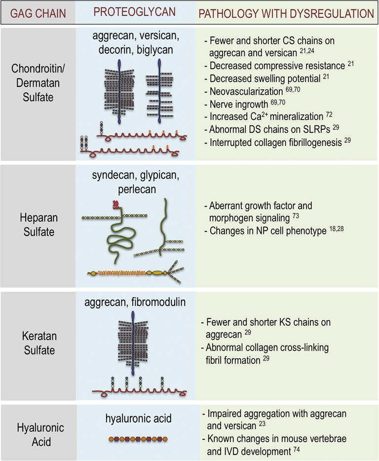
Pathologies associated with dysregulation of GAG synthesis in the intervertebral disc.
5. Conclusion and future studies
If the loss of expression or dysregulation of GAG biosynthesis enzymes represents an initiating factor in disc degeneration, then they represent possible therapeutic targets. Synthetic PGs have been explored for disc regeneration to restore biomechanical function. Most of these PG biomimetics attempt to replicate the functional properties of aggrecan without being vulnerable to proteolytic degradation. Aggrecan mimetics have already been developed for use in the field of articular cartilage repair. One such mimetic consists of CS chains functionalized with HA-binding sites. In model systems, this analog is resistant to proteolysis and is capable of up-regulating aggrecan production and down-regulating metalloproteinase production [80]. Another promising disc biomimetic is a GAG analog formed by polymerizing sulfonate groups in the presence of a crosslinking agent, providing the synthetic molecule with osmotic properties similar to the aggrecan molecule [81]. Importantly, the analog monomers are injected into the disc and polymerized in situ in a process that does not impair cell viability.
However, the complications associated with administering therapeutics into the disc space remains. At this point, it is not possible to deliver therapeutic mimetics to the disc in a way that does not cause structural damage; it has been shown that needle puncture alone causes disc degeneration [82]. For this reason, gene therapy is a promising alternative for the future of disc therapy. In an encouraging study, Venkatesan and colleagues have demonstrated that GlcAT-I gene delivery in cartilage explants provided recovery of PG synthesis and GAG content by increasing the number of CS chains [83]. They indicated that GlcAT-I gene therapy has a potential to treat patients suffering with osteoarthritis by targeting the biosynthetic pathway of GAGs and PGs. Perhaps administration of a transgene capable of stimulating GAG biosynthesis- such as XT-I, GlcAT-I, or ChSy/ChPF highlighted in this review- may provide a promising strategy to reverse the insidious loss of ECM molecules associated with the progression of disc degeneration.
Highlights.
Biomechanical function of the intervertebral disc is directly linked to the integrity of its extracellular matrix
Environmental factors unique to the intervertebral disc control expression of enzymes involved in GAG biosynthesis
Dysregulation of GAG biosynthesis may contribute to intervertebral disc degeneration through the loss of proteoglycan function
Acknowledgments
This work was supported by grants from the National Institutes of Health AR055655 and AR064733
Abbreviations
- NP
Nucleus Pulposus
- AF
Annulus Fibrosus
- CEP
Cartilaginous Endplate
- ECM
Extracellular Matrix
- PG
Proteoglycan
- CS/DS
Chondroitin Sulfate/Dermatan Sulfate
- HIF
Hypoxia Inducible Factor
- HS
Heparan Sulfate
- HA
Hyaluronic Acid
- GlcAT-I
β-1,3-Glucuronosyltransferase 3
- KS
Keratan-Sulfate
- MAPK
Mitogen Activated Protein Kinase
- XT-1
Xylosyltransferase 1
- TonEBP
Tonicity Enhancing Binding Protein
- ChSy
Chondroitin Synthase
- ChPF
Chondroitin Polymerizing Factor
- TGF-β
Transforming Growth Factor β
- SLRP
Small Leucine-rich Repeat Proteoglycan
- BMP-2
Bone Morphogenetic Protein 2
- Smad3
Mothers Against Decapentaplegic Homolog 3
Footnotes
Publisher's Disclaimer: This is a PDF file of an unedited manuscript that has been accepted for publication. As a service to our customers we are providing this early version of the manuscript. The manuscript will undergo copyediting, typesetting, and review of the resulting proof before it is published in its final citable form. Please note that during the production process errors may be discovered which could affect the content, and all legal disclaimers that apply to the journal pertain.
Conflict of Interest: none to declare
References
- 1.Shapiro IM, Vresilovic EJ, Risbud MV. Is the spinal motion segment a diarthrodial polyaxial joint: What a nice nucleus like you doing in a joint like this? Bone. 2012;50:771–776. doi: 10.1016/j.bone.2011.12.004. [DOI] [PMC free article] [PubMed] [Google Scholar]
- 2.Roberts S, Evans H, Trivedi J, Menage J. Histology and pathology of the human intervertebral disc. J Bone Joint Surg Am. 2006;88(Suppl 2):10–14. doi: 10.2106/JBJS.F.00019. [DOI] [PubMed] [Google Scholar]
- 3.Risbud MV, Shapiro IM. Role of cytokines in intervertebral disc degeneration: pain and disc content. Nat Rev Rheumatol. 2014;10:44–56. doi: 10.1038/nrrheum.2013.160. [DOI] [PMC free article] [PubMed] [Google Scholar]
- 4.Le Maitre CL, Pockert AP, Buttle DJ, Freemont AJ, Hoyland JA. Matrix synthesis and degradation in human intervertebral disc degeneration. Biochem Soc Trans. 2007;35:652–5. doi: 10.1042/BST0350652. [DOI] [PubMed] [Google Scholar]
- 5.Vo NV, Hartman RA, Patil PR, Risbud MV, Kletsas D, Iatridis JC, Hoyland JA, Le Maitre CL, Sowa GA, Kang JD. Molecular mechanisms of biological aging in intervertebral discs. J Orthop Res. 2016;34:1289–1306. doi: 10.1002/jor.23195. [DOI] [PMC free article] [PubMed] [Google Scholar]
- 6.Katz JN. Lumbar disc disorders and low-back pain: socioeconomic factors and consequences. J Bone Joint Surg Am. 2006;88(Suppl 2):21–24. doi: 10.2106/JBJS.E.01273. [DOI] [PubMed] [Google Scholar]
- 7.Miller JA, Schmatz C, Schultz AB. Lumbar disc degeneration: correlation with age, sex, and spine level in 600 autopsy specimens. Spine (Phila Pa 1976) 1988;13:173–8. [PubMed] [Google Scholar]
- 8.Rudert M, Tillmann B. Lymph and blood supply of the human intervertebral disc. Cadaver study of correlations to discitis. Acta Orthop Scand. 1993;64:37. doi: 10.3109/17453679308994524. [DOI] [PubMed] [Google Scholar]
- 9.Risbud MV, Guttapalli A, Stokes DG, Hawkins D, Danielson KG, Schaer TP, Albert TJ, Shapiro IM. Nucleus pulposus cells express HIF-1 alpha under normoxic culture conditions: a metabolic adaptation to the intervertebral disc microenvironment. J Cell Biochem. 2006;98:152–159. doi: 10.1002/jcb.20765. [DOI] [PubMed] [Google Scholar]
- 10.Agrawal A, Guttapalli A, Narayan S, Albert TJ, Shapiro IM, Risbud MV. Normoxic stabilization of HIF-1alpha drives glycolytic metabolism and regulates aggrecan gene expression in nucleus pulposus cells of the rat intervertebral disk. Am J Physiol Cell Physiol. 2007;293:C621–C631. doi: 10.1152/ajpcell.00538.2006. [DOI] [PubMed] [Google Scholar]
- 11.Fujita N, Chiba K, Shapiro IM, Risbud MV. HIF-1alpha and HIF-2alpha degradation is differentially regulated in nucleus pulposus cells of the intervertebral disc. J Bone Min Res. 2012;27:401–412. doi: 10.1002/jbmr.538. [DOI] [PMC free article] [PubMed] [Google Scholar]
- 12.Ishihara H, Warensjo K, Roberts S, Urban JP. Proteoglycan synthesis in the intervertebral disk nucleus: the role of extracellular osmolality. Am J Physiol. 1997;272:C1499–C1506. doi: 10.1152/ajpcell.1997.272.5.C1499. [DOI] [PubMed] [Google Scholar]
- 13.van Dijk B, Potier E, Ito K. Culturing Bovine Nucleus Pulposus Explants by Balancing Medium Osmolarity. Tissue Eng Part C Methods. 2011;17:1089–1096. doi: 10.1089/ten.TEC.2011.0215. [DOI] [PubMed] [Google Scholar]
- 14.Johnson ZI, Shapiro IM, Risbud MV. Extracellular osmolarity regulates matrix homeostasis in the intervertebral disc and articular cartilage: Evolving role of TonEBP. Matrix Biol. 2014;40:10–16. doi: 10.1016/j.matbio.2014.08.014. [DOI] [PMC free article] [PubMed] [Google Scholar]
- 15.Johnson ZI, Shapiro IM, Risbud MV. RNA sequencing reveals a role of TonEBP in regulation of pro-inflammatory genes in response to hyperosmolarity in healthy nucleus pulposus cells: A homeostatic response? J Biol Chem. 2016;291:26686–26697. doi: 10.1074/jbc.M116.757732. [DOI] [PMC free article] [PubMed] [Google Scholar]
- 16.Eyre D. Biochemistry of the Intervertebral Disc. Int Rev Connect Tissue Res. 1979;8:227–291. doi: 10.1016/b978-0-12-363708-6.50012-6. [DOI] [PubMed] [Google Scholar]
- 17.Hayes AJ, Benjamin M, Ralphs JR. Extracellular matrix in development of the intervertebral disc. Matrix Biol. 2001;20:107–121. doi: 10.1016/s0945-053x(01)00125-1. [DOI] [PubMed] [Google Scholar]
- 18.Melrose J, Roughley P. Proteoglycans of the intervertebral disc. Intervertebral Disc Mol Struct Stud Disc Heal Dis. 2014:53–77. [Google Scholar]
- 19.Tian Y, Yuan W, Li J, Wang H, Hunt MG, Liu C, Shapiro IM, Risbud MV. TGFβ regulates Galectin-3 expression through canonical Smad3 signaling pathway in nucleus pulposus cells: implications in intervertebral disc degeneration. Matrix Biol. 2016;50:39–52. doi: 10.1016/j.matbio.2015.11.008. [DOI] [PMC free article] [PubMed] [Google Scholar]
- 20.Kamper M, Hamann N, Prein C, Clausen-Schaumann H, Farkas Z, Aszodi A, Niehoff A, Paulsson M, Zaucke F. Early changes in morphology, bone mineral density and matrix composition of vertebrae lead to disc degeneration in aged collagen IX −/− mice. Matrix Biol. 2016;49:132–143. doi: 10.1016/j.matbio.2015.09.005. [DOI] [PubMed] [Google Scholar]
- 21.Melrose J, Ghosh P, Taylor TK. A comparative analysis of the differential spatial and temporal distributions of the large (aggrecan, versican) and small (decorin, biglycan, fibromodulin) proteoglycans of the intervertebral disc. J Anat. 2001;198:3–15. doi: 10.1046/j.1469-7580.2001.19810003.x. [DOI] [PMC free article] [PubMed] [Google Scholar]
- 22.Sivan SS, Wachtel E, Roughley P. Structure, function, aging and turnover of aggrecan in the intervertebral disc. Biochim Biophys Acta - Gen Subj. 2014;1840:3181–3189. doi: 10.1016/j.bbagen.2014.07.013. [DOI] [PubMed] [Google Scholar]
- 23.Zhang L. Chapter -1 Glycosaminoglycan (GAG) Biosynthesis and GAG-Binding Proteins. Mol Biol Transl Sci Glycosaminoglycans Dev Heal Dis. 2010;93:1–17. doi: 10.1016/S1877-1173(10)93001-9. [DOI] [PubMed] [Google Scholar]
- 24.Mörgelin M, Paulsson M, Hardingham TE, Heinegård D, Engel J. Cartilage proteoglycans. Assembly with hyaluronate and link protein as studied by electron microscopy. Biochem J. 1988;253:175–185. doi: 10.1042/bj2530175. [DOI] [PMC free article] [PubMed] [Google Scholar]
- 25.Sztrolovics R, Grover J, Cs-Szabo G, Shi SL, Zhang Y, Mort JS, Roughley PJ. The characterization of versican and its message in human articular cartilage and intervertebral disc. J Orthop Res. 2002;20:257–266. doi: 10.1016/S0736-0266(01)00110-3. [DOI] [PubMed] [Google Scholar]
- 26.Wight TN. Provisional matrix: A role for versican and hyaluronan. Matrix Biol. 2017;60–61:38–56. doi: 10.1016/j.matbio.2016.12.001. [DOI] [PMC free article] [PubMed] [Google Scholar]
- 27.Mwale F, Roughley P, Antoniou J, Alini M, Hollander A, Kirsch T, Stokes I. Distinction between the extracellular matrix of the nucleus pulposus and hyaline cartilage: A requisite for tissue engineering of intervertebral disc. Eur Cells Mater. 2004;8:58–64. doi: 10.22203/ecm.v008a06. [DOI] [PubMed] [Google Scholar]
- 28.Roberts S. Disc morphology in health and disease. Biochem Soc Trans. 2002;30:864–869. doi: 10.1042/bst0300864. [DOI] [PubMed] [Google Scholar]
- 29.Eyre DR, Muir H. Types I and II collagens in intervertebral disc. Interchanging radial distributions in annulus fibrosus. Biochem J. 1976;157:267–270. doi: 10.1042/bj1570267. [DOI] [PMC free article] [PubMed] [Google Scholar]
- 30.Binch ALA, Shapiro IM, Risbud MV. Syndecan-4 in intervertebral disc and cartilage: Saint or synner? Matrix Biol. 2016;52–54:355–362. doi: 10.1016/j.matbio.2016.01.005. [DOI] [PMC free article] [PubMed] [Google Scholar]
- 31.Iozzo RV, Schaefer L. Proteoglycan form and function: A comprehensive nomenclature of proteoglycans. Matrix Biol. 2015;42:11–55. doi: 10.1016/j.matbio.2015.02.003. [DOI] [PMC free article] [PubMed] [Google Scholar]
- 32.Paracuellos P, Kalamajski S, Bonna A, Bihan D, Farndale RW, Hohenester E. Structural and functional analysis of two small leucine-rich repeat proteoglycans, fibromodulin and chondroadherin. Matrix Biol. 2017;63:106–116. doi: 10.1016/j.matbio.2017.02.002. [DOI] [PMC free article] [PubMed] [Google Scholar]
- 33.Jackson RL, Busch SJ, Cardin AD. Glycosaminoglycans: molecular properties, protein interactions, and role in physiological processes. Physiol Rev. 1991;71:481–539. doi: 10.1152/physrev.1991.71.2.481. [DOI] [PubMed] [Google Scholar]
- 34.Hayes AJ, Hughes CE, Ralphs JR, Caterson B. Chondroitin sulphate sulphation motif expression in the ontogeny of the intervertebral disc. Eur Cells Mater. 2011;21:1–14. [PubMed] [Google Scholar]
- 35.Kuhn J, Götting C, Schnölzer M, Kempf T, Brinkmann T, Kleesiek K. First Isolation of Human UDP-D-Xylose: Proteoglycan Core Protein β-D-Xylosyltransferase Secreted from Cultured JAR Choriocarcinoma Cells. J Biol Chem. 2001;276:4940–4947. doi: 10.1074/jbc.M005111200. [DOI] [PubMed] [Google Scholar]
- 36.Venkatesan N, Barré L, Magdalou J, Mainard D, Netter P, Fournel-Gigleux S, Ouzzine M. Modulation of xylosyltransferase I expression provides a mechanism regulating glycosaminoglycan chain synthesis during cartilage destruction and repair. FASEB J. 2009;23:813–22. doi: 10.1096/fj.08-118166. [DOI] [PubMed] [Google Scholar]
- 37.Prante C, Milting H, Kassner A, Farr M, Ambrosius M, Schön S, Seidler DG, El Banayosy A, Körfer R, Kuhn J, Kleesiek K, Götting C. Transforming growth factor beta1-regulated xylosyltransferase I activity in human cardiac fibroblasts and its impact for myocardial remodeling. J Biol Chem. 2007;282:26441–9. doi: 10.1074/jbc.M702299200. [DOI] [PubMed] [Google Scholar]
- 38.Venkatesan N, Barré L, Bourhim M, Magdalou J, Mainard D, Netter P, Fournel-Gigleux S, Ouzzine M. Xylosyltransferase-i regulates glycosaminoglycan synthesis during the pathogenic process of human osteoarthritis. PLoS One. 2012;7 doi: 10.1371/journal.pone.0034020. [DOI] [PMC free article] [PubMed] [Google Scholar]
- 39.Faust I, Roch C, Kuhn J, Prante C, Knabbe C, Hendig D. Human xylosyltransferase-I - A new marker for myofibroblast differentiation in skin fibrosis. Biochem Biophys Res Commun. 2013;436:449–454. doi: 10.1016/j.bbrc.2013.05.125. [DOI] [PubMed] [Google Scholar]
- 40.Khair M, Bourhim M, Barré L, Li D, Netter P, Magdalou J, Fournel-Gigleux S, Ouzzine M. Regulation of xylosyltransferase i gene expression by interleukin 1β in human primary chondrocyte cells: Mechanism and impact on proteoglycan synthesis. J Biol Chem. 2013;288:1774–1784. doi: 10.1074/jbc.M112.419887. [DOI] [PMC free article] [PubMed] [Google Scholar]
- 41.Müller B, Prante C, Kleesiek K, Götting C. Identification and characterization of the human xylosyltransferase I gene promoter region. J Biol Chem. 2009;284:30775–30782. doi: 10.1074/jbc.M109.016592. [DOI] [PMC free article] [PubMed] [Google Scholar]
- 42.Ye W, Zhou J, Markova DZ, Tian Y, Li J, Anderson DG, Shapiro IM, Risbud MV. Xylosyltransferase-1 expression is refractory to inhibition by the inflammatory cytokines tumor necrosis factor α and IL-1β in nucleus pulposus cells: Novel regulation by AP-1, Sp1, and Sp3. Am J Pathol. 2014;185:485–495. doi: 10.1016/j.ajpath.2014.09.021. [DOI] [PMC free article] [PubMed] [Google Scholar]
- 43.Hiyama A, Gajghate S, Sakai D, Mochida J, Shapiro IM, Risbud MV. Activation of TonEBP by calcium controls β1,3-glucuronosyltransferase-I expression, a key regulator of glycosaminoglycan synthesis in cells of the intervertebral disc. J Biol Chem. 2009;284:9824–9834. doi: 10.1074/jbc.M807081200. [DOI] [PMC free article] [PubMed] [Google Scholar]
- 44.Hiyama A, Gogate SS, Gajghate S, Mochida J, Shapiro IM, Risbud MV. BMP-2 and TGF-β stimulate expression of β 1,3-glucuronosyl transferase 1 (GlcAT-1) in nucleus pulposus cells through AP1, TonEBP, and Sp1: Role of MAPKs. J Bone Miner Res. 2010;25:1179–1190. doi: 10.1359/jbmr.091202. [DOI] [PMC free article] [PubMed] [Google Scholar]
- 45.Wu Q, Wang J, Skubutyte R, Kepler CK, Huang Z, Anderson DG, Shapiro IM, Risbud MV. Smad3 controls beta-1,3-glucuronosyltransferase 1 expression in rat nucleus pulposus cells: implications of dysregulated expression in disc disease. Arthritis Rheum. 2012;64:3324–3333. doi: 10.1002/art.34570. [DOI] [PMC free article] [PubMed] [Google Scholar]
- 46.Gogate SS, Nasser R, Shapiro IM, Risbud MV. Hypoxic regulation of β-1,3-glucuronyltransferase 1 expression in nucleus pulposus cells of the rat intervertebral disc: Role of hypoxia-inducible factor proteins. Arthritis Rheum. 2011;63:1950–1960. doi: 10.1002/art.30342. [DOI] [PMC free article] [PubMed] [Google Scholar]
- 47.Alford AI, Yellowley CE, Jacobs CR, Donahue HJ. Increases in Cytosolic Calcium, but not Fluid Flow, Affect Aggrecan mRNA Levels in Articular Chondrocytes. J Cell Biochem. 2003;90:938–944. doi: 10.1002/jcb.10715. [DOI] [PubMed] [Google Scholar]
- 48.Parvizi J, Parpura V, Greenleaf JF, Bolander ME. Calcium signaling is required for ultrasound-stimulated aggrecan synthesis by rat chondrocytes. J Orthop Res. 2002;20:51–57. doi: 10.1016/S0736-0266(01)00069-9. [DOI] [PubMed] [Google Scholar]
- 49.Fagnen G, Phamantu NT, Bocquet J, Bonnamy PJ. Activation of protein kinase C increases proteoglycan synthesis in immature rat Sertoli cells. Biochim Biophys Acta - Gen Subj. 1999;1472:250–261. doi: 10.1016/s0304-4165(99)00128-2. [DOI] [PubMed] [Google Scholar]
- 50.Tsai TT, Danielson KG, Guttapalli A, Oguz E, Albert TJ, Shapiro IM, Risbud MV. TonEBP/OREBP Is a Regulator of Nucleus Pulposus Cell Function and Survival in the Intervertebral Disc. J Biol Chem. 2006;281:25416–25424. doi: 10.1074/jbc.M601969200. [DOI] [PubMed] [Google Scholar]
- 51.Moustakas A, Heldin CHH. The regulation of TGFbeta signal transduction. Development. 2009;136:3699–714. doi: 10.1242/dev.030338. [DOI] [PubMed] [Google Scholar]
- 52.Risbud MV, Di Martino A, Guttapalli A, Seghatoleslami R, Denaro V, Vaccaro AR, Albert TJ, Shapiro IM. Toward an optimum system for intervertebral disc organ culture: TGF-beta3 enhances nucleus pulposus and anulus fibrosus survival and function through modulation of TGF-B-R expression and ERK signaling. Spine (Phila Pa 1976) 2006;31:884–890. doi: 10.1097/01.brs.0000209335.57767.b5. [DOI] [PubMed] [Google Scholar]
- 53.Nerlich AG, Bachmeier BE, Boos N. Expression of fibronectin and TGF-β1 mRNA and protein suggest altered regulation of extracellular matrix in degenerated disc tissue. Eur Spine J. 2005;14:17–26. doi: 10.1007/s00586-004-0745-x. [DOI] [PMC free article] [PubMed] [Google Scholar]
- 54.Murakami H, Yoon ST, Attallah-Wasif ES, Tsai KJ, Fei Q, Hutton WC. The expression of anabolic cytokines in intervertebral discs in age-related degeneration. Spine (Phila Pa 1976) 2006;31:1770–1774. doi: 10.1097/01.brs.0000227255.39896.f3. [DOI] [PubMed] [Google Scholar]
- 55.Ogawa H, Hatano S, Sugiura N, Nagai N, Sato T, Shimizu K, Kimata K, Narimatsu H, Watanabe H. Chondroitin Sulfate Synthase-2 Is Necessary for Chain Extension of Chondroitin Sulfate but Not Critical for Skeletal Development. PLoS One. 2012;7:e43806. doi: 10.1371/journal.pone.0043806. [DOI] [PMC free article] [PubMed] [Google Scholar]
- 56.Hu B, Shi C, Tian Y, Zhang Y, Xu C, Chen H, Cao P, Yuan W. TGF-β Induces Up-Regulation of Chondroitin Sulfate Synthase 1 (CHSY1) in Nucleus Pulposus Cells Through MAPK Signaling. Cell Physiol Biochem. 2015;37:793–804. doi: 10.1159/000430396. [DOI] [PubMed] [Google Scholar]
- 57.Hu B, Xu C, Cao P, Tian Y, Zhang Y, Shi C, Xu J, Yuan W, Chen H. TGF-β Stimulates Expression of Chondroitin Polymerizing Factor in Nucleus Pulposus Cells Through the Smad3, RhoA/ROCK1, and MAPK Signaling Pathways. J Cell Biochem. 2018;119:566–579. doi: 10.1002/jcb.26215. [DOI] [PubMed] [Google Scholar]
- 58.Hu B, Xu C, Tian Y, Shi C, Zhang Y, Deng L, Zhou H, Cao P, Chen H, Yuan W. Inflammatory microRNA-194 and -515 attenuate the biosynthesis of chondroitin sulfate during human intervertebral disc degeneration. Oncotarget. 2017;8:49303–49317. doi: 10.18632/oncotarget.17571. [DOI] [PMC free article] [PubMed] [Google Scholar]
- 59.Tengblad A, Pearce RH, Grimmer BJ. Demonstration of link protein in proteoglycan aggregates from human intervertebral disc. Biochem J. 1984;222:85–92. doi: 10.1042/bj2220085. [DOI] [PMC free article] [PubMed] [Google Scholar]
- 60.Roughley P, Martens D, Rantakokko J, Alini M, Mwale F, Antoniou J. The involvement of aggrecan polymorphism in degeneration of human intervertebral disc and articular cartilage. Eur Cell Mater. 2006;11:1–7. discussion 7. [PubMed] [Google Scholar]
- 61.Collin EC, Carroll O, Kilcoyne M, Peroglio M, See E, Hendig D, Alini M, Grad S, Pandit A. Ageing affects chondroitin sulfates and their synthetic enzymes in the intervertebral disc. Signal Transduct Target Ther. 2017;2:17049. doi: 10.1038/sigtrans.2017.49. [DOI] [PMC free article] [PubMed] [Google Scholar]
- 62.Song Y-Q, Karasugi T, Cheung KM, Chiba K, Ho DW, Miyake A, Kao PY, Sze KLing, Yee A, Takahashi A, Kawaguchi Y, Mikami Y, Matsumoto M, Wu C, Tian W, Wang N, Leong JC, Luk KD, Yip S, Cherny SS, Wang J, Kubo M, Kimura T, Kubo T, Toyama Y, Mizuta H, Cheah KS, Tsunoda T, Sham P-C, Ikegawa S, Chan D. Lumbar disc degeneration is linked to a carbohydrate sulfotransferase 3 variant. J Clin Investig Jaro Karppinen Marjo-Riitta Järvelin. 2013;12321 doi: 10.1172/JCI69277. [DOI] [PMC free article] [PubMed] [Google Scholar]
- 63.Tsai TT, Ho NYJ, Fang HC, Lai PL, Niu CC, Chen LH, Chen WJ, Pang JHS. Increased sulfatase 1 gene expression in degenerative intervertebral disc cells. J Orthop Res. 2015;33:312–317. doi: 10.1002/jor.22766. [DOI] [PubMed] [Google Scholar]
- 64.Yasuda T, Mundy C, Kinumatsu T, Shibukawa Y, Shibutani T, Grobe K, Minugh-Purvis N, Pacifici M, Koyama E. Sulfotransferase Ndst1 is Needed for Mandibular and TMJ Development. J Dent Res. 2010;89:1111–1116. doi: 10.1177/0022034510373766. [DOI] [PMC free article] [PubMed] [Google Scholar]
- 65.Armstrong L, Tarailo-Graovac M, Sinclair G, Seath KI, Wasserman WW, Ross CJ, van Karnebeek CDM. A girl with developmental delay, ataxia, cranial nerve palsies, severe respiratory problems in infancy-Expanding NDST1 syndrome. Am J Med Genet Part A. 2017;173:712–715. doi: 10.1002/ajmg.a.37621. [DOI] [PubMed] [Google Scholar]
- 66.Reynard LN, Ratnayake M, Santibanez-Koref M, Loughlin J. Functional Characterization of the Osteoarthritis Susceptibility Mapping to CHST11-A Bioinformatics and Molecular Study. PLoS One. 2016;11:e0159024. doi: 10.1371/journal.pone.0159024. [DOI] [PMC free article] [PubMed] [Google Scholar]
- 67.Wilson DG, Phamluong K, Lin WY, Barck K, Carano RAD, Diehl L, Peterson AS, Martin F, Solloway MJ. Chondroitin sulfate synthase 1 (Chsy1) is required for bone development and digit patterning. Dev Biol. 2012;363:413–425. doi: 10.1016/j.ydbio.2012.01.005. [DOI] [PubMed] [Google Scholar]
- 68.Tian J, Ling L, Shboul M, Lee H, O’Connor B, Merriman B, Nelson SF, Cool S, Ababneh OH, Al-Hadidy A, Masri A, Hamamy H, Reversade B. Loss of CHSY1, a secreted FRINGE enzyme, causes syndromic brachydactyly in humans via increased NOTCH signaling. Am J Hum Genet. 2010;87:768–778. doi: 10.1016/j.ajhg.2010.11.005. [DOI] [PMC free article] [PubMed] [Google Scholar]
- 69.Wuyts W, Van Hul W, De Boulle K, Hendrickx J, Bakker E, Vanhoenacker F, Mollica F, Lüdecke HJ, Sayli BS, Pazzaglia UE, Mortier G, Hamel B, Conrad EU, Matsushita M, Raskind WH, Willems PJ. Mutations in the EXT1 and EXT2 genes in hereditary multiple exostoses. Am J Hum Genet. 1998;62:346–54. doi: 10.1086/301726. [DOI] [PMC free article] [PubMed] [Google Scholar]
- 70.Sgariglia F, Candela ME, Huegel J, Jacenko O, Koyama E, Yamaguchi Y, Pacifici M, Enomoto-Iwamoto M. Epiphyseal abnormalities, trabecular bone loss and articular chondrocyte hypertrophy develop in the long bones of postnatal Ext1-deficient mice. Bone. 2013;57:220–31. doi: 10.1016/j.bone.2013.08.012. [DOI] [PMC free article] [PubMed] [Google Scholar]
- 71.Jochmann K, Bachvarova V, Vortkamp A. Heparan sulfate as a regulator of endochondral ossification and osteochondroma development. Matrix Biol. 2014;34:55–63. doi: 10.1016/j.matbio.2013.11.003. [DOI] [PubMed] [Google Scholar]
- 72.Johnson WEB, Caterson B, Eisenstein SM, Roberts S. Human intervertebral disc aggrecan inhibits endothelial cell adhesion and cell migration in vitro. Spine (Phila Pa 1976) 2005;30:1139–1147. doi: 10.1097/01.brs.0000162624.95262.73. [DOI] [PubMed] [Google Scholar]
- 73.Johnson WEB, Caterson B, Eisenstein SM, Hynds DL, Snow DM, Roberts S. Human intervertebral disc aggrecan inhibits nerve growth in vitro. Arthritis Rheum. 2002;46:2658–64. doi: 10.1002/art.10585. [DOI] [PubMed] [Google Scholar]
- 74.Feng C, Yang M, Lan M, Liu C, Zhang Y, Huang B, Liu H, Zhou Y. ROS: Crucial Intermediators in the Pathogenesis of Intervertebral Disc Degeneration. Oxid Med Cell Longev. 2017;2017:5601593. doi: 10.1155/2017/5601593. [DOI] [PMC free article] [PubMed] [Google Scholar]
- 75.Hristova GI, Jarzem P, Ouellet JA, Roughley PJ, Epure LM, Antoniou J, Mwale F. Calcification in human intervertebral disc degeneration and scoliosis. J Orthop Res. 2011;29:1888–1895. doi: 10.1002/jor.21456. [DOI] [PubMed] [Google Scholar]
- 76.Fujita N, Hirose Y, Tran CM, Chiba K, Miyamoto T, Toyama Y, Shapiro IM, Risbud MV. HIF-1-PHD2 axis controls expression of syndecan 4 in nucleus pulposus cells. FASEB J. 2014;28:2455–2465. doi: 10.1096/fj.13-243741. [DOI] [PMC free article] [PubMed] [Google Scholar]
- 77.Robinson KA, Sun M, Barnum CE, Weiss SN, Huegel J, Shetye SS, Lin L, Saez D, Adams SM, Iozzo RV, Soslowsky LJ, Birk DE. Decorin and biglycan are necessary for maintaining collagen fibril structure, fiber realignment, and mechanical properties of mature tendons. Matrix Biol. 2017;64:81–93. doi: 10.1016/j.matbio.2017.08.004. [DOI] [PMC free article] [PubMed] [Google Scholar]
- 78.Huang Y, Askew EB, Knudson CB, Knudson W. CRISPR/Cas9 knockout of HAS2 in rat chondrosarcoma chondrocytes demonstrates the requirement of hyaluronan for aggrecan retention. Matrix Biol. 2016;56:74–94. doi: 10.1016/j.matbio.2016.04.002. [DOI] [PMC free article] [PubMed] [Google Scholar]
- 79.Roughley PJ, Lamplugh L, Lee ER, Matsumoto K, Yamaguchi Y. The Role of Hyaluronan Produced by Has2 Gene Expression in Development of the Spine. Spine (Phila Pa 1976) 2011;36:E914–E920. doi: 10.1097/BRS.0b013e3181f1e84f. [DOI] [PubMed] [Google Scholar]
- 80.Sharma S, Lee A, Choi K, Kim K, Youn I, Trippel SB, Panitch A. Biomimetic aggrecan reduces cartilage extracellular matrix from degradation and lowers catabolic activity in ex vivo and in vivo models. Macromol Biosci. 2013;13:1228–1237. doi: 10.1002/mabi.201300112. [DOI] [PubMed] [Google Scholar]
- 81.Sivan SS, Roberts S, Urban JPG, Menage J, Bramhill J, Campbell D, Franklin VJ, Lydon F, Merkher Y, Maroudas A, Tighe BJ. Injectable hydrogels with high fixed charge density and swelling pressure for nucleus pulposus repair: Biomimetic glycosaminoglycan analogues. Acta Biomater. 2014;10:1124–1133. doi: 10.1016/j.actbio.2013.11.010. [DOI] [PubMed] [Google Scholar]
- 82.Martin JT, Gorth DJ, Beattie EE, Harfe BD, Smith LJ, Elliott DM. Needle puncture injury causes acute and long-term mechanical deficiency in a mouse model of intervertebral disc degeneration. J Orthop Res. 2013;31:1276–1282. doi: 10.1002/jor.22355. [DOI] [PMC free article] [PubMed] [Google Scholar]
- 83.Venkatesan N, Barre L, Benani A, Netter P, Magdalou J, Fournel-Gigleux S, Ouzzine M. Stimulation of proteoglycan synthesis by glucuronosyltransferase-I gene delivery: A strategy to promote cartilage repair. Proc Natl Acad Sci. 2004;101:18087–18092. doi: 10.1073/pnas.0404504102. [DOI] [PMC free article] [PubMed] [Google Scholar]