


Figure 2. Overview of some dendritic spine signaling pathways of importance to this review.
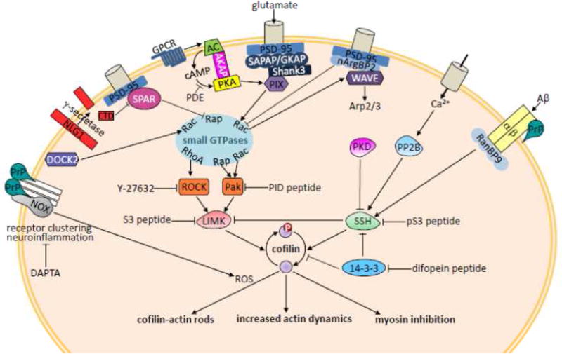
Cofilin is inhibited in binding to actin by phosphorylation and is activated by dephosphorylation. Active cofilin can sever F-actin, or bind cooperatively to saturate and stabilize pieces of F-actin, as elaborated in Figure 1. Cofilin binding to F-actin also competes with myosin II, inhibiting its contractile activity. The sites of inhibition by the cofilin-derived S3 and phosphoS3 (pS3) peptides are shown. LIM kinases (LIMK) and slingshot phosphatases (SSH) that regulate cofilin are themselves subject to regulation in response to many different receptors in dendritic spines, only some of which are shown here. The C-terminal domain (CTD) of NLG1 is generated by the activity of the same protease (γ-secretase) that cleaves APP to generate Aβ in AD. Abbreviations used in this figure and not defined elsewhere are: Pak-interacting exchange factor β (PIX), an activator of Rac1 and Rap1; Pak inhibitory domain (PID); G-protein coupled receptor (GPCR), which, when activated, stimulates nucleotide exchange on the alpha subunit of a heterotrimeric G protein, stimulating adenylcyclase (AC) to make cyclic AMP (cAMP), which activates protein kinase A (PKA) anchored via A kinase anchoring protein (AKAP), but whose response is limited by lowering cAMP via activation of a phosphodiesterase (PDE); ionotropic glutamate receptors and signaling proteins are organized by multidomain scaffolding proteins including postsynaptic density protein 95 (PSD-95), and synapse-associated protein PSD-95-associated protein/guanylate kinase-associated protein (SAPAP/GKAP); protein kinase D1 (PKD) phosphorylates and inactivates SSH; RanBP9 interacts with integrin dimers that contain the β1 subunit (α/β1).