



Abstract
Cofilin is a ubiquitous protein which cooperates with many other actin-binding proteins in regulating actin dynamics. Cofilin has essential functions in nervous system development including neuritogenesis, neurite elongation, growth cone pathfinding, dendritic spine formation, and the regulation of neurotransmission and spine function, components of synaptic plasticity essential for learning and memory. Cofilin’s phosphoregulation is a downstream target of many transmembrane signaling processes, and its misregulation in neurons has been linked in rodent models to many different neurodegenerative and neurological disorders including Alzheimer disease (AD), aggression due to neonatal isolation, autism, manic/bipolar disorder, and sleep deprivation. Cognitive and behavioral deficits of these rodent models have been largely abrogated by modulation of cofilin activity using viral-mediated, genetic, and/or small molecule or peptide therapeutic approaches. Neuropathic pain in rats from sciatic nerve compression has also been reduced by modulating the cofilin pathway within neurons of the dorsal root ganglia. Neuroinflammation, which occurs following cerebral ischemia/reperfusion, but which also accompanies many other neurodegenerative syndromes, is markedly reduced by peptides targeting specific chemokine receptors, which also modulate cofilin activity. Thus, peptide therapeutics offer potential for cost-effective treatment of a wide variety of neurological disorders. Here we discuss some recent results from rodent models using therapeutic peptides with a surprising ability to cross the rodent blood brain barrier and alter cofilin activity in brain. We also offer suggestions as to how neuronal-specific cofilin regulation might be achieved.
Keywords: dendritic spines, cofilin phosphoregulation, cognitive disorders, psychiatric disorders, neuropathic pain, sleep deprivation, rodent models
1. Introduction
Neurological and neurodegenerative disorders leading to cognitive and/or behavioral deficits are among the most devastating in terms of their financial impact on the economy and their disruption of family life. Alzheimer disease (AD) alone has a very high prevalence, and is currently listed as the 6th leading cause of death for all Americans, despite underreporting. Currently 5.4 million Americans, including 32% of people over the age of 85, are living with the disease. This has a huge economic impact, with payments in 2016 of $236 billion (from Medicare, other insurance providers, as well as out-of-pocket expenditures), plus assistance valued at $221 billion from unpaid caregivers, who provide 83% of care needed by those suffering from AD. By 2050, cases are projected to increase to 13.8 million. Despite this, AD has no pharmacological treatments which can prevent or reverse the loss of synapses and neuronal connectivity that underlies AD symptoms and fatality. Instead, all the current Food and Drug Administration-approved drugs for AD work to bolster cognition by increasing neurotransmitter signaling (Alzheimer’s Association, 2016). For other neurological disorders such as autism and manic/bipolar disorder, there are therapies that can help control symptoms, but none that truly help correct the molecular mechanisms that have been disrupted.
Genetic mutations associated with the less prevalent familial forms of AD result in the overproduction or lack of clearance of the amyloid-β (Aβ) peptide, which accumulates and is eventually deposited in amyloid plaques. Although the cognitive decline of AD subjects is not related directly to plaque burden (Tu et al., 2014), much of the current focus on therapeutics for AD has been on the development of monoclonal antibodies against Aβ or its aggregates, including the drugs aducanumab, gantenerumab, and solanezumab which are currently in phase III clinical trials (Panza et al., 2016; Cavanaugh et al., 2014). Interim results from double blind studies have suggested aducanumab reduces plaques and Aβ in human and AD mouse brain and slows somewhat the cognitive decline in humans (Sevigny et al., 2016). However, solanezumab, was recently reported to have failed in slowing the cognitive decline of patients with mild to moderate AD (Le Couteur et al., 2016). Although monoclonal antibodies have been very effective in some diseases and are important cancer treatments (Scott et al., 2012), their cost must be considered, especially for a chronic disease like AD that affects millions of people. The average annual cost per patient of the top 9 monoclonal antibody drugs was $200,000 in 2012 (Shaughnessy, 2012). At this amount, treating the more than 5 million AD sufferers in the US with monoclonal antibody therapy would cost over 1 trillion dollars per year.
Antibodies are not the only option for biologically-designed disease interventions. Peptide drugs offer opportunities for rational design and specificity, with relatively inexpensive production ($100/g), and a higher than average success rate at clinical trials (26% success rate for peptide drugs vs. 10% for all classes of drugs) (Otvos, 2014; Otvos and Wade, 2014). Although lower development and production costs do not always result in a sustainable cost to the patient (Hartung et al., 2015), currently available peptides drugs, although highly variable in cost, are still significantly less expensive than monoclonal antibody therapy (Otvos, 2014). There are peptide drugs in development for treatment of AD, but the focus continues to be on the disruption of Aβ aggregation (Sun et al., 2012; Funke et al., 2012). Focusing so exclusively on one approach (minimizing Aβ) seems far too risky for such a costly, common, and devastating disease. What about targeting other downstream processes?
2. Cofilin as a Therapeutic Target
The actin cytoskeleton is critical for all aspects of neuronal development, including formation of neurites, directed outgrowth of axons and dendrites, and the formation of synapses. In dendritic spines, the postsynaptic structures on excitatory synapses in the hippocampus and cortex, actin dynamics modulate the insertion of ion channels and the morphological changes associated with long-term potentiation (LTP) and long-term depression (LTD), electrophysiological correlates for memory formation. Morphological and functional alterations in dendritic spines are observed in human subjects and/or genetic mouse models of AD (Calabrese et al., 2006; Rust, 2015; Dorostkar et al., 2015), Down syndrome (Belichenko et al., 2004), Fragile X syndrome (Grossman et al., 2010), schizophrenia and autism (Blundell et al., 2010; Qiao et al., 2014; Foote et al., 2015; Copf, 2016; Liu et al., 2016), sleep deprivation (Havekes et al., 2016), and manic/bipolar disorder (Zhang Q, et al., 2016; Lee et al., 2016).
Cofilin, along with the less-abundant but related protein actin depolymerizing factor (ADF), is a critical regulator of the actin cytoskeleton in many systems, including dendritic spines, the major postsynaptic structures on excitatory synapses in brain. In vitro, ADF and cofilin sever actin filaments, but through their cooperative binding to F-actin, they can also stabilize and saturate short actin filaments (Adrianantoandro and Pollard, 2006; Chan et al., 2009; Chen and Pollard, 2011). Because cofilin is about 12 fold more abundant in neurons than ADF (Garvalov et al., 2007), we will henceforth exclusively refer to cofilin in this review. Dendritic spines undergo dynamic changes during learning and memory (Kennedy, 2016), and cofilin plays a major role in dendritic spine shape and volume (Rust, 2015; Bamburg and Bernstein, 2016; Lei et al., 2016). In vivo cofilin collaborates with many other actin binding proteins to dynamically remodel the actin cytoskeleton into actin meshworks, branched or linear filament arrays, or short F-actin networks, which through assembly dynamics and treadmilling alters membrane structure and organization (Figure 1). Mutations in some of these collaborating proteins or their regulatory factors may lead to developmental, neurological, or psychiatric disorders (Liu, 2011; Moon & Winshaw-Boris, 2013). Activation of cofilin under conditions of oxidative stress may lead to its dimerization and formation of linear bundles of 1:1 cofilin-actin (rods) within neurites, affecting transport and synapse function (Chen and Wang, 2015; Bamburg and Bernstein, 2016). Active cofilin also competes for actin filament binding with myosin II motors to alter contractility (Wiggan et al., 2012; Kanellos et al., 2015; Wiggan et al., 2017). Myosin II, the major myosin form associated with actin filament contraction and tension, is found in spines where it plays a role in maintenance of mushroom-shaped spine heads, as well as in the dynamics of spine protrusion and retraction, and in the clustering of glutamate (AMPA receptor) subunits important for proper spine function (Kneussel & Wagner, 2013). Thus, cofilin regulation of myosin II-F-actin interaction may be as important to spine function as its ability to disassemble and remodel the actin structures within spines. Although the levels of expression and/or regulation of other actin modulatory proteins can mediate alterations in dendritic spine shape and dynamics that occur in many neurodegenerative or neurological disorders, accumulated evidence suggests a central role for cofilin phosphoregulation in spine modulation (Zhou et al., 2004). Although alternative explanations may exist for many of the results reviewed below, our intention is to provide a unifying model rather than many divergent ones to explain the cognitive and behavioral deficits and how these are normalized in the animal models described. Thus, alternative explanations are not explored.
Figure 1. Modulation of actin and dendritic spine morphology by cofilin and ancillary proteins.
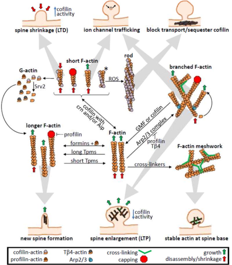
Actin dynamics and network formation depend upon nucleation factors, cross-linkers and G-actin subunit availability. Nucleation factors in spines include formins, involved in linear filament growth such as filopodial extensions for initial spine formation, and the Arp2/3 complex, which nucleates branched filament networks (Spence and Soderling, 2015). The role of cross-linkers, such as filamin, drebrin, and α-actinin, and the cross-linking and contractile motor myosin II, in creating the actin meshworks that contribute to spine morphology is still being elucidiated (Ivanov et al., 2009; Korobova & Svitkina, 2010; Chazeau & Giannone, 2016; Segura et al., 2016; Kneussel & Wagner, 2013). In addition to changes in dendritic spine shape and volume during LTP and LTD (structural plasticity), changes in ion channel surface expression and activity (functional plasticity) mediate synaptic transmission efficiency. Both processes rely on regulation of the actin cytoskeleton and myosin motors (Kneussel & Wagner, 2013; Spence & Soderling, 2015; Lei et al., 2016; Chazeau & Giannone, 2016). Cofilin works in concert with many other proteins to regulate actin filament dynamics, most notably as a factor to promote actin severing and depolymerization, allowing spine shrinkage for LTD, although cofilin activity may be transiently required for actin polymerization-dependent processes such as LTP (Gu et al., 2010). At saturating concentrations (*) cofilin promotes stabilization of F-actin but inhibits myosin II from binding actin and thus modulates myosin II-mediated contractile activity, which is necessary for synaptic functions including ion channel trafficking. In neurons undergoing oxidative stress, cofilin-saturated filaments can bundle into rods, sequestering cofilin and compromising synaptic function (Bamburg & Bernstein, 2016). Cofilin can also remove Arp2/3 complex-branched filament networks (Chan et al., 2009), although a cofilin-related protein (glial maturation factor, GMF) has evolved with greater efficiency in this function (Ydenberg et al., 2013; Poukkula et al., 2014). Cofilin competes for F-actin binding with some proteins, such as drebrin (Grintsevich & Reisler, 2014) and long isoforms of tropomyosins (Tpms), but short Tpms allow and may enhance cofilin effects (Bryce et al., 2003; Janco et al., 2016). Other proteins work in conjunction with cofilin to modulate actin dynamics: coronin1b (crn) enhances the recruitment of cofilin to F-actin (Mikati et al., 2015); actin interacting protein 1 (Aip) enhances the severing ability of cofilin and its complete disassembly of F-actin (Ono et al., 2004; Nadkarni and Brieher, 2014; Gressin et al., 2015; Jansen et al., 2015). Srv2 enhances the dissociation of cofilin from cofilin-actin monomers, or it can enhance cofilin-mediated filament depolymerization from both ends of uncapped filaments (Balcer et al., 2003; Chaudhry et al., 2014; Johnston et al., 2015). The source and maintenance of the G-actin pool in spines is less well understood. Monomer pools in brain are composed primarily of profilin-actin and Tβ4-actin complexes (Devineni et al., 1999), with profilin-actin being preferentially used for formin-mediated filament elongation (Lee et al., 2013). Profilin and Tβ4 inhibit Arp2/3 complex nucleation of filaments and thus serve as a gatekeeper for the switch between Arp2/3-branched networks and linear arrays (Suarez et al., 2015; Rotty et al., 2015; Vitriol et al., 2015; Pernier et al., 2016).
3. Cofilin Phosphoregulation
Dephosphorylation of cofilin phospho-ser3 is required for cofilin binding to actin and is mediated by the relatively specific but unrelated phosphatases, slingshot (SSH) and chronophin (CIN) (Huang et al., 2006; Mizuno, 2013) (Figure 2). Mammals express multiple isoforms of SSH (Ohta et al., 2003), one of which changes in expression along with actin dynamics and cofilin phosphorylation during hippocampal development (Lauterborn et al., 2016). Although phospho-cofilin is inactive in binding to actin, it does activate phospholipase D1 leading to production of membrane phosphatidic acid (Han et al., 2007). Such effects of phospho-cofilin are usually not investigated although this activity could dramatically alter transmembrane signaling since some upstream regulators of Rho family GTPases, such as DOCK2, need to bind phosphatidic acid before they can be activated by phosphatidylinositol phosphates to serve as guanine nucleotide exchange factors for Rho family GTPases (Nishikima et al., 2009; Ammar et al., 2014) (Figure 2). Several DOCK family proteins are expressed in neurons (Gadea and Blangy, 2014).
Figure 2. Overview of some dendritic spine signaling pathways of importance to this review.
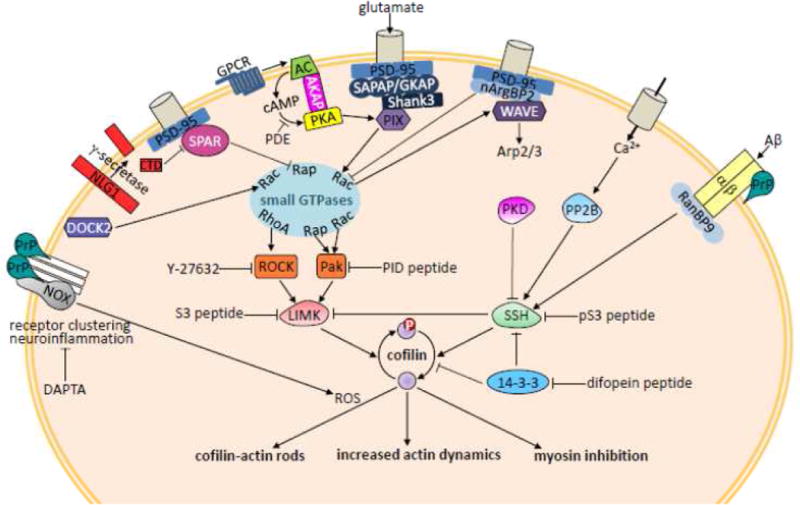
Cofilin is inhibited in binding to actin by phosphorylation and is activated by dephosphorylation. Active cofilin can sever F-actin, or bind cooperatively to saturate and stabilize pieces of F-actin, as elaborated in Figure 1. Cofilin binding to F-actin also competes with myosin II, inhibiting its contractile activity. The sites of inhibition by the cofilin-derived S3 and phosphoS3 (pS3) peptides are shown. LIM kinases (LIMK) and slingshot phosphatases (SSH) that regulate cofilin are themselves subject to regulation in response to many different receptors in dendritic spines, only some of which are shown here. The C-terminal domain (CTD) of NLG1 is generated by the activity of the same protease (γ-secretase) that cleaves APP to generate Aβ in AD. Abbreviations used in this figure and not defined elsewhere are: Pak-interacting exchange factor β (PIX), an activator of Rac1 and Rap1; Pak inhibitory domain (PID); G-protein coupled receptor (GPCR), which, when activated, stimulates nucleotide exchange on the alpha subunit of a heterotrimeric G protein, stimulating adenylcyclase (AC) to make cyclic AMP (cAMP), which activates protein kinase A (PKA) anchored via A kinase anchoring protein (AKAP), but whose response is limited by lowering cAMP via activation of a phosphodiesterase (PDE); ionotropic glutamate receptors and signaling proteins are organized by multidomain scaffolding proteins including postsynaptic density protein 95 (PSD-95), and synapse-associated protein PSD-95-associated protein/guanylate kinase-associated protein (SAPAP/GKAP); protein kinase D1 (PKD) phosphorylates and inactivates SSH; RanBP9 interacts with integrin dimers that contain the β1 subunit (α/β1).
Ser3-phosphorylated cofilin has been found to be associated with various isoforms of the 14-3-3 family of scaffolding proteins. Increasing expression of 14-3-3ζ increases the ser3 phosphorylated pool of cofilin (Gohla and Bokoch, 2002). Cofilin interaction with 14-3-3 is also mediated by a recognition motif that includes cofilin ser23/24, but mutation of these two residues to ala has no effect on either ser3 phosphorylation or 14-3-3 binding to the ser3 phosphorylated form (Gohla and Bokoch, 2002). However, either or both ser23/24 can be phosphorylated by PKCα, and this modification (or expression of a phosphomimetic form in which glutamates are substituted at these positions) results in preferential binding to 14-3-3ζ. In mast cells, PKCα phosphorylation of ser23/24 is used as a mechanism to inactivate cofilin through 14-3-3 binding and promote F-actin polymerization necessary for cessation of degranulation and histamine release (Sakuma et al., 2012). 14-3-3 also binds to a protein kinase D1 phosphorylated motif in SSH, helping to maintain SSH in its inactive state (Nagata-Ohashi et al., 2004; Soosairajah et al., 2005; Eiseler et al., 2009) (Figure 2).
Rapid activation of cofilin in response to ephrin A transmembrane signaling in neurons is dependent upon SSH, which is also localized to dendritic spines in mature neurons (Zhou et al., 2012). The requirement of the calcium/calmodulin-activated phosphatase calcineurin (aka PP2B) for SSH-mediated cofilin activation (Zhou et al., 2012), suggests dephosphorylation of either SSH or another SSH effector may be the regulated step (Wang et al., 2005; Mizuno, 2013). SSH activation and cofilin dephosphorylation may be transient, as occurs during early phases of high frequency stimulation leading to LTP (Gu et al., 2010), but spine shrinkage and retraction rather than enlargement will occur if SSH (and thus cofilin) remains active, such as during LTD (Zhou et al., 2004; Zhou et al., 2012). Indeed, the spine shrinkage that occurs in LTD is mechanistically separate from the depression in ion channel activity (functional LTD). Both are downstream of PP2B activation by calcium, but blocking the spine shrinkage by inhibiting cofilin activation does not block the ion channel depression (Zhou et al, 2004). Furthermore, spine growth may also be enhanced by actin polymerization stimulated by phospho-cofilin-dependent activation of phospholipase D1 (Han et al., 2007; Calabrese et al., 2014).
Although less well-studied than SSH, a major role for the phosphatase CIN in dendritic spine plasticity has been suggested by recent findings. CIN knock out mice have increased phospho-cofilin levels and greatly enlarged spines on glutamatergic neurons, whereas CIN overexpressing mice have more immature spines with small heads (Kim et al., 2016). Surprisingly, the CIN overexpressing mice with small spines show enhanced LTP and LTD induction (enhanced plasticity), whereas CIN knock out mice show large deficits in both LTP and LTD. These results suggest that receptor trafficking and insertion and not spine size may be the most functionally important aspect of cofilin-mediated actin dynamics in spines (Gu et al., 2010; Kim et al., 2016). The only known mechanism of CIN regulation is via its ATP-dependent binding and release by the chaperone heat shock protein 90 (Huang et al., 2008). This mechanism may operate in spines. The majority of ATP utilized by neurons maintains ionic homeostasis and cytoskeletal dynamics (Bernstein & Bamburg, 2003; Gisselsson et al., 2010). Since mitochondria are found in only ~20% of spines (Xu et al., 2014), it is quite likely that ATP levels may fluctuate rapidly in spines due to their small volume and highly active Na+/K+ and Ca2+ ATPases working to restore membrane potential after depolarization.
Cofilin is inactivated with respect to actin binding through phosphorylation by the relatively specific kinases LIM kinase (LIMK) 1 and 2 and the related TES kinase 1 and 2. Although TES kinases may be expressed in some neurons (Toshima et al., 2001), LIM kinases appear to be the major neuronal cofilin kinases. Whereas mice in which LIMK2 was knocked out had minimal abnormalities in dendritic spine structure and function, those in which LIMK1, or one of its upstream activators Rho kinase (ROCK2), was knocked out had deficits in spatial learning, alterations in LTP and spine structure abnormalities (Meng et al., 2002, 2003, 2004; Zhou et al., 2009). LIMK1 palmitoylation is critical for both targeting and function of LIMK1 in spines (George et al., 2015), as is its phosphorylation on thr508 by various isoforms of p21-activated kinase (Pak) and Rho kinase, suggesting LIMK1 activation and cofilin phosphorylation take place at the membrane. SSH can dephosphorylate LIMK1 as well as phospho-cofilin, thus having both direct and indirect effects on cofilin activation (Soosairajah et al., 2005) (Figure 2).
Cofilin phosphoregulation is significantly shifted from the norm in rodent models of many neurological disorders, including AD (Zhao et al., 2006; Woo et al., 2015a; Woo et al., 2015b), autism (Duffney et al., 2015; Liu et al., 2016), manic/bipolar disorder (Lee et al., 2016), sleep deprivation (Havekes et al., 2016), neonatal isolation (Tada et al., 2016), and neuropathic pain (Qiu et al., 2016) (Table 1). Dendritic spine shrinkage occurs as a result of cofilin hyperactivation (dephosphorylation) and has been observed in hippocampal neurons during establishment of long-term depression (Figure 2; Zhou et al., 2004), and in neurons of the hippocampus but not the prefrontal cortex of sleep deprived mice (Havekes et al., 2016). Neonatal rats subjected to social isolation exhibit increased aggression as juveniles, and this correlates with increased cofilin phosphorylation in the prefrontal cortex (Tada et al., 2016). Together, these findings demonstrate that cofilin activity within different CNS neuronal populations is uniquely responsive to the specific input. Furthermore, inhibition of the 14-3-3 family proteins in mice by expression of an isoform-independent inhibitory peptide (difopein; Figure 2) leads to a loss in dendritic complexity and spine density in forebrain excitatory neurons, and schizophrenia-related behavioral phenotypes (Qiao et al., 2014; Foote et al., 2015). Although downstream targets of 14-3-3 proteins are many, a significant decline in ser3 phospho-cofilin levels were observed in these mice, suggesting the possibility that cofilin hyperactivation leads to spine loss.
Table 1.
Disease/Disorder Models used in the Studies Discussed in this Review
| Disease Model | Intervention | Cofilin Response | Outcome | Reference |
|---|---|---|---|---|
| AD (APPsw/PS1ΔE9) | PrPc knockout (APPsw/PS1ΔE9xPrnp−/−) | ? | Rescued axonal degeneration, synaptic markers, learning & memory deficits, & early death | Gimbel et al., 2010 |
| AD (APPsw/PS1ΔE9) | Reduce RanBP9 (APPsw/PS1ΔE9xRanBP9+/−) | ↓ | Reduced synaptic damage, gliosis, Aβ accumulation. Rescued LTP & memory | Woo et al., 2015b |
| AD (APPsw/PS1ΔE9) | Reduce Cofilin (APPsw/PS1ΔE9xCofilin+/−) | ↓ | Reduced synaptic protein loss, gliosis. Rescued LTP & contextual memory | Woo et al., 2015a |
| AD (APPsw/flo/lon/PS1M146L/L286V) | TAT-pS3 peptide injection | ↓ | Improved e-phys, working memory (T-maze) & novel object recognition | Deng et al., 2016 |
| AD (Aβ injection into ventricle, 5ng in 2.5 μl) | Scyllo-inositol co-injection with Aβ | ? | Reduce synaptic loss & activation of microglia | Jin and Selkoe, 2015 |
| AD (APPsw,du,iow/NOS2−/−) | Oral RAP-310 peptide | ? | Improved learning & memory in a radial arm water maze | Ruff, 2014 |
| Aggression (neonatal isolation) | Expression of cofilin S3A in the medial prefrontal cortex | ↑ | Normalized aggressive behavior and glutamatergic transmission | Tada et al., 2016 |
| Autism (Shank3+/ΔE21) | TAT-pS3 peptide injection | ↓ | Rescued social deficits, reduced repetitive behaviors, normalized NMDAR, synaptic actin localization & e-phys | Duffney et al., 2015 |
| Autism (Shank3+/ΔE21) | CA Rac1 virus injection | ↓ | Rescued social deficits & normalized NMDAR e-phys | Duffney et al., 2015 |
| Autism (NLG1−/−) | TAT-NLG-CTD injection or slice treatment | ↓ | Restored cofilin phosphorylation & LTP | Liu et al., 2016 |
| Brain inflammation (LPS injection) | DAPTA peptide injection | ? | Decreased neuroinflammatory response (activated microglia & astrocytes) | Rosi et al., 2005 |
| Manic/Bipolar (Sorbs2ΔE12) Sorbs2 encodes nArgBP2 | None performed | ? | Impaired memory & dendritic development | Zhang Q et al., 2016 |
| Manic/Bipolar (nArgBP2 siRNA) | Expression of Pak inhibitory domain or active cofilin S3A | ↑ | Rescued dendritic spine morphology in culture | Lee et al., 2016 |
| Neuropathic pain (nerve constriction) | Oral RAP-103 (DAPTA analog) | ? | Reduced pain-related behaviors, microglial activation & monocyte infiltration | Padi et al., 2012 |
| Neuropathic pain (nerve constriction) | ROCK inhibitor Y-27632 injection | ↑ | Reduced pain-related behaviors & decreased LIMK & cofilin phosphorylation | Qiu et al., 2016 |
| Neuropathic pain (nerve constriction) | TAT-S3 peptide injection | ↑ | Reduced pain-related behaviors | Qiu et al., 2016 |
| Neuropathic pain (nerve constriction) | Simvastatin (HMG CoA reductase inhibitor) | ↑ | Reduced pain-related behaviors, Rho membrane targeting, & LIMK & cofilin phosphorylation | Qiu et al., 2016 |
| Schizophrenic behavior (14-3-3 inhibitory peptide) | None Performed | N/A | Reduced levels of phospho- cofilin & spine density | Foote et al., 2015 |
| Schizophrenic behavior (14-3-3ζ knockout) | Clozapine | ? | Improved behavior but does not reverse spine defects | Cheah et al., 2012; Jaehne et al., 2015 |
| Sleep deprivation | Expression of cofilinS3D or knock-out of cAMP phosphodiesterase-4A5 | ↓ | Reduced spine loss & deficits in synaptic plasticity (e-phys) and improved long-term memory | Havekes et al., 2016 |
| Stroke (ischemia-reperfusion) | DAPTA peptide injection | ? | Improved neurological behavior, & reduced neuronal apoptosis & ROCK expression | Li et al., 2016 |
| Traumatic brain injury (rat) | AcSDKP peptide from Thymosin β4 | ? | Improved sensimotor function, spatial learning & spine number | Zhang Y et al., 2016 |
Given the central role of cofilin in the regulation of actin dynamics, it is not surprising to find that cofilin activity is altered in AD and other neurological disorders. Although a shift in the phospho-cofilin pool is certainly not indicative of direct cofilin involvement, dysregulation of cofilin is an intriguing hypothesis for a common causative event in many seemingly unrelated neurological and degenerative disorders that provides a potential therapeutic target. As discussed below, the finding that the behavioral or cognitive deficits of disease model rodents can be ameliorated by modulating cofilin expression or activity strongly implicates cofilin dysregulation as an important effector of the deficits (Table 1).
4. Modulating Cofilin Activity Alleviates Deficits in Rodent Models of Neurological Disorders
Aβ interacts with many synaptic proteins including the cellular prion protein (PrPC), NMDA receptor, ephrin type B-2 receptor, immunoglobin constant domain gamma receptor IIb, paired immunoglobulin-like receptor B, metabotropic glutamate receptor 5 (mGluR5) and β1-integrin (Benilova and DeStrooper, 2013; Woo et al., 2015b; Um et al., 2013). Many of these synaptic proteins have been linked to effectors modulating cofilin phosphoregulation. Indeed, PrPC was found to be necessary for cognitive deficits in a mouse AD model (Table 1; Gimbel et al., 2010). PrPC is a required co-receptor for binding of Aβ to β1-integrin (Woo et al., 2015a) and mGluR5 (Haas et al., 2016). Furthermore, Aβ-binding to β1-integrin signals in a PrPC-dependent manner through RanBP9, a scaffolding protein and nuclear import factor, leading to cofilin dephosphorylation by SSH (Woo et al., 2015b). In a mouse model of AD (Table 1), decreasing RanBP9 or cofilin expression by crosses with RanBP9 or cofilin hemizygous mice results in protection from learning and memory defects in a contextual fear conditioning response, indicating the importance of cofilin activity levels in hippocampal learning and memory (Woo et al., 2015a; Woo et al., 2015b).
Small molecule inhibitors of cofilin function include the natural products, phalloidin and jasplakinolide, both of which inhibit cofilin binding to F-actin. However, these compounds are highly toxic to cells and thus cannot be used to interrogate the role of cofilin in cellular processes. Therefore, studies aimed at elucidating cofilin function have relied on modulating cofilin phosphoregulation by expression of constitutively active S3A mutants or by expression of an inactive phospho-mimetic form of cofilin, S3E or S3D, which is assumed to block dephosphorylation of endogenous phosphorylated cofilin (Konakahara et al., 2004; Gu et al., 2010; Popow-Wozniak et al., 2012). Peptides known to affect cofilin activity levels by modulating its phosphorylation have been in use for some time (Aizawa et al., 2001; Zhou et al., 2004). The S3 peptide containing the first 16 amino acids of cofilin inhibits cofilin phosphorylation by LIMK and thus increases cofilin activity in cells (Aizawa et al., 2001). Although not directly demonstrated, a ser3 phosphorylated version of this peptide (pS3), like the expressed phospho-mimetics of the protein, presumably inhibits the dephosphorylation of cofilin by phosphatases, thus inhibiting cofilin activation (Zhou et al., 2004). The presence of the pS3 peptide or expression of the S3E phosphomimetic mutant of cofilin-1 in neurons interferes with insertion of new AMPA receptors in dendritic spines subjected to chemical or electrical LTP, whereas the presence of the S3 peptide or expression of the constitutively active cofilin S3A interferes with spine enlargement that follows chemical or electrical LTP stimulation (Gu et al., 2010). Thus, unlike LTD in which channel depression is independent of cofilin, both components of LTP, the increase in functional glutamate receptors on spines and the increase in spine size, are cofilin-dependent processes.
Recently a therapeutic approach utilizing the pS3 peptide was applied to an AD mouse model (Table 1; Deng et al., 2016). The pS3 peptide was rendered cell permeable by inclusion of the highly basic protein transduction domain (GRKKRRQRRRPQ) of the human immunodeficiency virus (HIV) TAT protein. Administration of this peptide, but not a scrambled peptide or the TAT peptide alone, by i.v injection of 10 pmol/g once daily for 7 days significantly increased phospho-cofilin but not total cofilin in cortical slices of this AD mouse line (Deng et al., 2016). Surface expression of both NMDA and AMPA receptors was partially corrected by this treatment, as was the electrophysiological behavior (LTP) of brain slices from the TAT-pS3 peptide injected animals but not from animals injected with the TAT peptide alone. Improvements in working memory (T-maze) and novel object recognition also occurred exclusively in the TAT-pS3 peptide-injected group.
The cell permeable TAT-S3 and TAT-pS3 peptides also have been used to reduce or eliminate deficits in several other neurological disorder rodent models (Table 1). Mice with a hemizygous deficiency in the postsynaptic scaffolding protein Shank3 exhibit autism-like traits including social deficits and increased repetitive behaviors. These deficits were linked with decreased NMDA receptor localization to synapses due to misregulation of the synaptic actin cytoskeleton. Injecting Shank3+/− mice with TAT-pS3 rescued these autism-like defects and normalized NMDA receptor and synaptic actin localization (Duffney et al., 2015). Furthermore, wild type mice in which cofilin phosphorylation has been inhibited by blocking the LIMK signaling pathway develop the social deficits of the autistic mice, supporting the role of cofilin in the autistic behavior (Duffney et al., 2015). Other autistic mouse models have been developed by expression of mutations in, or gene knock out of, transmembrane proteins linking pre- and post-synaptic compartments (Lin et al., 2016). It remains to be determined if all of these have in common the downstream regulation of cofilin, although for one of these proteins, neuroligin 1, cofilin regulation plays a central role (see section 7 below).
Other psychiatric disorders also may arise from alterations in cofilin activity in dendritic spines. Deletion of neural Abelson-related gene-binding protein 2 (nArgBP2) in mice results in impaired spine development and memory along with manic/bipolar behaviors (Zhang Q, et al., 2016; Lee et al., 2016). nArgBP2 knock-down mice develop excitatory glutamatergic synapses along dendritic shafts but not in spine heads, suggesting altered synaptic receptor trafficking. These mice also demonstrate increased Pak phosphorylation and reduced cofilin activity, in conjunction with increased WAVE1 activity. WAVE1 enhances actin filament branching by Arp2/3 complex (Sloane and Vartanian, 2007), suggesting that excessive branched filament formation may be causing the defect. Normal spine function can be rescued in these mice by inhibiting Pak, or by expressing an active (non-phosphorylatable) cofilin (S3A) while sequestering WAVE1 by targeting it to mitochondria (Lee et al., 2016).
Although not yet tested in animal models, peptide modulation of cofilin activity may also be useful in the treatment of Down syndrome (DS; aka trisomy 21). Overexpression of a DS cell adhesion molecule has important consequences in neuronal proliferation, maturation, and synaptogenesis that appear to be mediated via a Pak-dependent pathway activating LIMK and inactivating cofilin (Perez-Nunez et al., 2016). Another chromosome 21 overexpressed protein, DS critical region 1 protein, a regulator of calcineurin, modulates axonal outgrowth and brain derived neurotrophic factor-induced growth cone guidance via local protein synthesis and cofilin phosphoregulation (Gehler et al., 2004; Wang W. et al., 2016). Furthermore, DS critical region 1 protein also interacts with Fragile X mental retardation protein, with which it may share a common pathway leading to intellectual disability (Wang et al., 2012).
Painful neuropathies result from a variety of neuronal injuries and metabolic diseases (Jensen and Finnerup, 2014). Many studies have shown that synaptic amplification of signals in the dorsal horn of the spinal cord enhances chronic neuropathic pain, and such signals are accompanied by changes in actin organization and spine morphology (Tan et al., 2011). In a study of neuropathic pain induced in rats by a chronic constriction injury of the sciatic nerve, pain alleviation, inferred through behavioral analysis, was provided by inhibiting ROCK and LIMK, and most interestingly, by the TAT-S3 peptide (Table 1). This suggests that increased cofilin activity reduces pain transmission signaling (Qiu et al., 2016).
The memory deficits associated with sleep deprivation have been associated with spine shrinkage and loss within the hippocampus. The deficits are modeled in mice deficient in the cyclicAMP-degrading phosphodiesterase-4A5, which reduces LIMK activation downstream of G-protein coupled receptors (Figure 2), and appear to arise from cofilin hyperactivation in the CA1 region of the hippocampus. Both memory and spine deficits can be blocked in sleep deprived mice as well as in cyclicAMP-degrading phosphodiesterase-4A5 knock out mice by hippocampal expression of S3D cofilin. This was achieved through microinjection of an adeno-associated virus expressing S3D driven by a promoter that is active in glutamatergic excitatory neurons in the hippocampus (Havekes et al., 2016). Similarly, the aggressive tendencies of neonatally-isolated rats can be corrected by modulating cofilin activity through virus-mediated local expression of full-length cofilin. In this case, hyperphosphorylation (reduced activity) of cofilin is counteracted by expression of constitutively active cofilin S3A (Tada et al., 2016). Although this approach is not peptide therapy per se, it suggests that local synthesis of cofilin activity-modulating peptides (such as S3 or a pS3 mimetic form such as S3D) within neuronal sub-populations might be applicable to a wide variety of neuro-behavioral issues. Our expanding knowledge of nanovehicles (restructured toxins and viruses) for delivery of genetic material into the CNS makes this approach one that could become viable for therapeutic delivery in the future (Ovsepian et al., 2016).
5. ROS and Neurodegeneration
In addition to phosphoregulation, changes in oxidation also alter cofilin and actin dynamics. The overproduction of reactive oxygen species (ROS) is associated with most neuroinflammatory and neurodegenerative disorders (Hsieh & Yang, 2013; Fischer and Maier, 2015). Mical, an actin-binding NADPH oxidase, produces ROS in response to the Semaphorin neuronal growth cone repellants (Hung et al. 2010). ROS produced by Mical is able to oxidize actin on methionine 44 and 47, which increases actin’s critical concentration for assembly 10 fold. Cofilin bound to F-actin represses the ability of Mical to oxidize F-actin but cofilin also greatly accelerates the depolymerization of Mical oxidized F-actin (Grintsevich et al., 2016). A specific methionine sulfoxide reductase is able to reverse the oxidation of actin, making this another pathway for localized cofilin-mediated actin assembly regulation (Hung et al., 2013).
ROS can also directly modulate cofilin in two different ways. Direct oxidation of cofilin cys 139 and 147 to sulfenic acid impairs its actin binding. This oxidation occurs in response to hydrogen peroxide production in migrating fibroblasts, and expression of a double cysteine mutant (C139,147A) that cannot be oxidized impairs cell spreading (Cameron et al., 2015). It is unclear whether this oxidation of cofilin occurs in dendritic spines, however, in neurons, cofilin activation under conditions of mild oxidative stress results in formation of an intermolecular disulfide bond between cys 147 and cys 39 of a second cofilin (Pfannstiel et al., 2001; Bernstein et al., 2012). This modification does not interfere with cofilin binding to F-actin and, in vivo, may actually occur between cofilin that is already bound to short pieces of F-actin, contributing to the generation of cofilin-actin rods (Chen and Wang, 2015; Bamburg and Bernstein, 2016), which have a cofilin:actin stoichiometry of 1:1 (Minamide et al., 2010).
At least two distinct pathways regulate the production of ROS required for cofilin sequestration into rods. One pathway is dependent upon one or more isoforms of membrane NADPH oxidase (NOX) and is mediated by PrPC working as a co-receptor with many other transmembrane receptors (Walsh et al., 2014a). This pathway for ROS generation is utilized by proinflammatory cytokines and Aβ peptides to induce rods with a time to half-maximum response of about 6 hrs. Overexpression of PrPC in the absence of an external inducer is sufficient for initiating this NOX-dependent rod formation. However, inhibition of NOX or using neurons from PrPC-null mice does not affect their ability to form rods induced by mitochondrial inhibitors, anoxia/hypoxia, or excitotoxic levels of glutamate. Rods formed by these latter inducers have a time to half-maximum response of about 10–15 min.
Based on the observation that anoxia and hypoxia induce cofilin-actin rods in neurons of cultured hippocampal slices (Davis et al., 2009), a role for cofilin-mediated synaptic deficits in cerebral ischemia (stroke) and in neuroinflammation has also been suggested. Indeed, a small peptide (AcSDKP) derived from the actin monomer sequestering protein thymosin β4 (Tβ4) was shown to improve sensimotor function and spine numbers in a rat model of traumatic brain injury, although its mechanism for doing so appears to involve its ability to decrease substance P-mediated neuronal injury (Zhang Y, et al., 2016; Table 1). Nevertheless, these findings beg the question: could cofilin modulatory peptides also be developed that are effective in treating neuroinflammation and stroke? The answer appears to be yes, although the discovery of such a peptide was quite serendipitous, arising from studies on HIV.
To develop specific reagents to inhibit HIV infection of cells, peptides derived from the HIV envelope protein gp120 were tested for inhibiting viral uptake. One peptide was further modified to enhance cell binding and peptide stability. This peptide, D-ala-Peptide T-Amide (DAPTA), an octapeptide with the sequence D-ala-STTTNYT-amide, was found to be an antagonist of G-protein-coupled chemokine receptors CCR2 and CCR5 (Polianova et al., 2005; Padi et al., 2012) used by HIV to gain cell entry. Activation of many chemokine receptors leads to a rapid dephosphorylation of cofilin and a corresponding decline in phalloidin stained F-actin (Cameron et al., 2010), whereas inhibition of cofilin activation through stimulation of the Rac1, LIMK, cofilin pathway blocks entry of CCR5-tropic HIV-1 viruses (Anand et al., 2013). CCR5 is also implicated in the inflammatory process in AD. Administration (i.v.) of DAPTA to rats that were chronically treated with lipopolysaccharide to induce brain inflammation dramatically inhibited the neuroinflammatory response (activated microglia and astrocytes) (Rosi et al., 2005). DAPTA also protects against focal cerebral ischemia-reperfusion injury in rats (Li et al., 2016; Table 1). Although the mechanism of this protection is not known, a similar peptide discussed below has been shown to inhibit and reverse cofilin-actin rods induced in neurons by proinflammatory cytokines (Walsh et al., 2014a; Ruff, 2014).
6. Challenges in Delivery of Peptide Therapeutics
The TAT fusion peptides used in the previously cited papers to alter phosphoregulation of cofilin affect biological processes in the brain, as evidenced by changes in electrophysiology and neuronal biochemistry after intravenous peptide injection. Nearly three decades after the discovery of TAT, there is still debate in the field about how cell penetrating peptides (CPPs) function, likely because multiple complex and variable mechanisms can take place simultaneously (Trabulo et al., 2010; Bechara and Sagan, 2013). Research has focused on various ways to improve or quantify the ability of CPPs to cross the blood brain barrier (Sancini et al., 2013; Zou et al., 2013; Zhang D, et al., 2016). Although TAT is used frequently, there may be CPPs superior in brain permeability, including the 18 amino acid vascular endothelial cadherin-derived peptide pVEC (Elmquist et al., 2006), which demonstrated improved influx characteristics and less brain efflux than TAT. Yet even pVEC shows extensive accumulation in organs other than brain, including spleen, liver and kidney (Stalmans et al., 2015). However, these measurements were made with the cell penetrating peptide alone. Each fusion of a CPP to a cargo can alter transport dynamics and must be assessed individually.
Another concern with peptide drugs is their stability. Most peptides are rapidly degraded by proteolysis. Modification of peptide drugs to overcome this is part of the drug design process. Possible modifications include incorporation of D-stereoisomer amino acids (such as in the peptide DAPTA), N-terminal acetylation, C-terminal amidylation, or fusion to a very stable protein such as human serum albumin (Weinstock et al., 2012; Wang et al., 2016). The peptides used in the above studies were i.v. injected, which is not an ideal delivery system for a drug treatment in chronic disease. There are additional hurdles to overcome to make a peptide drug orally-deliverable, including protecting the peptide from degradation in the digestive tract and ensuring sufficient absorption across the intestinal wall. These hurdles are so significant that as of 2015 there was only one clinically useful oral formulation of a peptide drug (desmopressin) (Choonara et al., 2014; Karsdal et al., 2015).
Despite these hurdles, DAPTA and related peptides under development offer some hope in the treatment of cognitive and motor disorders. RAP-310, an all D-amino acid version of DAPTA (ASTTTNYT) without the C-terminal amide, is reported to improve memory and learning in AD model mice (Ruff, 2014). Furthermore, this peptide is orally deliverable. A similar peptide, RAP-103 (all D-amino acid TTNYT), was able to attenuate rodent neuropathic pain, and like DAPTA, blocked G-protein-coupled chemokine receptors (Padi et al., 2012). For structured peptides, substituting D-amino acids for the naturally occurring L-amino acids gives a mirror image peptide that does not usually maintain high affinity interactions with natural binding partners of the L-amino acid peptide. The similar biological activity of DAPTA, RAP-103, and RAP-310 suggest that the multiple hydroxyl and amide side chains of the amino acids might be of more importance than the sequence. In this regard, it is of interest that scyllo-inositol, a six carbon sugar alcohol with alternating up and down orientations of the hydroxyl groups, inhibits Aβ-induced degenerative changes in neurons in vitro and in AD mice (McLaurin et al., 2000; Jin and Selkoe, 2015). Could the neuroprotective effects of DAPTA, RAP-310 and scyllo-inositol arise from their ability to bind a common target? Anti-inflammatory effects through cytokine receptor antagonism is one possibility (Li et al., 2016). More speculatively, all of these reagents might interact with PrPC or other proteins involved in ROS production and cofilin-actin rod formation (Walsh et al., 2014b). RAP-310 is able to block rod formation at picomolar levels (Ruff, 2014). Unfortunately, RAP-310 is currently mired in a legal battle, preventing its effective development as an AD or HIV therapeutic (Borchers, 2015).
7. Challenges in Cellular Specificity of Peptide Therapeutics
In the studies reported above, mice treated with the cofilin-modulating TAT peptides did not exhibit any acute or overt toxicity from short-term treatment, but there are still concerns that altering the activity of a protein critical for regulation of the actin cytoskeleton in all cells over the periods of time necessary to treat a chronic disease could have deleterious consequences. Gaining specificity by limiting the drug’s action to the cell type affected by AD is one approach to limiting the side effects of a new potential AD treatment. There are many neuron-specific signaling pathways that regulate cofilin phosphorylation levels, most of them mediated by proteins found localized at or near postsynaptic densities (Bellot et al., 2014; Spence and Soderling, 2015). Some of these pathways may be restricted to subsets of neurons in specific brain regions, such as the Pak3-mediated pathway involved in X-linked mental retardation (Allen et al., 1998). Theoretically, one could target pathways that either inhibit cofilin phosphatases or activate cofilin kinases. To date, no neuronal specific pathways are known that target slingshot, although such a regulatory pathway may exist for certain members of the protein kinase D family, which inactivate SSH by phosphorylation of ser402 (Barisic et al., 2011; Doppler et al., 2014). In contrast, there are many known neuronal specific pathways for the activation of LIMK (Spence and Soderling, 2015; Tejada-Simon, 2015). Many of these start 3 or 4 steps upstream of LIMK, although the further upstream the pathway is modulated, the less specific that modulation will be.
Mice that are deficient in neuroligin 1 (NLG1), the postsynaptic binding partner of neurexin (Song et al., 1999; Lise and El-Husseini, 2006), have deficiencies in synapse development and spine function that have been linked to autism and other psychiatric disorders (Blundell et al., 2010; Phillips and Pozzo-Miller, 2015). Surprisingly, these deficits in NLG1 knock out mice appear to be independent of the expression of the NLG1 transmembrane domain but arise through increased activity of cofilin in spines due to the loss of signaling from the proteolytically cleaved 126 amino acid NLG1 C-terminal domain (CTD) (Suzuki et al., 2012; Liu et al., 2016) (Figure 2). The NLG1-CTD enhances the phosphorylation of cofilin by binding to and downregulating SPAR, a GTPase-activating protein. Loss of SPAR leads to increased activity of Rap1/Rac1 in spines, which, presumably via Pak and LIMK1, increases cofilin phosphorylation (Figure 2). Delivery of TAT-NLG1-CTD to neurons in hippocampal slices of NLG1 knock out mouse brain restores phosphorylation of cofilin and alleviates most of the electrophysiological deficits in LTP and LTD (Liu et al., 2016) (Table 1). This rescue is not achieved when the NLG1-CTD is truncated by 4 amino acids, removing a known PDZ-binding motif, a common structural domain shared by the major postsynaptic density protein PSD-95 (Romero et al., 2011). SPAR is a PDZ domain-containing protein and through its PDZ domain interacts with the 7 C-terminal residues of the NLG1-CTD (Li et al., 2006). Although further studies are needed to develop higher affinity NLG1-CTD peptides for binding to and causing SPAR degradation, such a peptide might provide a neuron-specific enhancer of cofilin phosphorylation, working upstream of LIMK activation.
8. Conclusions
The neuronal cytoskeleton, consisting of actin filaments, intermediate filaments and microtubules, is dynamic and regulates virtually every aspect of neuronal development and function. Neurofilaments have been deemed important in motor neuron disease (Bruijn et al., 2004), and microtubules play major roles in organization of neurite polarity and compartmentalization, as well as in vesicular transport (Romaniello et al., 2015). Several drugs that function by stabilizing microtubules have made it into clinical trials for treatment of neurodegenerative diseases (Eira et al., 2016). Actin filaments, which are the major dynamic structural element within postsynaptic densities, have been understudied as potential targets for modulating synaptic function. Here we bring to attention recent studies which demonstrate that control of actin dynamics through modulation of cofilin activity has the potential to decrease or eliminate functional synaptic deficits in neurodegenerative, psychiatric, and pain disorders. Peptide reagents, with their potential for high target and cell type specificity, low cost, and possibly long term stability through use of D-amino acids, might be the future direction for research in this area.
Acknowledgments
The time and effort in preparation of this article was supported by the National Institutes of Health grant AG049668 to JRB and from generous donations to our research program. We thank Laurie Minamide, Tyler Wallace and O’Neil Wiggan for their input during the writing of this manuscript.
List of Abbreviations
- Aβ
β-amyloid peptide produced from amyloid precursor protein in AD
- AD
Alzheimer disease
- ADF
actin depolymerizing factor
- AMPA
α-amino-3-hydroxy-5-methyl-4-isoxazolepropionic acid
- APP
amyloid precursor protein
- APPsw
human APP with Swedish mutation
- Arp2/3 complex
actin-related proteins 2/3 complex
- CCR2 and CCR5
G-protein coupled chemokine receptors
- CIN
chronophin, a cofilin phosphatase (aka pyridoxal-5′-phosphate phosphatase)
- CNS
central nervous system
- CPP
cell penetrating peptide
- CTD
C-terminal domain
- DAPTA
D-ala peptide T-amide (D-ala-STTTNYT-amide)
- DOCK
dedicator of cytokinesis
- DS
Down syndrome
- F-actin
filamentous actin
- G-actin
globular actin (monomeric)
- HIV
human immunodeficiency virus
- LIMK
a cofilin kinase with a LIM domain
- LTD
long-term depression
- LTP
long-term potentiation
- nArgBP2
neural Abelson-related gene-binding protein 2, product of the Sorbs2 gene
- NLG1
neuroligin 1
- NMDA
N-methyl-D-aspartate
- NOX
NADPH oxidase
- Pak
p21-activated kinase
- PDZ
a structural domain first identified in the proteins PSD-95, Dlg1 and ZO-1
- PKCα
protein kinase C alpha
- PP2B
protein phosphatase 2B (aka calcineurin)
- PrPC
cellular prion protein
- PS12
presenilin 1 with M146L and L286V mutations
- PS1ΔE9
human presenilin 1 missing exon 9
- pS3 peptide
the S3 peptide phosphorylated on serine 3
- RanBP9
Ran binding protein 9
- RAP-310
an all D-amino acid version of DAPTA
- ROCK
Rho kinase
- ROS
reactive oxygen species
- S3 peptide
The N-terminal 16 amino acids of mammalian cofilin
- Shank3
SH3 and multiple Ankyrin repeat domains 3, also known as ProSAP2
- SPAR
spine-associated Rap GTPase-activating protein
- Srv2
named for suppressor of RAS2-val19 allele 2. Also known as cyclase associated protein (CAP or Srv2/CAP)
- SSH
slingshot phosphatase
- TAT
transactivator of transcription from the human immunodeficiency virus
- Tβ4
thymosin β4
- Tpm
tropomyosin
- WAVE1
Wiskott-Aldrich Syndrome protein-family verprolin homologous protein 1
Footnotes
Publisher's Disclaimer: This is a PDF file of an unedited manuscript that has been accepted for publication. As a service to our customers we are providing this early version of the manuscript. The manuscript will undergo copyediting, typesetting, and review of the resulting proof before it is published in its final citable form. Please note that during the productionprocess errors may be discovered which could affect the content, and all legal disclaimers that apply to the journal pertain.
Conflict of Interest Statement: Alisa E. Shaw has no conflict of interest. James R. Bamburg briefly served on the scientific advisory board of Rapid Pharmaceuticals, the company that developed DAPTA and RAP-310. He has no financial interest or any current relationship with the company or individuals within it.
Reference List
- Aizawa H, Wakatsuki S, Ishii A, Moriyama K, Sasaki Y, Ohashi K, et al. Phosphorylation of cofilin by LIM-kinase is necessary for semaphorin 3A-induced growth cone collapse. Nature Neuroscience. 2001;4:367–373. doi: 10.1038/86011. [DOI] [PubMed] [Google Scholar]
- Allen KM, Gleeson JG, Bagrodia S, Partington MW, MacMillan JC, Cerione RA, et al. PAK3 mutation in non-syndromic X-linked mental retardation. Nature Genetics. 1998;20:25–30. doi: 10.1038/1675. [DOI] [PubMed] [Google Scholar]
- Alzheimer’s Association. Alzheimer’s disease facts and figures. Alzheimer’s & Dementia. 2016;12:459–509. doi: 10.1016/j.jalz.2016.03.001. [DOI] [PubMed] [Google Scholar]
- Ammar MR, Kassas N, Bader MF, Vitale N. Phosphatidic acid in neuronal development: a node for membrane and cytoskeletal rearrangements. Biochemie. 2014;107:51–57. doi: 10.1016/j.biochi.2014.07.026. [DOI] [PubMed] [Google Scholar]
- Anand AR, Zhao H, Nagaraja T, Robinson LA, Ganju RK. N-terminal Slit2 inhibits HIV01 replication by regulating the actin cytoskeleton. Retrovirology. 2013;10:2. doi: 10.1186/1742-4690-10-2. 2013. [DOI] [PMC free article] [PubMed] [Google Scholar]
- Andrianantoandro E, Pollard TD. Mechanism of actin filament turnover by severing and nucleation at different concentrations of ADF/cofilin. Molecular Cell. 2006;24:13–23. doi: 10.1016/j.molcel.2006.08.006. [DOI] [PubMed] [Google Scholar]
- Balcer HI, Goodman AL, Rodal AA, Smith E, Kugler J, Heuser JE, et al. Coordinated regulation of actin filament turnover by a high-molecular-weight Srv2/CAP complex, cofilin, profilin, and Aip1. Current Biology. 2003;13:2159–2169. doi: 10.1016/j.cub.2003.11.051. [DOI] [PubMed] [Google Scholar]
- Bamburg JR, Bernstein BW. Actin dynamics and cofilin-actin rods in Alzheimer disease. Cytoskeleton (Hoboken) 2016;73:477–497. doi: 10.1002/cm.21282. [DOI] [PMC free article] [PubMed] [Google Scholar]
- Barisic S, Nagel AC, Franz-Wachtel M, Macek B, Preiss A, Link G, et al. Phosphorylation of Ser402 impedes phosphatase activity of slingshot 1. EMBO Reports. 2011;12:527–533. doi: 10.1038/embor.2011.53. [DOI] [PMC free article] [PubMed] [Google Scholar]
- Bechara C, Sagan S. Cell-penetrating peptides: 20 years later, where do we stand? FEBS Letters. 2013;587:1693–1702. doi: 10.1016/j.febslet.2013.04.031. [DOI] [PubMed] [Google Scholar]
- Belichenko PV, Masliah E, Kleschevnikov AM, Villar AJ, Epstein CJ, Salehi A, et al. Synaptic structural abnormalities in the Ts65Dn mouse model of Down Syndrome. Journal of Comparative Neurology. 2004;480:281–298. doi: 10.1002/cne.20337. [DOI] [PubMed] [Google Scholar]
- Bellot A, Guivernau B, Tajes M, Bosch-Morató M, Valls-Comamala V, Muñoz FJ. The structure and function of actin cytoskeleton in mature glutamatergic dendritic spines. Brain Research. 2014;573:1–16. doi: 10.1016/j.brainres.2014.05.024. [DOI] [PubMed] [Google Scholar]
- Benilova I, DeStrooper B. Promiscuous Alzheimer’s amyloid: yet another partner. Science. 2013;341:1354–1355. doi: 10.1126/science.1244166. [DOI] [PubMed] [Google Scholar]
- Bernstein BW, Bamburg JR. Actin-ATP hydrolysis is a major energy drain for neurons. Journal of Neuroscience. 2003;23:1–6. doi: 10.1523/JNEUROSCI.23-01-00002.2003. [DOI] [PMC free article] [PubMed] [Google Scholar]
- Bernstein BW, Shaw AE, Minamide LS, Pak CW, Bamburg JR. Incorporation of cofilin into rods depends on disulfide intermolecular bonds: implications for actin regulation and neurodegenerative disease. Journal of Neuroscience. 2012;32:6670–6681. doi: 10.1523/JNEUROSCI.6020-11.2012. [DOI] [PMC free article] [PubMed] [Google Scholar]
- Blundell J, Blaiss CA, Etherton MR, Espinosa F, Tabuchi K, Walz C, et al. Neuroligin-1 deletion results in impaired spatial memory and increased repetitive behavior. Journal of Neuroscience. 2010;30:2115–2129. doi: 10.1523/JNEUROSCI.4517-09.2010. [DOI] [PMC free article] [PubMed] [Google Scholar]
- Borchers C. Dispute over HIV treatment featured in movie sparks lawsuit. Boston Globe, September. 2015;17:2015. [Google Scholar]
- Bruijn LI, Miller TM, Cleveland DW. Unraveling the mechanisms involved in motor neuron degeneration in ALS. Annual Review of Neuroscience. 2004;27:723–749. doi: 10.1146/annurev.neuro.27.070203.144244. [DOI] [PubMed] [Google Scholar]
- Bryce NS, Schevzov G, Ferguson V, Percival JM, Lin JJ, Matsumura F, et al. Specification of actin filament function and molecular composition by tropomyosin isoforms. Molecular Biology of the Cell. 2003;14:1002–1016. doi: 10.1091/mbc.E02-04-0244. [DOI] [PMC free article] [PubMed] [Google Scholar]
- Calabrese B, Wilson MS, Halpain S. Development and regulation of dendritic spine synapses. Physiology. 2006;21:38–47. doi: 10.1152/physiol.00042.2005. [DOI] [PubMed] [Google Scholar]
- Calabrese B, Saffin JM, Halpain S. Activity-dependent dendritic spine shrinkage and growth involve downregulation of cofilin via distinct mechanisms. PLoS One. 2014;9(4):e94787. doi: 10.1371/journal.pone.0094787. 2014. [DOI] [PMC free article] [PubMed] [Google Scholar]
- Cameron JM, Gabrielsen M, Chim YH, Munro J, McGhee EJ, Sumpton D, et al. Polarized cell motility induces hydrogen peroxide to inhibit cofilin via cysteine oxidation. Current Biology. 2015;25:1520–1525. doi: 10.1016/j.cub.2015.04.020. [DOI] [PMC free article] [PubMed] [Google Scholar]
- Cameron PU, Saleh S, Sallmann G, Solomon A, Wightman F, Evans VA, et al. Establishment of HIV-1 latency in resting CD4+ T-cells depends on chemokine-induced changes in the actin cytoskeleton. Proceedings of the National Academy of Sciences USA. 2010;107:16934–16939. doi: 10.1073/pnas.1002894107. [DOI] [PMC free article] [PubMed] [Google Scholar]
- Cavanaugh SE, Pippin JJ, Barnard ND. Animal models of Alzheimer disease: historical pitfalls and a path forward. ALTEX. 2014;31:279–302. doi: 10.14573/altex.1310071. [DOI] [PubMed] [Google Scholar]
- Chan C, Beltzner CC, Pollard TD. Cofilin dissociates Arp2/3 complex and branches from actin filaments. Current Biology. 2009;19:537–545. doi: 10.1016/j.cub.2009.02.060. [DOI] [PMC free article] [PubMed] [Google Scholar]
- Chaudhry F, Jansen S, Little K, Suarez C, Boujemaa-Paterski R, Blanchoin L, et al. Autonomous and in trans functions for the two halves of Srv2/CAP in promoting actin turnover. Cytoskeleton (Hoboken) 2014;71:351–360. doi: 10.1002/cm.21170. [DOI] [PMC free article] [PubMed] [Google Scholar]
- Chazeau A, Giannone G. Organization and dynamics of the actin cytoskeleton during dendrite spine morphological remodeling. Cellular and Molecular Life Sciences. 2016;73:3053–3073. doi: 10.1007/s00018-016-2214-1. [DOI] [PMC free article] [PubMed] [Google Scholar]
- Cheah PS, Ramshaw HS, Thomas PQ, Toyo-Oka K, Xu X, Martin S, et al. Neurodevelopmental and neuropsychiatric behavior defects arise from 14-3-3ζ deficiency. Molecular Psychiatry. 2012;17:451–466. doi: 10.1038/mp.2011.158. [DOI] [PubMed] [Google Scholar]
- Chen B, Wang Y. Cofilin rod formation in neurons impairs neuronal structure and function. CNS Neurological Disorders - Drug Targets. 2015;14:554–560. doi: 10.2174/1871527314666150225144052. [DOI] [PubMed] [Google Scholar]
- Chen Q, Pollard TD. Actin filaments severing by cofilin is more important for assembly than constriction of the cytokinetic contractile ring. Journal of Cell Biology. 2011;195:485–498. doi: 10.1083/jcb.201103067. [DOI] [PMC free article] [PubMed] [Google Scholar]
- Choonara BF, Choonara YE, Kumar P, Bijukumar D, du Toit LC, Pillay V. A review of advanced oral drug delivery technologies facilitating the protection and absorption of protein and peptide molecules. Biotechnical Advances. 2014;32:1269–1282. doi: 10.1016/j.biotechadv.2014.07.006. [DOI] [PubMed] [Google Scholar]
- Copf T. Impairments in dendrite morphogenesis as etiology for neurodevelopmental disorders and implications for therapeutic treatments. Neuroscience and Biobehavioral Reviews. 2016;68:946–978. doi: 10.1016/j.neubiorev.2016.04.008. [DOI] [PubMed] [Google Scholar]
- Davis RC, Maloney MT, Minamide LS, Flynn KC, Stonebraker MA, Bamburg JR. Mapping cofilin-actin rods in stressed hippocampal slices and the role of cdc42 in amyloid-beta-induced rods. Journal of Alzheimer’s Disease. 2009;18:35–50. doi: 10.3233/JAD-2009-1122. [DOI] [PMC free article] [PubMed] [Google Scholar]
- Deng Y, Wei J, Cheng J, Zhong P, Xiong Z, Liu A, et al. Partial amelioration of synaptic and cognitive deficits by inhibiting cofilin dephosphorylation in an animal model of Alzheimer’s disease. Journal of Alzheimer’s Disease. 2016;53:1419–1432. doi: 10.3233/JAD-160167. [DOI] [PubMed] [Google Scholar]
- Devineni N, Minamide LS, Niu M, Safer D, Verma R, Bamburg JR, et al. A quantitative analysis of G-actin bidning proteins and the G-actin pool in developing chick brain. Brain Research. 1999;823:129–140. doi: 10.1016/s0006-8993(99)01147-6. [DOI] [PubMed] [Google Scholar]
- Doppler H, Bastea LI, Borges S, Spratley SJ, Pearce SE, Storz P. Protein kinase d isoforms differentially modulate cofilin-driven directed cell migration. PLoS One. 2014;9(5):e98090. doi: 10.1371/journal.pone.0098090. 2014. [DOI] [PMC free article] [PubMed] [Google Scholar]
- Dorostkar MM, Zou C, Blazquez-Llorca L, Herms J. Analyzing dendritic spine pathology in Alzheimer’s disease: problems and opportunities. Acta Neuropathologica. 2015;130:1–19. doi: 10.1007/s00401-015-1449-5. [DOI] [PMC free article] [PubMed] [Google Scholar]
- Duffney LJ, Zhong P, Wei J, Matas E, Cheng J, Qin L, et al. Autism-like deficits in Shank3-deficient mice are rescued by targeting actin regulators. Cell Reports. 2015;11:1400–1413. doi: 10.1016/j.celrep.2015.04.064. [DOI] [PMC free article] [PubMed] [Google Scholar]
- Eira J, Silva CS, Sousa MM, Liz MA. The cytoskeleton as a novel therapeutic target for old neurodegenerative disorders. Progress in Neurobiology. 2016;141:61–82. doi: 10.1016/j.pneurobio.2016.04.007. [DOI] [PubMed] [Google Scholar]
- Eiseler T, Doppler H, Yan IK, Kitatani K, Mizuno K, Storz P. Protein kinase D1 regulates cofilin-mediated F-actin reorganization and cell motility through slingshot. Nature Cell Biology. 2009;11:545–556. doi: 10.1038/ncb1861. [DOI] [PMC free article] [PubMed] [Google Scholar]
- Elmquist A, Hansen M, Langel U. Structure-activity relationship study of the cell-penetrating peptide pVEC. Biochimica Biophysica Acta. 2006;1758:721–729. doi: 10.1016/j.bbamem.2006.05.013. [DOI] [PubMed] [Google Scholar]
- Fischer R, Maier O. Interrelation of oxidative stress and inflammation in neurodegenerative disease: role of TNF. Oxidative Metabolism and Cell Longevity. 2015;2015:610813. doi: 10.1155/2015/610813. [DOI] [PMC free article] [PubMed] [Google Scholar]
- Foote M, Qiao H, Graham K, Wu Y, Zhou Y. Inhibition of 14-3-3 proteins leads to Schizophrenia-related behavioral phenotypes and synaptic defects in mice. Biological Psychiatry. 2015;78:386–395. doi: 10.1016/j.biopsych.2015.02.015. [DOI] [PMC free article] [PubMed] [Google Scholar]
- Funke SA, Willbold D. Peptides for therapy and diagnosis of Alzheimer’s disease. Current Pharmaceutical Design. 2012;18:755–767. doi: 10.2174/138161212799277752. [DOI] [PMC free article] [PubMed] [Google Scholar]
- Gadea G, Blangy A. Dock-family exchange factors in cell migration and disease. European Journal of Cell Biology. 2014;93:466–477. doi: 10.1016/j.ejcb.2014.06.003. [DOI] [PubMed] [Google Scholar]
- Garvalov BK, Flynn KC, Neukirchen D, Meyn L, Teusch N, Wu X, et al. Cdc42 regulates cofilin during the establishment of neuronal polarity. Journal of Neuroscience. 2007;28:13117–13129. doi: 10.1523/JNEUROSCI.3322-07.2007. [DOI] [PMC free article] [PubMed] [Google Scholar]
- Gehler S, Shaw AE, Sarmiere PD, Bamburg JR, Letourneau PC. Brain-derived neurotrophic factor regulation of retinal growth cone filopodial dynamics is mediated through actin depolymerizing factor/cofilin. Journal of Neuroscience. 2004;24:10741–10749. doi: 10.1523/JNEUROSCI.2836-04.2004. [DOI] [PMC free article] [PubMed] [Google Scholar]
- George J, Soares C, Montersino A, Beique JC, Thomas GM. Palmitoylation of LIM Kinase-1 ensures spine-specific actin polymerization and morphological plasticity. eLlife. 2015;4:e06327. doi: 10.7554/eLife.06327. 2015. [DOI] [PMC free article] [PubMed] [Google Scholar]
- Gimbel DA, Nygaard HB, Coffey EE, Gunther EC, Laurén J, Gimbel ZA, et al. Memory impairment in transgenic Alzheimer mice requires cellular prion protein. Journal of Neuroscience. 2010;30:6367–6374. doi: 10.1523/JNEUROSCI.0395-10.2010. [DOI] [PMC free article] [PubMed] [Google Scholar]
- Gisselsson L, Toresson H, Ruscher K, Wieloch T. Rho kinase inhibition protects CA1 cells in organotypic hippocampal slices during in vitro ischemia. Brain Research. 2010;1316:92–100. doi: 10.1016/j.brainres.2009.11.087. [DOI] [PubMed] [Google Scholar]
- Gohla A, Bokoch GM. 14-3-3 regulates actin dynamics by stabilizing phosphorylated cofilin. Current Biology. 2002;12:1704–1710. doi: 10.1016/s0960-9822(02)01184-3. [DOI] [PubMed] [Google Scholar]
- Gressin L, Guillotin A, Guérin C, Blanchoin L, Michelot A. Architecture dependence of actin filament network disassembly. Current Biology. 2015;25:1437–1447. doi: 10.1016/j.cub.2015.04.011. [DOI] [PubMed] [Google Scholar]
- Grintsevich EE, Reisler E. Drebrin inhibits cofilin-induced severing of F-actin. Cytoskeleton (Hoboken) 2014;71:472–483. doi: 10.1002/cm.21184. [DOI] [PMC free article] [PubMed] [Google Scholar]
- Grintsevich EE, Yesilyurt HG, Rich SK, Hung RJ, Terman JR, Reisler E. F-actin dismantling through a redox-driven synergy between Mical and cofilin. Nature Cell Biology. 2016;18:876–885. doi: 10.1038/ncb3390. [DOI] [PMC free article] [PubMed] [Google Scholar]
- Grossman AW, Aldridge GM, Lee KJ, Zeman MK, Jun CS, Azam HS, et al. Developmental characteristics of dendritic spines in the dentate gyrus of Fmr1 knockout mice. Brain Research. 2010;1355:221–227. doi: 10.1016/j.brainres.2010.07.090. [DOI] [PMC free article] [PubMed] [Google Scholar]
- Gu J, Lee CW, Fan Y, Komios D, Tang X, Sun C, et al. ADF/cofilin-mediated actin dynamics regulate AMPA receptor trafficking during synaptic plasticity. Nature Neuroscience. 2010;13:1208–1215. doi: 10.1038/nn.2634. [DOI] [PMC free article] [PubMed] [Google Scholar]
- Haas LT, Salazar SV, Kostylev MA, Um JW, Kaufman AC, Strittmatter SM. Metabotropic glutamate receptor 5 couples cellular prion protein to intracellular signalling in Alzheimer’s disease. Brain. 2016;139(Pt 2):526–546. doi: 10.1093/brain/awv356. [DOI] [PMC free article] [PubMed] [Google Scholar]
- Han L, Stope MB, de Jesus ML, Oude Weernink PA, Urban M, Wieland T. Direct stimulation of receptor-controlled phospholipase D1 by phospho-cofilin. The EMBO Journal. 2007;26:4189–4202. doi: 10.1038/sj.emboj.7601852. [DOI] [PMC free article] [PubMed] [Google Scholar]
- Hartung DM, Bourdette DN, Ahmed SM, Whitham RH. The cost of multiple sclerosis drugs in the US and the pharmaceutical industry. Neurology. 2015;84:2185–2192. doi: 10.1212/WNL.0000000000001608. [DOI] [PMC free article] [PubMed] [Google Scholar]
- Havekes R, Park AJ, Tudor JC, Luczak VG, Hansen RT, Ferri SL, et al. Sleep deprivation causes memory deficits by negatively impacting neuronal connectivity in hippocampal area CA1. eLIFE. 2016;5:e13424. doi: 10.7554/eLife.13424. 2016. [DOI] [PMC free article] [PubMed] [Google Scholar]
- Hsieh HL, Yang CM. Role of redox signaling in neuroinflammation and neurodegenerative diseases. Biomedical Research International. 2013;2013:484613. doi: 10.1155/2013/484613. 2013. [DOI] [PMC free article] [PubMed] [Google Scholar]
- Huang TY, Minamide LS, Bamburg JR, Bokoch GM. Chronophin mediates an ATP-sensing mechanism for cofilin dephosphorylation and neuronal cofilin-actin rod formation. Developmental Cell. 2008;15:691–703. doi: 10.1016/j.devcel.2008.09.017. [DOI] [PMC free article] [PubMed] [Google Scholar]
- Huang TY, DerMardirossian C, Bokoch GM. Cofilin phosphatases and regulation of actin dynamics. Current Opinion in Cell Biology. 2006;18:26–31. doi: 10.1016/j.ceb.2005.11.005. [DOI] [PubMed] [Google Scholar]
- Hung RJ, Yazdani U, Yoon J, Wu H, Yang T, Gupta N. Mical links semaphorins to F-actin disassembly. Nature. 2010;463:823–827. doi: 10.1038/nature08724. [DOI] [PMC free article] [PubMed] [Google Scholar]
- Hung RJ, Spaeth CS, Yesilyurt HG, Terman JR. SelR reverses Mical-mediated oxidation of actin to regulate F-actin dynamics. Nature Cell Biology. 2013;15:1445–1454. doi: 10.1038/ncb2871. [DOI] [PMC free article] [PubMed] [Google Scholar]
- Ivanov A, Esclapez M, Pellegrino C, Shirao T, Ferhat L. Drebrin A regulates dendritic spine plasticity and synaptic function in mature cultured hippocampal neurons. Journal of Cell Science. 2009;122(Pt 4):524–534. doi: 10.1242/jcs.033464. [DOI] [PubMed] [Google Scholar]
- Jaehne EJ, Ramshaw H, Xu X, Saleh E, Clark SR, Schubert KO, et al. In-vivo administration of clozapine affects behavior but does not reverse dendritic spine deficits in the 14-3-3ζ KO mouse model of schizophrenia-like disorders. Pharmacology and Biochemistry of Behavior. 2015;138:1–8. doi: 10.1016/j.pbb.2015.09.006. [DOI] [PubMed] [Google Scholar]
- Janco M, Bonello TT, Byun A, Coster AC, Lebhar H, Dedova I, et al. The impact of tropomyosins on actin filament assembly is isoform specific. Bioarchitecture. 2016;6:61–75. doi: 10.1080/19490992.2016.1201619. [DOI] [PMC free article] [PubMed] [Google Scholar]
- Jansen S, Collins A, Chin SM, Ydenberg CA, Gelles J, Goode BL. Single-molecule imaging of a three-component ordered actin disassembly mechanism. Nature Communications. 2015;6:7202. doi: 10.1038/ncomms8202. [DOI] [PMC free article] [PubMed] [Google Scholar]
- Jensen TS, Finnerup NB. Allodynia and hyperalgesia in neuropathic pain: clinical manifestations and mechanisms. Lancet Neurology. 2014;13:924–935. doi: 10.1016/S1474-4422(14)70102-4. [DOI] [PubMed] [Google Scholar]
- Jin M, Selkoe DJ. Systematic analysis of time-dependent neural effects of soluble amyloid β oligomers in culture and in vivo: Prevention by scyllo-inositol. Neurobiology of Disease. 2015;82:152–163. doi: 10.1016/j.nbd.2015.05.020. [DOI] [PMC free article] [PubMed] [Google Scholar]
- Johnston AB, Collins A, Goode BL. High-speed depolymerization at actin filament ends jointly catalysed by Twinfilin and Srv2/CAP. Nature Cell Biology. 2015;17:1504–1511. doi: 10.1038/ncb3252. [DOI] [PMC free article] [PubMed] [Google Scholar]
- Kanellos G, Zhou J, Patel H, Ridgway RA, Huels D, Gurniak CB, et al. ADF and cofilin1 control actin stress fibers, nuclear integrity, and cell survival. Cell Reports. 2015;13:1949–1964. doi: 10.1016/j.celrep.2015.10.056. [DOI] [PMC free article] [PubMed] [Google Scholar]
- Karsdal MA, Riis BJ, Mehta N, Stern W, Arbit E, Christiansen C, et al. Lessons learned from the clinical development of oral peptides. British Journal of Clinical Pharmacology. 2015;79:720–732. doi: 10.1111/bcp.12557. [DOI] [PMC free article] [PubMed] [Google Scholar]
- Kennedy MB. Synaptic signaling in learning and memory. Cold Spring Harbor Perspectives in Biology. 2016;8:a016824. doi: 10.1101/cshperspect.a016824. 2016. [DOI] [PMC free article] [PubMed] [Google Scholar]
- Kim JE, Kim YJ, Lee DS, Kim JY, Ko AR, Hyun HW, et al. PLPP/CIN regulates bidirectional synaptic plasticity via GluN2A interaction with postsynaptic proteins. Scientific Reports. 2016;6:26576. doi: 10.1038/srep26576. [DOI] [PMC free article] [PubMed] [Google Scholar]
- Kneussel M, Wagner W. Myosin motors at neuronal synapses: drivers of membrane transport and actin dynamics. Nature Reviews of Neuroscience. 2013;14:233–247. doi: 10.1038/nrn3445. [DOI] [PubMed] [Google Scholar]
- Konakahara S, Ohashi K, Mizuno K, Itoh K, Tsuji T. CD29 integrin- and LIMK1/cofilin-mediated actin reorganization regulates the migration of haematopoietic progenitor cells underneath bone marrow stromal cells. Genes to Cells. 2004;9:345–358. doi: 10.1111/j.1356-9597.2004.00726.x. [DOI] [PubMed] [Google Scholar]
- Korobova F, Svitkina T. Molecular architecture of synaptic actin cytoskeleton in hippocampal neurons reveals a mechanism of dendritic spine morphogenesis. Molecular Biology of the Cell. 2010;21:165–178. doi: 10.1091/mbc.E09-07-0596. [DOI] [PMC free article] [PubMed] [Google Scholar]
- Lauterborn JC, Kramár EA, Rice JD, Babayan AH, Cox CD, Karsten CA, et al. Cofilin activation is temporally associated with the cessation of growth in the developing hippocampus. Cerebral Cortex. 2016 Apr 12; doi: 10.1093/cercor/bhw088. (2016) pii: bhw088. [Epub ahead of print] [DOI] [PMC free article] [PubMed] [Google Scholar]
- Le Couteur DG, Hunter S, Brayne C. Solanezumab and the amyloid hypothesis for Alzheimer disease. BMJ. 2016;355:i6771. doi: 10.1136/bmj.i6771. 2016. [DOI] [PubMed] [Google Scholar]
- Lee CW, Vitriol EA, Shim S, Wise AL, Velayutham RP, Zheng JQ. Dynamic localization of G-actin during membrane protrusion in neuronal motility. Current Biology. 2013;23:1046–1056. doi: 10.1016/j.cub.2013.04.057. [DOI] [PMC free article] [PubMed] [Google Scholar]
- Lee SE, Kim Y, Han JK, Park H, Lee U, Na M, et al. nArgBP2 regulates excitatory synapse formation by controlling dendritic spine morphology. Proceedings of the National Academy of Sciences U S A. 2016;113:6749–6754. doi: 10.1073/pnas.1600944113. [DOI] [PMC free article] [PubMed] [Google Scholar]
- Lei W, Omotade O, Myers KR, Zheng JQ. Actin cytoskeleton in dendritic spine development and plasticity. Current Opinion in Neurobiology. 2016;39:86–92. doi: 10.1016/j.conb.2016.04.010. [DOI] [PMC free article] [PubMed] [Google Scholar]
- Li L, Zhi D, Shen Y, Liu K, Li H, Chen J. Effects of CC-chemokine receptor 5 on ROCK2 and P-MLC2 expression after focal cerebral ischaemia-reperfusion injury in rats. Brain Injury. 2016;30:468–473. doi: 10.3109/02699052.2015.1129557. [DOI] [PubMed] [Google Scholar]
- Li X, Zhang J, Cao Z, Wu J, Shi Y. Solution structure of GOPC PDZ domain and its interaction with the C-terminal motif of neuroligin. Protein Science. 2006;15:2149–2158. doi: 10.1110/ps.062087506. [DOI] [PMC free article] [PubMed] [Google Scholar]
- Lin YC, Frei JA, Kilander MBC, Shen W, Blatt GJ. A subset of autism-associated genes regulate the structural stability of neurons. Frontiers in Cellular Neuroscience. 2016;10 doi: 10.3389/fncel.2016.00263. Article 263. [DOI] [PMC free article] [PubMed] [Google Scholar]
- Lisé MF, El-Husseini A. The neuroligin and neurexin families: from structure to function at the synapse. Cell and Molecular Life Science. 2006;63:1833–1849. doi: 10.1007/s00018-006-6061-3. [DOI] [PMC free article] [PubMed] [Google Scholar]
- Liu JS. Molecular genetics of neuronal migration disorders. Current Neurology and Neuroscience Reports. 2011;11:171–178. doi: 10.1007/s11910-010-0176-5. [DOI] [PubMed] [Google Scholar]
- Liu A, Zhou Z, Dang R, Zhu Y, Qi J, He G, et al. Neuroligin 1 regulates spines and synaptic plasticity via LIMK1/cofilin-mediated actin reorganization. Journal of Cell Biology. 2016;212:449–463. doi: 10.1083/jcb.201509023. [DOI] [PMC free article] [PubMed] [Google Scholar]
- McLaurin J, Golomb R, Jurewicz A, Antel JP, Fraser PE. Inositol stereoisomers stabilize an oligomeric aggregate of Alzheimer amyloid beta peptide and inhibit Abeta -induced toxicity. Journal of Biological Chemistry. 2000;275:18495–18502. doi: 10.1074/jbc.M906994199. [DOI] [PubMed] [Google Scholar]
- Meng Y, Zhang Y, Tregoubov V, Janus C, Cruz L, Jackson M, et al. Abnormal spine morphology and enhanced LTP in LIMK-1 knockout mice. Neuron. 2002;35:121–133. doi: 10.1016/s0896-6273(02)00758-4. [DOI] [PubMed] [Google Scholar]
- Meng Y, Zhang Y, Tregoubo V, Falls DL, Jia Z. Regulation of spine morphology and synaptic function by LIMK and the actin cytoskeleton. Review of Neuroscience. 2003;14:233–240. doi: 10.1515/revneuro.2003.14.3.233. [DOI] [PubMed] [Google Scholar]
- Meng Y, Takahashi H, Meng J, Zhang Y, Lu G, Asrar S, et al. Regulation of ADF/cofilin phosphorylation and synaptic function by LIM-kinase. Neuropharmacology. 2004;47:746–754. doi: 10.1016/j.neuropharm.2004.06.030. [DOI] [PubMed] [Google Scholar]
- Mikati MA, Breitsprecher D, Jansen S, Reisler E, Goode BL. Coronin enhances actin filament severing by recruiting cofilin to filament sides and altering F-actin conformation. Journal of Molecular Biology. 2015;427:3137–3147. doi: 10.1016/j.jmb.2015.08.011. [DOI] [PMC free article] [PubMed] [Google Scholar]
- Minamide LS, Maiti S, Boyle JA, Davis RC, Coppinger JA, Bao Y, et al. Isolation and characterization of cytoplasmic cofilin-actin rods. Journal of Biological Chemistry. 2010;285:5450–5460. doi: 10.1074/jbc.M109.063768. [DOI] [PMC free article] [PubMed] [Google Scholar]
- Mizuno K. Signaling mechanisms and functional roles of cofilin phosphorylation and dephosphorylation. Cellular Signaling. 2013;25:457–469. doi: 10.1016/j.cellsig.2012.11.001. [DOI] [PubMed] [Google Scholar]
- Moon HM, Winshaw-Boris A. Cytoskeleton in action: Lissencephaly, a neuronal migration disorder. Interdisciplinary Reviews in Developmental Biology. 2013;2:229–245. doi: 10.1002/wdev.67. [DOI] [PMC free article] [PubMed] [Google Scholar]
- Nadkarni AV, Brieher WM. Aip1 destabilizes cofilin-saturated actin filaments by severing and accelerating monomer dissociation from ends. Current Biology. 2014;24:2749–2757. doi: 10.1016/j.cub.2014.09.048. [DOI] [PMC free article] [PubMed] [Google Scholar]
- Nagata-Ohashi K, Ohta Y, Goto K, Chiba S, Mori R, Nishita M, et al. A pathway of neuregulin-induced activation of cofilin-phosphatase Slingshot and cofilin in lamellipodia. Journal of Cell Biology. 2004;165:465–471. doi: 10.1083/jcb.200401136. [DOI] [PMC free article] [PubMed] [Google Scholar]
- Nishikima A, Fukuhara H, Su W, Hongu T, Takasuga S, Mihara H, et al. Sequential regulation of DOCK2 dynamics by two phospholipids during neutrophil chemotaxis. Science. 2009;324:384–387. doi: 10.1126/science.1170179. [DOI] [PMC free article] [PubMed] [Google Scholar]
- Ohta Y, Kousaka K, Nagata-Ohashi K, Ohashi K, Muramoto A, Shima Y, et al. Differential activities, subcellular distribution and tissue expression patterns of three members of Slingshot family phosphatases that dephosphorylate cofilin. Genes and Cells. 2003;8:811–824. doi: 10.1046/j.1365-2443.2003.00678.x. [DOI] [PubMed] [Google Scholar]
- Ono S, Mohri K, Ono K. Microscopic evidence that actin-interacting protein 1 actively disassembles actin-depolymerizing factor/Cofilin-bound actin filaments. Journal of Biological Chemistry. 2004;279:14207–14212. doi: 10.1074/jbc.M313418200. [DOI] [PubMed] [Google Scholar]
- Otvos L., Jr Peptide-based drug research and development: relative costs, comparative value. Pharmaceutical Outsourcing. 2014;15:16–20. [Google Scholar]
- Otvos L, Jr, Wade JD. Current challenges in peptide-based drug discovery. Frontiers in Chemistry. 2014 Aug 8;2:62. doi: 10.3389/fchem.2014.00062. (2014) [DOI] [PMC free article] [PubMed] [Google Scholar]
- Ovsepian SV, O’Leary VB, Ntziachristos V, Dolly JO. Circumventing brain barriers: nanovehicles for retroaxonal therapeutic delivery. Trends in Molecular Medicine. 2016;22:983–993. doi: 10.1016/j.molmed.2016.09.004. [DOI] [PubMed] [Google Scholar]
- Padi SS, Shi XQ, Zhao YQ, Ruff MR, Baichoo N, Pert CB, et al. Attenuation of rodent neuropathic pain by an orally active peptide, RAP-103, which potently blocks CCR2- and CCR5-mediated monocyte chemotaxis and inflammation. Pain. 2012;153:95–106. doi: 10.1016/j.pain.2011.09.022. [DOI] [PubMed] [Google Scholar]
- Panza F, Seripa D, Solfrizzi V, Imbimbo BP, Lozupone M, Leo A, et al. Emerging drugs to reduce abnormal β-amyloid protein in Alzhemier’s disease patients. Expert Opinion on Emerging Drugs. 2016 Oct 6;:1–15. doi: 10.1080/14728214.2016.1241232. (2016) [Epub ahead of print] [DOI] [PubMed] [Google Scholar]
- Perez-Nunez R, Barraza N, Gonzalez-Jamett A, Cardenas AM, Barnier JV, Caviedes P. Overexpressed Down syndrome cell adhesion molecule (DSCAM) deregulates 21-activated kinase (PAK) activity in an in vitro neuronal model of Down syndrome: consequences on cell process formation and extension. Neurotoxicity Research. 2016;30:76–87. doi: 10.1007/s12640-016-9613-9. [DOI] [PubMed] [Google Scholar]
- Pernier J, Shekhar S, Jegou A, Guichard B, Carlier MF. Profilin interaction with actin filament barbed ends controls dynamic instability, capping, branching and motility. Developmental Cell. 2016;36:201–214. doi: 10.1016/j.devcel.2015.12.024. [DOI] [PMC free article] [PubMed] [Google Scholar]
- Pfannstiel J, Cyrklaff M, Habermann A, Stoeva S, Griffiths G, Shoeman R, et al. Human cofilin forms oligomers exhibiting actin binding activity. Journal of Biological Chemistry. 2001;276:49476–49484. doi: 10.1074/jbc.M104760200. [DOI] [PubMed] [Google Scholar]
- Phillips M, Pozzo-Miller L. Dendritic spine dysgenesis in autism related disorders. Neuroscience Letters. 2015;601:30–40. doi: 10.1016/j.neulet.2015.01.011. [DOI] [PMC free article] [PubMed] [Google Scholar]
- Polianova MT, Ruscetti FW, Pert CB, Ruff MR. Chemokine receptor-5 (CCR5) is a receptor for the HIV entry inhibitor peptide T (DAPTA) Antiviral Research. 2005;67:83–92. doi: 10.1016/j.antiviral.2005.03.007. [DOI] [PubMed] [Google Scholar]
- Popow-Wozniac A, Mazur AJ, Mannherz HG, Malicka-Blaszkiewicz M, Nowak D. Cofilin overexpression affects actin cytoskeleton organization and migration of human colon adenocarcinoma cells. Histochemistry and Cell Biology. 2012;138:725–736. doi: 10.1007/s00418-012-0988-2. [DOI] [PMC free article] [PubMed] [Google Scholar]
- Poukkula M, Hakala M, Pentinmikko N, Sweeney MO, Jansen S, Mattila J, et al. GMF promotes leading-edge dynamics and collective cell migration in vivo. Current Biology. 2014;24:2533–2540. doi: 10.1016/j.cub.2014.08.066. [DOI] [PMC free article] [PubMed] [Google Scholar]
- Qiao H, Foote M, Graham K, Wu Y, Zhou Y. 14-3-3 Proteins are required for hippocampal long-term potentiation and associative learning and memory. Journal of Neuroscience. 2014;34:4801–4808. doi: 10.1523/JNEUROSCI.4393-13.2014. [DOI] [PMC free article] [PubMed] [Google Scholar]
- Qiu Y, Chen WY, Wang ZY, Liu F, Wei M, Ma C, et al. Simvastatin attenuates neuropathic pain by inhibiting the RhoA/LIMK/Cofilin pathway. Neurochemical Research. 2016;41:2457–2469. doi: 10.1007/s11064-016-1958-1. [DOI] [PubMed] [Google Scholar]
- Romaniello R, Arrigoni F, Bassi MT, Borgatti R. Mutations in α- and β-tubulin encoding genes: implications in brain malformations. Brain Development. 2015;37:273–280. doi: 10.1016/j.braindev.2014.06.002. [DOI] [PubMed] [Google Scholar]
- Romero G, von Zastrow M, Friedman PA. Role of PDZ proteins in regulating trafficking, signaling, and function of GPCRs: means, motif, and opportunity. Advances in Pharmacology. 2011;62:279–314. doi: 10.1016/B978-0-12-385952-5.00003-8. [DOI] [PMC free article] [PubMed] [Google Scholar]
- Rosi S, Pert CB, Ruff MR, McGann-Gramling K, Wenk GL. Chemokine receptor 5 antagonist D-Ala-peptide T-amide reduces microglia and astrocyte activation within the hippocampus in a neuroinflammatory rat model of Alzheimer’s disease. Neuroscience. 2005;134:671–676. doi: 10.1016/j.neuroscience.2005.04.029. [DOI] [PubMed] [Google Scholar]
- Rotty JD, Wu C, Haynes EM, Suarez C, Winkelman JD, Johnson HE, et al. Profilin-1 serves as a gatekeeper for actin assembly by Arp2/3-dependent and -independent pathways. Developmental Cell. 2015;32:54–67. doi: 10.1016/j.devcel.2014.10.026. [DOI] [PMC free article] [PubMed] [Google Scholar]
- Ruff MR. Peptides for treating Alzheimer’s disease and related conditions. (Pub. No.: US 2014/0322252 A1).United States Patent Application. 2014 Pub. Date: Oct. 30, 2014, App#14/263,560. [Google Scholar]
- Rust MB. Novel functions for ADF/cofilin in excitatory synapses - lessons from gene-targeted mice. Communicative and Integrative Biology. 2015;8:e1114194. doi: 10.1080/19420889.2015.1114194. [DOI] [PMC free article] [PubMed] [Google Scholar]
- Sakuma M, Shirai Y, Yoshino KI, Kuramasu M, Nakamura T, Yanagita T, et al. Novel PKCα-mediated phosphorylation site(s) on cofilin and their potential role in terminating histamine release. Molecular Biology of the Cell. 2012;23:3707–3721. doi: 10.1091/mbc.E12-01-0053. [DOI] [PMC free article] [PubMed] [Google Scholar]
- Sancini G, Gregori M, Salvati E, Cambianica I, Re F, Ornaghi F, et al. Functionalization with TAT-peptide enhances blood-brain barrier crossing in vitro of nanoliposomes carrying a curcumin-derivative to bind Amyloid-β peptide. Journal of Nanomedicine and Nanotechology. 2013;4:3. [Google Scholar]
- Scott AM, Wolchok JD, Old LJ. Antibody therapy of cancer. Nature Reviews Cancer. 2012;12:278–287. doi: 10.1038/nrc3236. [DOI] [PubMed] [Google Scholar]
- Segura I, Lange C, Knevels E, Moskalyuk A, Pulizzi R, Eelen G, et al. The oxygen sensor HD2 controls dendritic spines and synapses via modification of filamin A. Cell Reports. 2016;14:2653–2667. doi: 10.1016/j.celrep.2016.02.047. [DOI] [PMC free article] [PubMed] [Google Scholar]
- Sevigny J, Chiao P, Bussiere T, Weinreb PH, Williams L, Maier M, et al. The antibody aducanumab reduces Aβ plaques in Alzheimer’s disease. Nature. 2016;537:50–56. doi: 10.1038/nature19323. [DOI] [PubMed] [Google Scholar]
- Shaughnessy AF. Monoclonal antibodies: magic bullets with a hefty price tag. BMJ. 2012;345:e8346. doi: 10.1136/bmj.e8346. 2012. [DOI] [PubMed] [Google Scholar]
- Sloane JA, Vartanian TK. WAVE1 and regulation of actin nucleation in myelination. Neuroscientist. 2007;13:486–491. doi: 10.1177/1073858407299423. [DOI] [PubMed] [Google Scholar]
- Song JY, Ichtchenko K, Sudhof TC, Brose N. Neuroligin 1 is a postsynaptic cell-adhesion molecule of excitatory synapses. Proceedings of the National Academy of Sciences USA. 1999;96:1100–1105. doi: 10.1073/pnas.96.3.1100. [DOI] [PMC free article] [PubMed] [Google Scholar]
- Sooairajah J, Maiti S, Wiggan O, Sarmiere P, Moussi N, Sarevic B, et al. Interplay between components of a novel LIM kinase-slingshot phosphatase complex regulates cofilin. The EMBO Journal. 2005;24:473–486. doi: 10.1038/sj.emboj.7600543. [DOI] [PMC free article] [PubMed] [Google Scholar]
- Spence EF, Soderling SH. Actin Out: regulation of the synaptic cytoskeleton. Journal of Biological Chemistry. 2015;290:28613–28622. doi: 10.1074/jbc.R115.655118. [DOI] [PMC free article] [PubMed] [Google Scholar]
- Stalmans S, Bracke N, Wynendaele E, Gevaert B, Peremans K, Burvenich C, et al. Cell-penetrating peptides selectively cross the blood-brain barrier in vivo. PLoS ONE. 2015;10:e0139652. doi: 10.1371/journal.pone.0139652. 2015. [DOI] [PMC free article] [PubMed] [Google Scholar]
- Suarez C, Carroll RT, Burke TA, Christensen JR, Bestul AJ, Sees JA, et al. Profilin regulates F-actin network homeostasis by favoring formin over Arp2/3 complex. Developmental Cell. 2015;32:43–53. doi: 10.1016/j.devcel.2014.10.027. [DOI] [PMC free article] [PubMed] [Google Scholar]
- Sun N, Funke SA, Willbold D. A survey of peptides with effective therapeutic potential in Alzheimer’s disease rodent models or in human clinical studies. Mini-Reviews in Medicinal Chemistry. 2012;12:388–398. doi: 10.2174/138955712800493942. [DOI] [PMC free article] [PubMed] [Google Scholar]
- Suzuki K, Hayashi Y, Nakahara S, Kumazaki H, Prox J, Horiuchi K, et al. Activity-dependent proteolytic cleavage of neuroligin-1. Neuron. 2012;76:410–422. doi: 10.1016/j.neuron.2012.10.003. [DOI] [PubMed] [Google Scholar]
- Tada H, Miyazaki T, Takemoto K, Takase K, Jitsuki S, Nakajima W, et al. Neonatal isolation augments social dominance by altering actin dynamics in the medial prefrontal cortex. Proceedings of the National Academy of Sciences USA. 2016;113:7097–7105. doi: 10.1073/pnas.1606351113. [DOI] [PMC free article] [PubMed] [Google Scholar]
- Tan AM, Chang YW, Zhao P, Hains BC, Waxman SG. Rac1-regulated dendritic spine remodeling contributes to neuropathic pain after peripheral nerve injury. Experimental Neurology. 2011;232:222–233. doi: 10.1016/j.expneurol.2011.08.028. [DOI] [PubMed] [Google Scholar]
- Tejada-Simon MV. Modulation of actin dynamics by Rac1 to target cognitive function. Journal of Neurochemistry. 2015;133:767–779. doi: 10.1111/jnc.13100. [DOI] [PubMed] [Google Scholar]
- Toshima J, Toshima JY, Suzuki M, Noda T, Mizuno K. Cell-type-specific expression of a TESK1 promoter-linked lacZ gene in transgenic mice. Biochemical Biophysical Research Communication. 2001;286:566–573. doi: 10.1006/bbrc.2001.5404. [DOI] [PubMed] [Google Scholar]
- Trabulo S, Cardoso AL, Mano M, Pedroso de Lima MC. Cell-penetrating peptides— mechanisms of cellular uptake and generation of delivery systems. Pharmaceuticals. 2010;3:961–993. doi: 10.3390/ph3040961. [DOI] [PMC free article] [PubMed] [Google Scholar]
- Tu S, Okamoto S, Lipton SA, Xu H. Oligomeric Aβ-induced synaptic dysfunction in Alzheimer’s disease. Molecular Neurodegeneration. 2014;9:48. doi: 10.1186/1750-1326-9-48. [DOI] [PMC free article] [PubMed] [Google Scholar]
- Um JW, Kaufman AC, Kostylev M, Heiss JK, Stagi M, Takahashi H, et al. Metabotropic glutamate receptor 5 is a coreceptor for Alzheimer Aβ oligomer bound to cellular prion protein. Neuron. 2013;79:887–902. doi: 10.1016/j.neuron.2013.06.036. [DOI] [PMC free article] [PubMed] [Google Scholar]
- Vitriol EA, McMillen LM, Kapustina M, Gomez SM, Vavylonis D, Zheng JQ. Two functionally distinct sources of actin monomers supply the leading edge of lamellipodia. Cell Reports. 2015;11:433–445. doi: 10.1016/j.celrep.2015.03.033. [DOI] [PMC free article] [PubMed] [Google Scholar]
- Walsh KP, Minamide LS, Kane SJ, Shaw AE, Brown DR, Pulford B, et al. Amyloid-β and proinflammatory cytokines utilize a prion protein-dependent pathway to activate NADPH oxidase and induce cofilin-actin rods in hippocampal neurons. PLoS One. 2014a;9:e95995. doi: 10.1371/journal.pone.0095995. [DOI] [PMC free article] [PubMed] [Google Scholar]
- Walsh KP, Kuhn TB, Bamburg JR. Cellular prion protein: A co-receptor mediating neuronal cofilin-actin rod formation induced by β-amyloid and proinflammatory cytokines. Prion. 2014b;8:375–380. doi: 10.4161/pri.35504. [DOI] [PMC free article] [PubMed] [Google Scholar]
- Wang M, Zhi D, Wang H, Ru Y, Ren H, Wang N, et al. TAT-HSA-α-MSH fusion protein with extended half-life inhibits tumor necrosis factor-α in brain inflammation of mice. Applied Microbiology and Biotechnology. 2016;100:5353–5361. doi: 10.1007/s00253-015-7251-4. [DOI] [PubMed] [Google Scholar]
- Wang W, Zhu JZ, Chang KT, Min KT. DSCR1 interacts with FMRP and is required for spine morphogenesis and local protein synthesis. The EMBO Journal. 2012;31:3655–3666. doi: 10.1038/emboj.2012.190. [DOI] [PMC free article] [PubMed] [Google Scholar]
- Wang W, Rai A, Hur EM, Smilansky Z, Chang KT, Min KT. DSCR1 is required for both axonal growth cone extension and steering. Journal of Cell Biology. 2016;213:451–462. doi: 10.1083/jcb.201510107. [DOI] [PMC free article] [PubMed] [Google Scholar]
- Wang Y, Shibasaki F, Mizuno K. Calcium signal-induced cofilin dephosphorylation is mediated by Slingshot via calcineurin. Journal of Biological Chemistry. 2005;280:12683–12689. doi: 10.1074/jbc.M411494200. [DOI] [PubMed] [Google Scholar]
- Weinstock MT, Francis JN, Redman JS, Kay MS. Protease-resistant peptide design. Biopolymers. 2012;98:431–442. doi: 10.1002/bip.22073. [DOI] [PMC free article] [PubMed] [Google Scholar]
- Wiggan O, Shaw AE, DeLuca JG, Bamburg JR. ADF/cofilin regulates actomyosin assembly through competative inhibition of myosin II binding to F-actin. Developmental Cell. 2012;22:530–543. doi: 10.1016/j.devcel.2011.12.026. [DOI] [PMC free article] [PubMed] [Google Scholar]
- Wiggan O, Schroder B, Krapf D, Bamburg JR, DeLuca JG. Cofilin regulates nuclear architecture through a myosin-II dependent mechanotransduction module. Scientific Reports. 2017;7:40953. doi: 10.1038/srep40953. (2017) [DOI] [PMC free article] [PubMed] [Google Scholar]
- Woo JA, Zhao X, Khan H, Penn C, Wang X, Joly-Amado A, et al. Slingshot-cofilin activation mediates mitochondrial and synaptic dysfunction via Aβ ligation to β1-integrin conformers. Cell Death and Differentiation. 2015a;22:921–934. doi: 10.1038/cdd.2015.5. [DOI] [PMC free article] [PubMed] [Google Scholar]
- Woo JA, Boggess T, Uhlar C, Wang X, Khan H, Cappos G, et al. RanBP9 at the intersection between cofilin and Aβ pathologies: rescue of neurodegenerative changes by RanBP9 reduction. Cell Death and Disease. 2015b;6:1676. doi: 10.1038/cddis.2015.37. 2015. [DOI] [PMC free article] [PubMed] [Google Scholar]
- Xu S, Liu G, Bao X, Wu J, Li S, Zheng B, et al. Rosiglitazone prevents amyloid-β oligomer-induced impairment of synapse formation and plasticity via increasing dendrite and spine mitochondrial number. Journal of Alzheimer’s Disease. 2014;39:239–251. doi: 10.3233/JAD-130680. [DOI] [PubMed] [Google Scholar]
- Ydenberg CA, Padrick SB, Sweeney MO, Gandhi M, Sokolova O, Goode BL. GMF severs actin-Arp2/3 complex branch junctions by a cofilin-like mechanism. Current Biology. 2013;23:1037–1045. doi: 10.1016/j.cub.2013.04.058. [DOI] [PMC free article] [PubMed] [Google Scholar]
- Zhang D, Wang J, Xu D. Cell-penetrating peptides as noninvasive transmembrane vectors for the development of novel multifunctional drug-delivery systems. Journal of Controlled Release. 2016;229:130–139. doi: 10.1016/j.jconrel.2016.03.020. [DOI] [PubMed] [Google Scholar]
- Zhang Q, Gao X, Li C, Feliciano C, Wang D, Zhou D, et al. Impaired dendritic development and memory in Sorbs2 knock-out mice. Journal of Neuroscience. 2016;36:2247–2260. doi: 10.1523/JNEUROSCI.2528-15.2016. [DOI] [PMC free article] [PubMed] [Google Scholar]
- Zhang Y, Zhang ZG, Chopp M, Meng Y, Zhang L, Mahmood A, et al. Treatment of traumatic brain injury in rats with N-acetyl-seryl-aspartyl-lysyl-proline. Journal of Neurosurgery. 2016 May 20;:1–14. doi: 10.3171/2016.3.JNS152699. (2016) [Epub ahead of print] [DOI] [PMC free article] [PubMed] [Google Scholar]
- Zhao L, Ma QL, Calon F, Harris-White ME, Yang F, Lim GP, et al. Role of p21-activated kinase pathway defects in the cognitive deficits of Alzheimer disease. Nature Neuroscience. 2006;9:234–242. doi: 10.1038/nn1630. [DOI] [PubMed] [Google Scholar]
- Zhou L, Jones EV, Murai KK. EphA signaling promotes actin-based dendritic spine remodeling through slingshot phosphatase. Journal of Biological Chemistry. 2012;287:9346–9359. doi: 10.1074/jbc.M111.302802. [DOI] [PMC free article] [PubMed] [Google Scholar]
- Zhou Q, Homma KJ, Poo M. Shrinkage of dendritic spines associated with long-term depression of hippocampal synapses. Neuron. 2004;44:749–757. doi: 10.1016/j.neuron.2004.11.011. [DOI] [PubMed] [Google Scholar]
- Zhou Z, Meng Y, Asrar S, Todorovski Z, Jia Z. A critical role of Rho-kinase ROCK2 in the regulation of spine and synaptic function. Neuropharmacology. 2009;56:81–89. doi: 10.1016/j.neuropharm.2008.07.031. [DOI] [PubMed] [Google Scholar]
- Zou LL, Ma JL, Wang T, Yang TB, Liu CB. Cell-penetrating peptide-mediated therapeutic molecule delivery into the central nervous. Current Neuropharmacology. 2013;11:197–208. doi: 10.2174/1570159X11311020006. [DOI] [PMC free article] [PubMed] [Google Scholar]