



Abstract
Background
Corticotropin releasing factor (CRF) and urocortin play an important role in many stress responses and also can regulate ethanol intake. Adaptations in CRF signaling in the central amygdala promote ethanol consumption after long-term ethanol intake in dependent animals and also after brief periods of binge ethanol intake. Thus, even brief episodes of ethanol consumption can alter the function of the CRF system, allowing CRF to regulate ethanol intake. Here, we examined whether brief binge ethanol consumption leads to CRF receptor adaptations within the ventral tegmental area (VTA), a structure involved in signaling rewarding and aversive events and important in the development and expression of drug and alcohol addiction.
Methods
We utilized a mouse model of binge drinking known as drinking in the dark (DID), where C57BL/6J mice drink approximately ~6 gkg/4 hours and achieve blood ethanol concentrations of approximately 100 mg/dL, which is equivalent to binge drinking in humans. We used ex vivo whole-cell recordings from putative VTA dopamine neurons to examine CRF regulation of NMDA receptor (NMDAR) currents. We also examined the impact of CRF receptor antagonist injection in the VTA on binge ethanol intake.
Results
Ex vivo whole-cell recordings from putative VTA dopamine neurons showed enhanced CRF–mediated potentiation of NMDA receptor (NMDAR) currents in juvenile mice that consumed ethanol in the DID procedure. CRF-induced potentiation of NMDAR currents in ethanol drinking mice was blocked by administration of CP-154,526 (3 μM), a selective CRF1 receptor antagonist. Furthermore, intra-VTA infusion of CP-154,526 (1 μg) significantly reduced binge ethanol consumption in adult mice. These results were not due to alterations of VTA NMDAR number or function, suggesting that binge drinking may enhance signaling through VTA CRF1 receptors onto NMDARs.
Conclusion
Altered CRF1R-mediated signaling in the VTA promotes binge-like ethanol consumption in mice, which supports the idea that CRF1Rs may therefore be a promising pharmacological target for reducing binge drinking in humans.
Keywords: corticotropin releasing factor (CRF), ethanol, binge drinking, ventral tegmental area (VTA), NMDA
INTRODUCTION
Corticotropin releasing factor (CRF), a 41 amino acid peptide, has been implicated in a number of ethanol-related behaviors. Stress-induced reinstatement of ethanol self-administration is reduced by a systemic CRF antagonist and increased by intracerebreventricular (i.c.v.) infusion of CRF (Le et al., 2000; Stewart, 2000). In addition, long-term ethanol intake and ethanol dependence enhance CRF function in the central amygdala, and i.c.v. and intra-amygdalar administration of a CRF receptor antagonist reduces ethanol self-administration after a period of ethanol deprivation in ethanol-dependent rats (Funk et al., 2006; Valdez et al., 2002). Recent evidence has also shown that CRF is a crucial neuromodulator during brief periods of binge ethanol drinking, as four days of DID ethanol intake also enhances CRF function in the central amygdala, and systemic, i.c.v., and intra-amygdalar administration of a CRF1 antagonist reduces binge DID intake (Lowery et al., 2010; Lowery-Gionta et al., 2012; Sparta et al., 2008). Additionally, the CRF1 receptor also contributes to acute tolerance to ethanol in mice (Jee et al., 2012). Finally, urocortins also bind to the CRF R1 and R2 receptors with a high affinity and have been implicated in mediating alcohol consumption (Reul and Holsboer, 2002; Ryabinin et al., 2012), and thus urocortins could mediate the CRF receptor activation that has been shown to promote excessive ethanol intake.
Although CRF immunoreactive fibers are evident in many brain areas, the physiology of CRF in the VTA is also particularly interesting in relation to motivated behaviors (Swanson et al., 1983). The VTA is considered part of the reward/reinforcement system of the brain, and is critically involved in the etiology of drug and alcohol abuse (Hahn et al., 2009; Luscher and Malenka, 2011; Wang et al., 2005; 2007; Wise and Morales, 2010). At a cellular level, CRF potentiates NMDAR currents in VTA dopamine (DA) neurons in vitro (Ungless et al., 2003), and the CRF enhancement of VTA NMDAR currents is greater after cocaine exposure (Hahn et al., 2009). In addition, CRF receptors in the VTA mediate stress-induced reinstatement for cocaine (Wang et al., 2005; 2007) and ethanol intake under longer-term intermittent access (Hwa et al., 2012). Furthermore, inactivation of the VTA reduces DID-induced binge drinking (Linsenbardt and Boehm, 2009; Melon and Boehm, 2011; Moore et al., 2007; Moore and Boehm, 2009). Thus, the purpose of the current studies was to determine if altered CRF R1 receptor signaling in the VTA could contribute to the molecular mechanisms underlying binge drinking of ethanol.
MATERIALS AND METHODS
Animals
Male C57BL/6J mice (Jackson Laboratory, Bar Harbor, ME) were used in all experiments. For electrophysiology experiments, mice were between 21-35 days old. For in vivo microinjection and biochemistry experiments, mice were between 6-8 weeks old at the start of the experiment. All mice were single housed, with standard rodent chow available at all times. The vivarium rooms were maintained at an ambient temperature of 22°C with a 12-hour/12-hour light/dark cycle. All experiments were performed in accordance with the guidelines of the National Institutes of Health Guide for the Care and Use of Laboratory Animals, and were approved by the Institutional Animal Care and Use Committee of the Ernest Gallo Clinic and Research Center and the University of California, San Francisco.
Drinking in the Dark Procedures
All mice underwent a modified DID protocol (Rhodes et al., 2005; 2007; Sparta et al., 2008). The home cage water bottle was replaced with a single bottle of 20% (v/v) ethanol, 3 hours into the start of the dark phase. Mice had access to ethanol for 2 hours/day for 3 consecutive days (Training Days). On Day 4 (Test Day), all mice had access to 20% ethanol for 4 hours, starting 3 hours into the dark cycle. Our control ethanol-naïve mice were handled and weighed daily during DID training (Days 1-4). Additionally, these mice had water bottles replaced 3 hr into the dark cycle with another water bottle to mimic the same behavioral training parameters that the binge-drinking ethanol group received.
Reagents
CRF and CP-154,526 were obtained from Tocris (Ellisville, MO). For all ex vivo patch clamp recordings, CRF and CP-154,526 were dissolved in DMSO. For all in vivo experiments, CP-154,526 was dissolved in 10% DMSO, 90% saline. Antibodies used included anti-NR1 (Cell Signaling Technology, Danvers, MA, 4204, 1:1000), anti-NR2A (Santa Cruz, Santa Cruz, CA, sc-1568, 1:1000), anti-NR2B (Santa Cruz, sc-1469, 1:1000), and anti-GAPDH (Santa Cruz, sc-25778, 1:50,000).
Electrophysiology
Because the DID procedure models binge drinking, and binges occur several hours into withdrawal from a prior drinking session, we examined the effect of CRF on evoked NMDA receptor (NMDAR)-mediated currents in vitro (and biochemical measurements, described below) at ~16 hours after the final drinking session (Day 4). These results were compared with ethanol-naïve mice that received the same behavioral training parameters except with a water bottle instead of an alcohol bottle. Mice were euthanized and brain slices prepared (Madhavan et al., 2010; Wanat et al., 2008). VTA neurons in brain slice were visualized using infrared differential contrast illumination, and whole-cell voltage-clamp recordings were performed with an Axopatch 1D amplifier (Axon Instruments, Sunnyvale, CA). 3-4 MΩ electrodes contained (in mM): 117 cesium methansulfonic acid, 20 HEPES, 0.4 EGTA, 2.8 NaCl, 5 TEA-Cl, 2.5 MgATP, 0.25 MgGTP; pH 7.2-7.4, 275-285 mOsm. A bipolar stimulating electrode was placed ~100 μm rostral to the recording electrode and was used to stimulate excitatory afferents at 0.1 Hz. Glutamatergic EPSCs were recorded using IgorPro software (Wavemetrics, Portland, OR), and filtered at 2 kHz and digitized at 5-10 kHz. Putative VTA DA neurons were identified by the presence of an Ih current (Johnson and North, 1992), using a series of 500 ms hyperpolarizing steps from a −70 mV holding potential. Although Margolis et al. (2006) have shown that Ih current is also present in nondopaminergic VTA neurons, >90% of Ih positive neurons contain tyrosine hydroxylase when recordings are made just medial to the medial terminal nucleus of the accessory optic tract (Madhavan et al., 2010; Wanat et al., 2008), where patching was performed here. NMDAR-mediated EPSCs were recorded at +40 mV and measured 25 ms after the stimulation artifact, when the EPSC is primarily NMDAR-mediated. Picrotoxin (100 μM) was present throughout the experiments to block GABAA receptor-mediated IPSCs.
In vivo VTA microinjections
Mice were surgically implanted with a 26-gauge bilateral cannula (Plastics One, Roanoke, VA) aimed at the VTA using the following stereotaxic coordinates: 3.2 mm posterior to bregma, +/− 0.5 mm lateral to the midline, and 4.7 mm ventral to the skull surface. After surgery, mice recovered for approximately two weeks before the start of DID training. One week before behavioral testing, mice were handled daily and given 2 sham injections consisting of insertion of the injector needle into the cannula (to 0.5 mm beyond the end of the cannula) for approximately 10 min, the same amount of time the animal was restrained and infused on Test Day. During behavioral training (Days 1-3) all mice were handled and give sham injections to habituate them to restraint and stress. On Day 4, mice were given intra-VTA injections of CP-154,526, a selective CRF1 antagonist (1 μg, 0.5 μl per side) or vehicle (90% saline/10% DMSO). We chose a volume of 0.5 μl in order to minimize any injection issues due to the viscosity of the drug. We have used injection volumes of 0.5 μl in mice in order to consistently deliver the light activated opsin ChR2 to selective brain regions (Adamantidis et al., 2011; Stuber et al., 2011), and have observed little or no overflow to adjacent brain regions. Nonetheless, even though our injections targeted the VTA (Fig. 6b), we may not be able to completely rule out effects through regions near to the VTA such as the PAG (Miguel et al., 2011). Also, we chose a dose of the CRF1 antagonist to get a maximal pharmacological effect (see Lowery et al., 2010).
Figure 6. Intra-VTA infusion of a CRF1 receptor antagonist reduces binge ethanol drinking.

(A) Mean ethanol consumption (g/kg) on Day 4 after intra-VTA infusion of CP-154,526 (1 μg) or vehicle 15 min before access to ethanol bottles. We observed a significant reduction of ethanol intake in mice pretreated with CP-154,526 compared with vehicle. Numbers in each bar graph correspond to the n per group. (B) Histology of injector track targeting in the VTA. * p < 0.05.
Following behavioral experimentation, mice were euthanized and brains were processed to verify placements of injector tracks. 200 μm coronal sections of the VTA were taken and were stained in cresyl violet, and injector site verified using histology. Mice that had injector tracks that did not terminate within the VTA were excluded from analysis.
Biochemistry
Mice (6-8 week old) were euthanized and VTA samples were collected approximately 16 hours after the last DID session (Day 4) in binge-drinking DID mice, and also 16 hr after the last intake session in ethanol-naïve control mice that drank water under a DID schedule (see above in “Drinking in the Dark Procedures”). Tissue from 3 mice was combined for each control or binge-drinking replicate in western blot experiments comparing NMDAR subunit levels. VTA samples were frozen in liquid nitrogen and resolved by SDS-PAGE on 4-12% BIS-Tris gel and transferred overnight at 4°C on nitrocellulose membranes. Membranes were blocked in milk solution (5% milk in Tris Buffered Saline with Tween-20 (TBST)), and were then incubated with primary antibodies diluted in TBST with 1% milk for 1 hour. Following washes in TBST, membranes were incubated with horseradish peroxidase-conjugated secondary antibodies for 1 hour. Immunoreactivity was detected using enhanced chemiluminescence (GE healthcare, Buckinghamshire, UK) and captured using CL-X Posure™ X-ray film (Thermo Scientific, Rockford, IL). Results were quantified using NIH ImageJ 1.41.
Data Analysis
All data are presented as means ± SEM. For ex vivo patch clamp experiments, one way or two way analyses of variance was used, with Tukey post-hoc tests. Since CRF produced a delayed increase in NMDAR currents that persisted beyond washout of CRF, as previously observed (Ungless et al., 2003), we calculated the CRF-related change in NMDAR currents by averaging the 10 min of data after ending exposure to CRF; data were expressed relative to the average of the 8 min of baseline before CRF expression which were normalized to 100%. For in vivo microinjection and biochemistry experiments, unpaired t-tests were used. In all experiments, differences between mean values were considered significant when p < 0.05.
RESULTS
We allowed mice to drink under the DID paradigm before preparation of brain slices for in vitro electrophysiology in VTA neurons. Mice increased their drinking over the course of the DID procedure and on Day 4 (Test Day) they consumed approximately 10.4 g/kg of ethanol, which was significantly greater than during the three previous days [Fig. 1; F(3,83) = 13.15, p < 0.001]. We examined the effect of CRF on evoked NMDA receptor (NMDAR)-mediated currents in vitro ~16 hours after the final drinking session (Day 4); we compared results with those from an age-matched, ethanol-naive control group. We utilized a concentration of CRF (100 nM) that previously was shown to be the lowest dose tested that had the possibility of enhancing VTA NMDAR currents (Ungless et al., 2003). CRF (100 nM) significantly enhanced NMDAR currents (Fig. 2A,B) in putative VTA DA neurons from binge-drinking mice, but had no effect in neurons from naïve mice [t(14) = 2.159, p = 0.049 naïve vs binge-drinker]. CRF potentiation of VTA NMDAR currents in the binge-drinking ethanol group was blocked by application of CP-154,526 (3 μM), a selective CRF1 receptor antagonist [Fig. 3A,B; t(17) = 2.138, p = 0.047].
Figure 1. Ethanol consumption during the DID procedure in mice used for in vitro electrophysiology experiments.
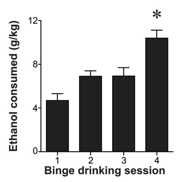
Mean consumption (g/kg) of 20% ethanol. On Days 1-3, mice had 2 hours access to the ethanol solution. On Day 4, mice had 4 hours access to the ethanol solution. In vitro electrophysiology was performed on Day 5. * p < 0.05 compared with days 1-3.
Figure 2. CRF potentiates NMDAR currents after binge ethanol drinking.

(A,B) (A) Example traces (inset) and data across time, and (B) averaged data showing enhancement of evoked NMDAR currents by 100 nM CRF in VTA neurons from binge-drinking mice but not from naïve mice. Scale bars in (A) represent 150 pA and 150 msec. “Baseline” and “CRF-wash” indicate that example traces were taken from the pre-CRF baseline and from 10 min after washout of CRF, a time point when the NMDAR currents were still enhanced. Numbers in each bar correspond to the n per group. * p < 0.05.
Figure 3. CRF1 receptor antagonists reduce the CRF-induced potentiation of NMDAR currents in binge-ethanol-drinking mice.

(A) Data across time and (B) averaged data showing the effect of 100 nM CRF on the evoked NMDAR current in the binge-drinking ethanol group in the presence or absence of CP-154,526, a potent CRF1 antagonist. Data without antagonist are the same as in Fig. 2A,B. Putative VTA DA neurons were administered CP-154,526 8 min before and also during the 100 nM CRF application. CP-154,526 prevented the CRF-induced potentiation of NMDAR currents in neurons from binge-drinking mice. Numbers in each bar graph corresponds to the n per group. * p < 0.05.
Next, we determined whether the levels of the NMDAR subunits were altered after binge ethanol drinking. Fig 4 depicts the total VTA protein levels of the NMDAR subunits (NR1, NR2A, NR2B) in binge-drinking and naive mice. Western blot analyses revealed no differences in total protein levels of the three subunits between the two groups (Fig. 4; t(4) < 0.5 and p > 0.1 for naïve versus binge-drinker for each NMDAR subunit). To examine whether NMDAR function was altered after binge ethanol drinking, we evoked NMDAR currents at 4 different stimulus intensities to generate an input-output curve. Analysis revealed no difference of evoked NMDAR currents at any of the intensities tested between the binge-drinking ethanol (n = 5) and naïve (n = 6) mice [Fig. 5; stimulation intensity: F(4,36) = 11.076, p < 0.001; group: F(1,9) = 0.110, p = 0.747; interaction: F(4,36) = 0.579, p = 0.680].
Figure 4. No changes in VTA total protein levels of NMDAR subunits in binge-ethanol-drinking mice.

(A) Examples and (B) grouped data of Western blot analyses of VTA protein levels of NMDAR subunits (NR1, NR2A, NR2B) in binge-drinking and naive mice. GAPDH levels were measured to verify equal sample loading. Numbers in each bar correspond to the n per group. O.D.: optical density.
Figure 5. No change in input-output relationship of NMDAR currents in binge-ethanol-drinking mice.
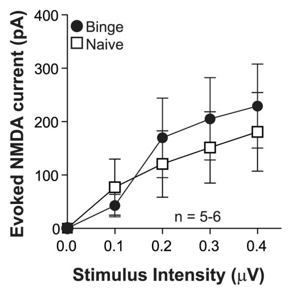
Evoked NMDA current at 4 different stimulus intensities (0-0.4 pA) in neurons from binge-drinking (n = 5) and naïve mice (n = 6).
Finally, since our in vitro experiments suggested that the greater CRF enhancement of NMDAR currents in VTA neurons occurred through CRF1 receptors, we examined whether intra-VTA blockade of the CRF1 receptor would attenuate binge ethanol drinking on the Test Day (Day 4). Mice were given an intra-VTA infusion of CP 154,526 (1 μg, 0.5 μl/side) approximately 15 min before access to the 20% ethanol bottle. CP-154,526 significantly reduced ethanol consumption compared with vehicle [Fig. 6A; n = 12 for each group; t(22) = 2.143, p = 0.043]. Together these results suggest that binge ethanol drinking is sustained by increased VTA CRF1 receptor signaling.
DISCUSSION
Here, we used the DID model of binge drinking in mice to show that binge ethanol intake is associated with CRF1 receptor enhancement of NMDAR currents in the VTA, and that VTA CRF1 receptors are necessary for high levels of binge ethanol intake. CRF at 100 nM significantly enhanced NMDAR currents in VTA DA neurons in the binge-drinking ethanol group, with no effect in ethanol-naive control mice. CRF enhancement of NMDAR currents in DID mice was blocked by co-application of CP-154,526, a selective CRF1 receptor antagonist. Accordingly, intra-VTA administration of CP-154,526 significantly reduced binge ethanol consumption. Finally, our biochemical and electrophysiological analyses suggest that binge ethanol intake enhances CRF1 receptor signaling at VTA NMDARs receptors without increasing the number of NMDARs or their baseline function. Together, these data expand and add to a growing body of literature implicating altered CRF signaling as an important regulator of ethanol consumption (for review see Lowery and Thiele, 2010).
Recent studies have suggested that the CRF system modulates binge-like ethanol drinking in mice (Kaur et al., 2012; Lowery et al., 2010; Lowery-Gionta et al., 2012; Ryabinin et al., 2008; Sharpe and Phillips, 2009; Sparta et al., 2008), although not in rats (Ji et al., 2008), though this study noted that longer duration of intake might allow CRF regulation of binge intake in rats. Koob and colleagues have proposed that CRF function is increased within the “extended amygdala” after chronic ethanol administration and dependence (Koob, 2003), in particular the central amygdala (Funk et al., 2006), and recent work has shown that CRF within the central amygdala regulates binge ethanol intake under the DID model (Lowery-Gionta et al., 2012). Urocortins also bind to CRF receptors with high affinity (Reul and Holsboer, 2002; Ryabinin et al., 2012) and thus could mediate the CRF receptor activation that can promote ethanol intake.
The VTA has been previously implicated as a critical regulator of binge ethanol drinking, as intra-VTA infusions of agents that may inhibit VTA function, including an endocannabinoid agonist and a GABAB receptor agonist, reduce DID ethanol consumption (Lisenbardt and Boehm, 2009; Moore et al., 2007; Moore and Boehm, 2009). Here, we show a novel finding, that CRF1-receptor-mediated signaling within the VTA promotes binge-like ethanol intake in mice. Also, although we did not measure the effects of intra-VTA blockade of CRF1 receptor on general fluid intake, CRF1 antagonists can reduce alcohol consumption without altering non-binge ethanol intake or sucrose or water intake (Hwa et al., 2013; Lowery-Gionta et al., 2012; Sparta et al., 2008), and CRF1 receptor knockout mice drink less ethanol but more sucrose than wild type controls (Kaur et al., 2012), suggesting that the CP-154,526 effects observed here were not mediated through non-selective motor or intake effects.
Since NMDAR level or function were not altered after binge ethanol consumption, we speculate that binge drinking caused an upregulation of CRF1 receptors on VTA dopamine neurons, although it is also possible that increased levels of VTA CRF after DID training (Lowery-Gionta et al., 2012) could contribute. We did not examine CRF1 receptor levels due to relatively poor CRF receptor-selective antibodies. Also, previous studies have implicated CRF2 receptor in the effects of CRF in the VTA including CRF modulation of NMDARs (Hahn et al., 2009; Ungless et al., 2003; Wang et al., 2007), although the CRF2 receptor antagonist did not reduce the effects of CRF in the VTA in vitro (not shown). This agrees with a recent report showing no change in DID drinking in the CRF2 knockout mouse (Kaur et al., 2012). Thus, our in vitro and behavioral results instead implicate the CRF1 receptor. The VTA receives three main sources of CRF input: the bed nucleus of the stria terminalis, the central nucleus of the amygdala, and the paraventricular nucleus of the hypothalamus (Rodaros et al., 2007). Future studies will be required to examine which of these CRF pathways into the VTA are activated after binge ethanol drinking, as well as the molecular basis of the CRF-related adaptation with ethanol intake. In addition, our NMDAR results contrast with previous work showing that ethanol withdrawal (Spanagel, 2009), intermittent ethanol exposure in culture (Qiang et al., 2010) and longer-term ethanol intoxication in rodents (Chen et al., 1997; Haugbøl et al., 2005) can all be associated with increased NMDAR levels. Our results differ in that we examine NMDARs in a specific region, the VTA, rather than in the whole brain. Also, our studies involved a relatively brief period of ethanol intake relative to previous work (Chen et al., 1997; Haugbøl et al., 2005).
We should note that we used younger, juvenile (p21-35) mice for patch-clamp experiments than were used for in vivo CRF1 receptor antagonist experiments (~8 wk old). However, our results suggest that CRF1 receptors are related to binge ethanol intake in both ages of mice. In particular, we found that juvenile mice given an i.p. injection of CP-154,526 (10 mg/kg) consumed significantly less ethanol than the vehicle-treated group (Vehicle: 6.86 ± 0.59 g/kg, n = 8; 10 mg/kg CP-154,526: 4.67 ± 0.82 g/kg, n = 8; t(14) = 2.194, p = 0.045), which are in line with the finding in Sparta et al. (2008) which showed a reduction in Day 4 ethanol consumption after systemic administration of the same CRF1 receptor antagonist in adult mice, and our own results in Fig. 6 in ~8 wk old young adult mice. Nonetheless, differences in glutamatergic and dopaminergic systems have been noted between juvenile and adult animals (Liu et al., 2004; Walker et al., 2004; Zhou and Baudry, 2006 but see Cao et al., 2000) and could be present in the VTA also. Thus, a CRF1 receptor blocker may decrease DID intake in both juvenile and adult mice but could do so by a different mechanism. Further, younger mice in our studies consumed more ethanol (10.4 g/kg) in the DID paradigm than what has previously been reported for adult mice (5-8 g/kg) (Lowery et al., 2010; Lyons et al., 2008; Rhodes et al., 2005; 2007; Sparta et al., 2008). Although we did have a dummy cage to control for spillage, we believe that the amount of ethanol spillage was not a true reflection of total ethanol loss. The juvenile mice were extremely active during the dark phase (unpublished observations) and could have disturbed the ethanol bottle during testing. However, many other studies have shown that juvenile mice consume more alcohol than adults in the DID (Moore et al., 2010) as well as other self-administration models (Doremus et al., 2005; Maldonado et al., 2008; Strong et al., 2010; Vetter et al., 2007). Additionally, adult mouse intake on Day 4 in experiments in Fig. 6A might have been reduced by the microinjection procedure (see Lowery et al., 2010). Binge alcohol drinking has been identified as an important component in the development of alcoholism and is linked to increased cardiovascular disease and dysfunction of other organs (Bonomo et al., 2004; Hingson 2005; 2007; Jennison, 2004; Miller et al., 2007; NIAAA, 2004; van de Wiel et al., 2008). Thus, any factor that reduces binge intake could have multiple health benefits. Taken together, our results suggest that binge ethanol intake in mice is associated with greater CRF1 receptor enhancement of NMDAR currents in the VTA, without a change in NMDAR number or function, and that VTA CRF1 receptors promote binge ethanol intake. We propose that the VTA CRF1 receptor remains an intriguing candidate for the treatment of binge drinking.
ACKNOWLEDGEMENTS
We thank A. Koopmans, L. Wang, R. Kermarrec, and A. Kisch-Hancock for assistance with behavioral experiments. This work was supported by NIH through 7F32AA018610 (DRS), NIH/NIAAA R01A/MH13438 (DR), and DA016782-06 (AB), and funds provided by the State of California for medical research on alcohol and substance abuse through the University of California, San Francisco (ROM, DR, AB).
REFERENCES
- Adamantidis AR, Tsai HC, Boutrel B, Zhang F, Stuber GD, Budygin EA, Touriño C, Bonci A, Deisseroth K, de Lecea L. Optogenetic interrogation of dopaminergic modulation of the multiple phases of reward-seeking behavior. J Neurosci. 2011;31:10829–10835. doi: 10.1523/JNEUROSCI.2246-11.2011. [DOI] [PMC free article] [PubMed] [Google Scholar]
- Bonomo YA, Bowes G, Coffey C, Carlin JB, Patton GC. Teenage drinking and the onset of alcohol dependence: a cohort study over seven years. Addiction. 2004;12:1520–1528. doi: 10.1111/j.1360-0443.2004.00846.x. [DOI] [PubMed] [Google Scholar]
- Cao Z, Lickey ME, Liu L, Kirk E, Gordon B. Postnatal development of NR1, NR2A and NR2B immunoreactivity in the visual cortex of the rat. Brain Res. 2000;859:26–37. doi: 10.1016/s0006-8993(99)02450-6. [DOI] [PubMed] [Google Scholar]
- Chen X, Michaelis ML, Michaelis EK. Effects of chronic ethanol treatment on the expression of calcium transport carriers and NMDA/glutamate receptor proteins in brain synaptic membranes. J Neurochem. 1997;69:1559–1569. doi: 10.1046/j.1471-4159.1997.69041559.x. [DOI] [PubMed] [Google Scholar]
- Doremus TL, Brunell SC, Rajendran P, Spear LP. Factors influencing elevated ethanol consumption in adolescent relative to adult rats. Alcohol Clin Exp Res. 2005;29:1796–1808. doi: 10.1097/01.alc.0000183007.65998.aa. [DOI] [PubMed] [Google Scholar]
- Funk CK, O’Dell LE, Crawford EF, Koob GF. Corticotropin-releasing factor within the central nucleus of the amygdala mediates enhanced ethanol self-administration in withdrawn, ethanol-dependent rats. J Neurosci. 2006;26:11324–11332. doi: 10.1523/JNEUROSCI.3096-06.2006. [DOI] [PMC free article] [PubMed] [Google Scholar]
- Hahn J, Hopf FW, Bonci A. Chronic cocaine enhances corticotropin-releasing factor-dependent potentiation of excitatory transmission in ventral tegmental area dopamine neurons. J Neurosci. 2009;29:6535–6544. doi: 10.1523/JNEUROSCI.4773-08.2009. [DOI] [PMC free article] [PubMed] [Google Scholar]
- Haugbøl SR, Ebert B, Ulrichsen J. Upregulation of glutamate receptor subtypes during alcohol withdrawal in rats. Alcohol Alcohol. 2005;40:89–95. doi: 10.1093/alcalc/agh117. [DOI] [PubMed] [Google Scholar]
- Hingson R, Heeren T, Winter M, Wechsler H. Magnitude of alcohol-related mortality and morbidity among U.S. college students ages 18–24: changes from 1998 to 2001. Ann Rev Public Health. 2005;26:259–279. doi: 10.1146/annurev.publhealth.26.021304.144652. [DOI] [PubMed] [Google Scholar]
- Hingson RW, Heeren T, Winter MR. Age of alcohol-dependence onset: associations with severity of dependence and seeking treatment. Pediatrics. 2007;118:e755–e763. doi: 10.1542/peds.2006-0223. [DOI] [PubMed] [Google Scholar]
- Hwa LS, Debold JF, Miczek KA. Alcohol in excess: CRF(1) receptors in the rat and mouse VTA and DRN. Psychopharmacology (Berl) 2012 Aug 11; doi: 10.1007/s00213-012-2820-z. [Epub ahead of print] [DOI] [PMC free article] [PubMed] [Google Scholar]
- Jee C, Lee J, Lim JP, Parry D, Messing RO, McIntire SL. SEB-3, a CRF receptor-like GPCR, regulates locomotor activity states, stress responses, and ethanol tolerance in C. elegans. Genes Brain Behav. 2012 Jul 31; doi: 10.1111/j.1601-183X.2012.00829.x. doi: 10.1111/j.1601-183X.2012.00829.x. [Epub ahead of print] [DOI] [PMC free article] [PubMed] [Google Scholar]
- Jennison KM. The short-term effects and unintended long-term consequences of binge drinking in college: a 10-year follow-up study. Am J Drug Alcohol Abuse. 2004;30:659–684. doi: 10.1081/ada-200032331. [DOI] [PubMed] [Google Scholar]
- Ji D, Gilpin NW, Richardson HN, Rivier CL, Koob GF. Effects of naltrexone, duloxetine, and a corticotropin-releasing factor type 1 receptor antagonist on binge-like alcohol drinking in rats. Behav Pharmacol. 2008;19:1–12. doi: 10.1097/FBP.0b013e3282f3cf70. [DOI] [PMC free article] [PubMed] [Google Scholar]
- Johnson SW, North RA. Two types of neurone in the rat ventral tegmental area and their synaptic inputs. J Physiol. 1992;450:455–468. doi: 10.1113/jphysiol.1992.sp019136. [DOI] [PMC free article] [PubMed] [Google Scholar]
- Kaur S, Li J, Stenzel-Poore MP, Ryabinin AE. Corticotropin-releasing factor acting on corticotropin-releasing factor receptor type 1 is critical for binge alcohol drinking in mice. Alcohol Clin Exp Res. 2012;36:369–376. doi: 10.1111/j.1530-0277.2011.01610.x. [DOI] [PMC free article] [PubMed] [Google Scholar]
- Koob GF. Alcoholism: allostasis and beyond. Alcohol Clin Exp Res. 2003;27:232–243. doi: 10.1097/01.ALC.0000057122.36127.C2. [DOI] [PubMed] [Google Scholar]
- Lange JE, Voas RB. Defining binge drinking quantities through resulting BACs. Annu Proc Assoc Adv Automot Med. 2000;44:389–404. [PMC free article] [PubMed] [Google Scholar]
- Lê AD, Harding S, Juzytsch W, Watchus J, Shalev U, Shaham Y. The role of corticotrophin-releasing factor in stress-induced relapse to alcohol-seeking behavior in rats. Psychopharm. 2000;150:317–324. doi: 10.1007/s002130000411. [DOI] [PubMed] [Google Scholar]
- Linsenbardt DN, Boehm SL., 2nd Agonism of the endocannabinoid system modulates binge-like alcohol intake in male C57BL/6J mice: involvement of the posterior ventral tegmental area. Neuroscience. 2009;164:424–434. doi: 10.1016/j.neuroscience.2009.08.007. [DOI] [PMC free article] [PubMed] [Google Scholar]
- Liu XB, Murray KD, Jones EG. Switching of NMDA receptor 2A and 2B subunits at thalamic and cortical synapses during early postnatal development. J Neurosci. 2004;24:8885–8895. doi: 10.1523/JNEUROSCI.2476-04.2004. [DOI] [PMC free article] [PubMed] [Google Scholar]
- Lowery EG, Spanos M, Navarro M, Lyons AM, Hodge CW, Thiele TE. CRF-1 antagonist and CRF-2 agonist decrease binge-like ethanol drinking in C57BL/6J mice independent of the HPA axis. Neuropsychopharm. 2010;35:1241–1252. doi: 10.1038/npp.2009.209. [DOI] [PMC free article] [PubMed] [Google Scholar]
- Lowery EG, Thiele TE. Pre-clinical evidence that corticotropin-releasing factor (CRF) receptor antagonists are promising targets for pharmacological treatment of alcoholism. CNS Neurol Disord Drug Targets. 2010;9:77–86. doi: 10.2174/187152710790966605. [DOI] [PMC free article] [PubMed] [Google Scholar]
- Lowery-Gionta EG, Navarro M, Li C, Pleil KE, Rinker JA, Cox BR, Sprow GM, Kash TL, Thiele Corticotropin releasing factor signaling in the central amygdala is recruited during binge-like ethanol consumption in C57BL/6J mice. J Neurosci. 2012;32:3405–3413. doi: 10.1523/JNEUROSCI.6256-11.2012. [DOI] [PMC free article] [PubMed] [Google Scholar]
- Luscher C, Malenka RC. Drug-evoked synaptic plasticity in addiction: from molecular changes to circuit remodeling. Neuron. 2011;69:650–663. doi: 10.1016/j.neuron.2011.01.017. [DOI] [PMC free article] [PubMed] [Google Scholar]
- Lyons AM, Lowery EG, Sparta DR, Thiele TE. Effects of food availability and administration of orexigenic and anorectic agents on elevated ethanol drinking associated with drinking in the dark procedures. Alcohol Clin Exp Res. 2008;32:1962–1968. doi: 10.1111/j.1530-0277.2008.00784.x. [DOI] [PMC free article] [PubMed] [Google Scholar]
- Madhavan A, He L, Stuber GD, Bonci A, Whistler JL. micro-Opioid receptor endocytosis prevents adaptations in ventral tegmental area GABA transmission induced during naloxone-precipitated morphine withdrawal. J Neurosci. 2010;30:3276–3286. doi: 10.1523/JNEUROSCI.4634-09.2010. [DOI] [PMC free article] [PubMed] [Google Scholar]
- Maldonado AM, Finkbeiner LM, Alipour KK, Kirstein CL. Voluntary ethanol consumption differs in adolescent and adult male rats using a modified sucrose-fading paradigm. Alcohol Clin Exp Res. 2008;32:1574–1582. doi: 10.1111/j.1530-0277.2008.00733.x. [DOI] [PubMed] [Google Scholar]
- Margolis EB, Lock H, Hjelmstad GO, Fields HL. The ventral tegmental area revisited: is there an electrophysiological marker for dopaminergic neurons? J Physiol. 2006;577:907–924. doi: 10.1113/jphysiol.2006.117069. [DOI] [PMC free article] [PubMed] [Google Scholar]
- Melón LC, Boehm SL., 2nd GABA(A) receptors in the posterior, but not anterior, ventral tegmental area mediate Ro15-4513-induced attenuation of binge-like ethanol consumption in C57BL/6J female mice. Behav Brain Res. 2011;220:230–237. doi: 10.1016/j.bbr.2011.02.014. [DOI] [PMC free article] [PubMed] [Google Scholar]
- Miguel TT, Nunes-de-Souza RL. Anxiogenic and antinociceptive effects induced by corticotropin-releasing factor (CRF) injections into the periaqueductal gray are modulated by CRF1 receptor in mice. Horm Behav. 2011;60:292–300. doi: 10.1016/j.yhbeh.2011.06.004. [DOI] [PubMed] [Google Scholar]
- Miller JW, Naimi TS, Brewer RD, Jones SE. Binge drinking and associated health risk behaviors among high school students. Pediatrics. 2007;119:76–85. doi: 10.1542/peds.2006-1517. [DOI] [PubMed] [Google Scholar]
- Moore EM, Boehm SL., 2nd Site-specific microinjection of baclofen into the anterior ventral tegmental area reduces binge-like ethanol intake in male C57BL/6J mice. Behav Neurosci. 2009;123:555–563. doi: 10.1037/a0015345. [DOI] [PMC free article] [PubMed] [Google Scholar]
- Moore EM, Mariani JN, Linsenbardt DN, Melón LC, Boehm SL., 2nd Adolescent C57BL/6J (but not DBA/2J) mice consume greater amounts of limited-access ethanol compared to adults and display continued elevated ethanol intake into adulthood. Alcohol Clin Exp Res. 2010;34:734–742. doi: 10.1111/j.1530-0277.2009.01143.x. [DOI] [PMC free article] [PubMed] [Google Scholar]
- Moore EM, Serio KM, Goldfarb KJ, Stepanovska S, Linsenbardt DN, Boehm SL., 2nd GABAergic modulation of binge-like ethanol intake in C57BL/6J mice. Pharmacol Biochem Behav. 2007;88:105–113. doi: 10.1016/j.pbb.2007.07.011. [DOI] [PMC free article] [PubMed] [Google Scholar]
- National Institute on Alcohol and Alcoholism Advisory Council NIAAA council approves definition of binge drinking. NIAAA Newsletter. 2004 Winter. [Google Scholar]
- Qiang M, Denny A, Chen J, Ticku MK, Yan B, Henderson G. The site specific demethylation in the 5′-regulatory area of NMDA receptor 2B subunit gene associated with CIE-induced up-regulation of transcription. PLoS One. 2010;5:e8798. doi: 10.1371/journal.pone.0008798. [DOI] [PMC free article] [PubMed] [Google Scholar]
- Reul JM, Holsboer F. Corticotropin-releasing factor receptors 1 and 2 in anxiety and depression. Curr Opin Pharmacol. 2002;2:23–33. doi: 10.1016/s1471-4892(01)00117-5. [DOI] [PubMed] [Google Scholar]
- Rhodes JS, Best K, Belknap JK, Finn DA, Crabbe JC. Evaluation of a simple model of ethanol drinking to intoxication in C57BL/6J mice. Physiol Behav. 2005;84:53–63. doi: 10.1016/j.physbeh.2004.10.007. [DOI] [PubMed] [Google Scholar]
- Rhodes JS, Ford MM, Yu CH, Brown LL, Finn DA, Garland T, Jr, Crabbe JC. Mouse inbred strain differences in ethanol drinking to intoxication. Genes Brain Behav. 2007;6:1–18. doi: 10.1111/j.1601-183X.2006.00210.x. [DOI] [PubMed] [Google Scholar]
- Rodaros D, Caruana DA, Amir S, Stewart J. Corticotropin-releasing factor projections from limbic forebrain and paraventricular nucleus of the hypothalamus to the region of the ventral tegmental area. Neuroscience. 2007;150:8–13. doi: 10.1016/j.neuroscience.2007.09.043. [DOI] [PubMed] [Google Scholar]
- Ryabinin AE, Tsoory MM, Kozicz T, Thiele TE, Neufield-Cohen A, Chen A, Lowery-Gionta EG, Giardino WJ, Kaur S. Urocoritns: CRF’s siblings and their potential role in anxiety, depression and alcohol drinking behavior. Alcohol. 2012;46:349–357. doi: 10.1016/j.alcohol.2011.10.007. [DOI] [PMC free article] [PubMed] [Google Scholar]
- Ryabinin AE, Yoneyama N, Tanchuck MA, Mark GP, Finn DA. Urocortin 1 microinjection into the mouse lateral septum regulates the acquisition and expression of alcohol consumption. Neuroscience. 2008;151:780–790. doi: 10.1016/j.neuroscience.2007.11.014. [DOI] [PMC free article] [PubMed] [Google Scholar]
- Sharpe AL, Phillips TJ. Central urocortin 3 administration decreases limited-access ethanol intake in nondependent mice. Behav Pharmacol. 2009;20:346–351. doi: 10.1097/FBP.0b013e32832f01ba. [DOI] [PMC free article] [PubMed] [Google Scholar]
- Spanagel R. Alcoholism: a systems approach from molecular physiology to addictive behavior. Physiol Rev. 2009;89:649–705. doi: 10.1152/physrev.00013.2008. [DOI] [PubMed] [Google Scholar]
- Sparta DR, Sparrow AM, Lowery EG, Fee JR, Knapp DJ, Thiele TE. Blockade of the corticotropin releasing factor type 1 receptor attenuates elevated ethanol drinking associated with drinking in the dark procedures. Alcohol Clin Exp Res. 2008;32:259–265. doi: 10.1111/j.1530-0277.2007.00575.x. [DOI] [PMC free article] [PubMed] [Google Scholar]
- Sprow GM, Thiele TE. The neurobiology of binge-like ethanol drinking: Evidence from rodent models. Physiol Behav. 2012;106:325–331. doi: 10.1016/j.physbeh.2011.12.026. [DOI] [PMC free article] [PubMed] [Google Scholar]
- Stewart J. Pathways to relapse: the neurobiology of drug- and stress-induced relapse to drug-taking. J Psychiatry Neurosci. 2000;25:125–136. [PMC free article] [PubMed] [Google Scholar]
- Strong MN, Yoneyama N, Fretwell AM, Snelling C, Tanchuck MA, Finn DA. “Binge” drinking experience in adolescent mice shows sex differences and elevated ethanol intake in adulthood. Horm Behav. 2010;58:82–90. doi: 10.1016/j.yhbeh.2009.10.008. [DOI] [PMC free article] [PubMed] [Google Scholar]
- Stuber GD, Sparta DR, Stamatakis AM, van Leeuwen WA, Hardjoprajitno JE, Cho S, Tye KM, Kempadoo KA, Zhang F, Deisseroth K, Bonci A. Excitatory transmission from the amygdala to nucleus accumbens facilitates reward seeking. Nature. 2011;475:377–380. doi: 10.1038/nature10194. [DOI] [PMC free article] [PubMed] [Google Scholar]
- Swanson LW, Sawchenko PE, Rivier J, Vale WW. Organization of ovine corticotropin-releasing factor immunoreactive cells and fibers in the rat brain: an immunohistochemical study. Neuroendocrinology. 1983;36:165–186. doi: 10.1159/000123454. [DOI] [PubMed] [Google Scholar]
- Ungless MA, Singh V, Crowder TL, Yaka R, Ron D, Bonci A. Corticotropin-releasing factor requires CRF binding protein to potentiate NMDA receptors via CRF receptor 2 in dopamine neurons. Neuron. 2003;39:401–407. doi: 10.1016/s0896-6273(03)00461-6. [DOI] [PubMed] [Google Scholar]
- Valdez GR, Roberts AJ, Chan K, Davis H, Brennan M, Zorrilla EP, Koob GF. Increased ethanol self-administration and anxiety-like behavior during acute ethanol withdrawal and protracted abstinence: regulation by corticotropin-releasing factor. Alcohol Clin Exp Res. 2002;26:1494–1501. doi: 10.1097/01.ALC.0000033120.51856.F0. [DOI] [PubMed] [Google Scholar]
- van de Wiel A, de Lange DW. Cardiovascular risk is more related to drinking pattern than to the type of alcoholic drinks. Neth J Med. 2008;66:467–473. [PubMed] [Google Scholar]
- Vetter CS, Doremus-Fitzwater TL, Spear LP. Time course of elevated ethanol intake in adolescent relative to adult rats under continuous, voluntary-access conditions. Alcohol Clin Exp Res. 2007;31:1159–1168. doi: 10.1111/j.1530-0277.2007.00417.x. [DOI] [PMC free article] [PubMed] [Google Scholar]
- Walker QD, Morris SE, Arrant AE, Nagel JM, Parylak S, Zhou G, Caster JM, Kuhn CM. Dopamine uptake inhibitors but not dopamine releasers induce greater increases in motor behavior and extracellular dopamine in adolescent rats than in adult male rats. J Pharmacol Exp Ther. 2010;335:124–132. doi: 10.1124/jpet.110.167320. [DOI] [PMC free article] [PubMed] [Google Scholar]
- Wanat MJ, Hopf FW, Stuber GD, Phillips PE, Bonci A. Corticotropin-releasing factor increases mouse ventral tegmental area dopamine neuron firing through a protein kinase C-dependent enhancement of Ih. J Physiol. 2008;586:2157–2170. doi: 10.1113/jphysiol.2007.150078. [DOI] [PMC free article] [PubMed] [Google Scholar]
- Wang B, Shaham Y, Zitzman D, Azari S, Wise RA, You ZB. Cocaine experience establishes control of midbrain glutamate and dopamine by corticotropin-releasing factor: a role in stress-induced relapse to drug seeking. J Neurosci. 2005;25:5389–5396. doi: 10.1523/JNEUROSCI.0955-05.2005. [DOI] [PMC free article] [PubMed] [Google Scholar]
- Wang B, You ZB, Rice KC, Wise RA. Stress-induced relapse to cocaine seeking: roles for the CRF(2) receptor and CRF-binding protein in the ventral tegmental area of the rat. Psychopharmacology. 2007;193:283–294. doi: 10.1007/s00213-007-0782-3. [DOI] [PubMed] [Google Scholar]
- Wise RA, Morales M. A ventral tegmental CRF-glutamate-dopamine interaction in addiction. Brain Res. 2010;1314:38–43. doi: 10.1016/j.brainres.2009.09.101. [DOI] [PMC free article] [PubMed] [Google Scholar]
- Zhou M, Baudry M. Developmental changes in NMDA neurotoxicity reflect developmental changes in subunit composition of NMDA receptors. J Neurosci. 2006;26:2956–2963. doi: 10.1523/JNEUROSCI.4299-05.2006. [DOI] [PMC free article] [PubMed] [Google Scholar]