


Figure 2. MRI of the effect of intetumumab on the formation of breast cancer brain metastases.
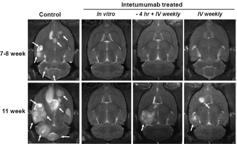
MRI was performed approximately 49–56 and 77 d after intra-carotid cell infusion. One representative rat is shown from each of four randomized treatment groups: 1) Control; 2) INT_in-vitro, cells were pretreated in vitro with intetumumab (0.5 mg/ml) prior to infusion and received no further treatment; 3) INT_-4h+IV, rats received intetumumab (10 mg/kg IV) 4 h prior to cell infusion plus weekly after infusion; 4) Rats received intetumumab (10 mg/kg IV) starting 7 d after cell infusion. The presence of brain lesions (metastases) in MRI was indicated by arrows.