



Abstract
Brain metastases commonly occur in patients with breast, lung and melanoma systemic cancers. The anti-αV integrin monoclonal antibody intetumumab binds cell surface proteins important for adhesion, invasion and angiogenesis in the metastatic cascade. The objective of this study was to investigate the anti-metastatic effect of intetumumab in a hematogenous breast cancer brain metastasis model. Female nude rats received intra-carotid infusion of human brain-seeking metastatic breast cancer cells (231BR-HER2) and were randomly assigned into 4 groups: 1) control; 2) intetumumab mixed with cells in vitro 5 min before infusion without further treatment; 3) intetumumab intravenously 4 h before and weekly after cell infusion; 4) intetumumab intravenously weekly starting 7 d after cell infusion. Brain metastases were detected by magnetic resonance imaging (MRI) and immunohistochemistry. Comparisons were made using the Kruskal-Wallis test and Dunnett’s test. Survival times were estimated using Kaplan-Meier analysis. All control rats with brain tissue available for histology (9 of 11 rats) developed multiple brain metastases (median=14). Intetumumab treatment either in vitro prior to cell infusion or intravenous before or after cell infusion prevented metastasis formation on MRI and decreased the number of metastases on histology (median=2, p=0.0055), including 30% of animals without detectable tumors at the end of the study. The overall survival was improved by intetumumab compared to controls (median 77+ versus 52 d, p=0.0277). Our results suggest that breast cancer patients at risk of metastases might benefit from early intetumumab treatment.
Keywords: integrin, intetumumab, breast cancer, brain metastasis, MRI
Introduction
Brain metastasis occurs in as many as one third of breast cancer patients, and is associated with high mortality [1]. Methods to prevent or delay formation of breast cancer brain metastases would have a significant health benefit. A HER2-overexpressing clone of a brain-seeking derivative of MDA-MB-231 human breast cancer cells (231BR-HER2 cells) consistently forms multiple brain hematogenous metastases and provides a model for testing anti-metastatic therapies [2–4]. The hematogenous metastatic breast cancer model used in this study mimics the clinical situation: successful metastasis of high HER2 breast cancer cells that travel through the bloodstream, adhere to the blood vessels in the brain, invade the brain parenchyma, and grow in the new environment [5].
Integrins are a large family of heterodimeric integral membrane proteins comprising at least 24 combinations of 18 α and 8 β subunits. These receptors are involved in cell–cell and cell–extracellular matrix (ECM) interactions, cytoskeleton organization, and cell signaling. In cancer, integrins promote the proliferation of tumor cells and tumor vascular endothelial cells [6–9], and appear to be involved in multiple aspects of metastasis, including tumor cell binding, invasion, growth and angiogenesis [9–12]. The involvement of integrins in multiple steps of the metastatic process and their differential expression between tumors and normal tissue makes them a promising therapeutic target [13–16].
Intetumumab (INT; CNTO 95) is a fully human IgG1k monoclonal antibody (mAb) that binds αV integrins with broad specificity, with a dissociation constant of Kd 1–24 nmol [17, 18]. It has a serum half life of 8–9 d and was found to be well tolerated with no or relatively low adverse effects at 10 mg/kg in clinical phase I and II trials [17, 19–21]. The objective of this study was to investigate the anti-metastatic effect of intetumumab in a hematogenous breast cancer brain metastasis model in nude rats.
Materials and Methods
The care and use of the animals was approved by the Institutional Animal Care and Use Committee and was under the supervision of the Department of Comparative Medicine at OHSU. Intetumumab (fully human anti-αV integrin mAb) was provided by Ortho Biotech Oncology R&D (Radnor, PA). Other antibodies used were trastuzumab (anti-HER2) and rituximab (anti-CD20) (Genentech, San Francisco, CA), anti-human αV, α5, β3, β5 integrin and HER2 (Cell Signaling Technology, Danvers, MA), anti-human mitochondrial antigen (Chemicon/Millipore Temecula, CA), and anti-tubulin (Sigma, St. Louis, MO). Cyclophosphamide (Cytoxan ®) was from Bristol-Myers Squibb (Princeton, NJ).
Cell culture and in vitro studies
Human metastatic breast cancer cells (MDA-MB-231BR-HER2; 231BR-HER2) and a matched plasmid-transfected cell line without HER2 protein overexpression (231BR-vector) were provided by Dr. Pat Steeg (NCI, Bethesda, MD) and were cultured with DME medium supplemented with serum and antibiotics. Cellular integrin protein expression in cultured cells was characterized using the Alpha/Beta Integrin-Mediated Cell Adhesion Assay Combo Kit (Chemicon/Millipore, Temecula, CA) and immunobloting analysis. Western immunobloting was performed as described previously [22]. For the cell trafficking study, 231BR-HER2 cells were labeled in vitro with ferumoxides-protamine sulfate (FE-Pro) complex (100 μg/mL; w/w; 10:1 ratio) in serum free medium for 1 h, then rinsed in serum-free medium prior to intra-carotid infusion [23]. For the in vitro adhersion assay, 105 231BR-HER2 cells were treated with 0.5 mg/mL intetumumab, trastuzumab or rituximab antibody in complete medium. After 1 h incubation at 37 oC, the unattached cells were gently pipetted and counted after trypan blue staining.
Study designs
Hematogenous breast cancer brain metastasis model
Female nude rats (rnu/rnu, 200–250 g, from the OHSU Blood-Brain Barrier Program in-house colony) were used for all studies. Rats were pretreated with cyclophosphamide (100 mg/kg IP) 1 d before cell infusion to reduce innate immunity and 14 d after tumor cell inoculation to enhance VEGF production [4]. The cyclophosphamide treatments are necessary for consistency in the hematogenous metastasis model. Rats were anesthetized with isoflurane, and a catheter filled with heparinized saline was tied into the right external carotid artery. 231BR-HER2 cells (106) were infused into the right internal carotid artery.
Hematogenous tumor cell localization
For the MRI brain localization study, FE-Pro labeled 231BR-HER2 breast cancer cells were pretreated with either Intetumumab or normal human IgG (0.5 mg/ml; n=4 per group) in vitro for 5 min prior to intra-carotid infusion. Iron labeled cells were detected as signal dropout on T2-weighted MR images acquired 1 d after cell infusion.
Hematogenous metastasis imaging and therapy study
Rats were randomly assigned into 4 groups: 1) control treated weekly with normal human IgG (10 mg/kg IV) (n=11); 2) INT_in-vitro: intetumumab (0.5 mg/ml) mixed with cells in vitro 5 min before infusion and no further treatment (n=8); 3) INT_-4h+IV: intetumumab (10 mg/kg IV) 4 h before and weekly after cell infusion (n=11); 4) INT_IV: intetumumab (10 mg/kg IV) weekly starting 7 d after cell infusion (n=8). The presence of brain metastases was monitored by MRI at 49–56 d after cell infusion and 77 d if rats were still alive. Rats were euthanized at 20% weight loss or poor clinical condition, or at the pre-determined study end point of 77 d after tumor cell infusion.
Effect of intetumumab on intracerebral xenograft growth
For intracerebral tumor implantation, female nude rats were anesthetized with ketamine (60 mg/kg IP) and diazepam (7.5 mg/kg IP) and 106 231BR-HER2 cells were stereotactically injected in the right caudate putamen (vertical, bregma 6 mm; lateral, bregma 3 mm). To evaluate the effect of in vitro intetumumab treatment, 231BR-HER2 cells were pretreated with intetumumab (0.5 mg/ml) or saline for 5 min before implantation (n=3 per group) and rats received no further treatment. Rat brains were harvested for tumor volume analysis at 21 d after implantation. To further assess if intetumumab had tumoricidal effects, other rats with intracerebral xenografts of untreated 231BR-HER2 cells were randomized on d 3 after tumor implantation to receive either no treatment (n=13) or a single dose of intetumumab (30 mg/kg IV, n=12) and were followed for survival.
Magnetic Resonance Imaging (MRI), histology and immunohistochemistry
Standard protocols for MRI, histology and immunohistochemistry have been published previously [24]. T2* MRI signal intensity in regions of interest (ROI) in the right hemisphere versus matched ROI was measured using NIH ImageJ in n=3 each for control and intetumumab-treated rats. For immunohistochemistry analysis, brains were formalin fixed, sectioned at 100 μm, and every sixth section stained for human mitochondrial antigen. Possible metastases in other tissues were not evaluated. Tumors were manually outlined, including interior holes in large tumors in 3 control rats and 5 intetumumab-treated rats. The volume of individual metastases and total tumor burden was assessed using NIH ImageJ software.
Statistical analysis
Summary statistics are presented as medians, and means for numbers and volumes of metastases. The number of metastases at the first MRI or histology, and for volume of metastases by histology was compared using the Kruskal-Wallis test. Survival time was estimated using Kaplan-Meier estimates from first d of treatment until death or euthanasia. In the latter case, the survival time was censored. The four groups were compared using the log-rank test and, if there were an overall difference between the groups, pair-wise comparisons were made among all pairs using the log-rank test with adjustment based on Tukey’s studentized range test. MRI signal intensity measurements were compared using one-way analysis of variance with Tukey’s test. Metastasis volume and location in all three intetumumab treatment groups were combined and compared with control metastases using one-way analysis of variance with Dunnett’s test. The in vitro adhesion assay and intracerebral xenograft studies were compared by Students t-test. Significance was determined at the 5% level, two-sided, using Microsoft Excel or SAS® version 9.2 (SAS Institute, 2002–2008).
Results
Human 231BR-HER2 cells expressed αV, α5 and β3 integrin but no β5 integrin by immunoblotting and ELISA and showed abundant αVβ3 but lower αVβ5 integrin (Figure 1A). In an in vitro adhesion assay, 89.0 ± 2.9% of untreated 231BR-HER2 cells adhered to culture plates at 1 h. Incubation with intetumumab (0.5 mg/mL) significantly (p < 0.05) decreased adhesion to 24.8 ± 17.8% at 1 h. This inhibitory effect was not found with either trastuzumab or rituximab treatment (Figure 1B). Cell viability was 97–100% regardless of antibody treatment.
Figure 1. Cellular integrin characterization and effect of Intetumumab treatment on breast cancer cell adhesion in vitro and cell localization in the brain.
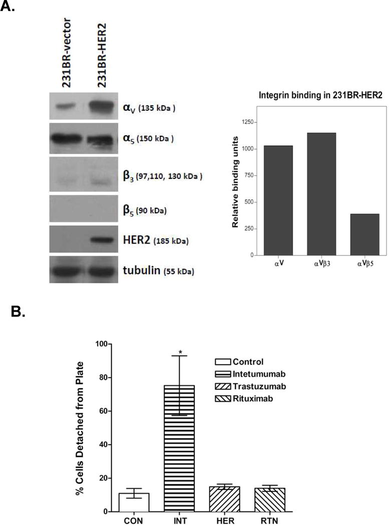
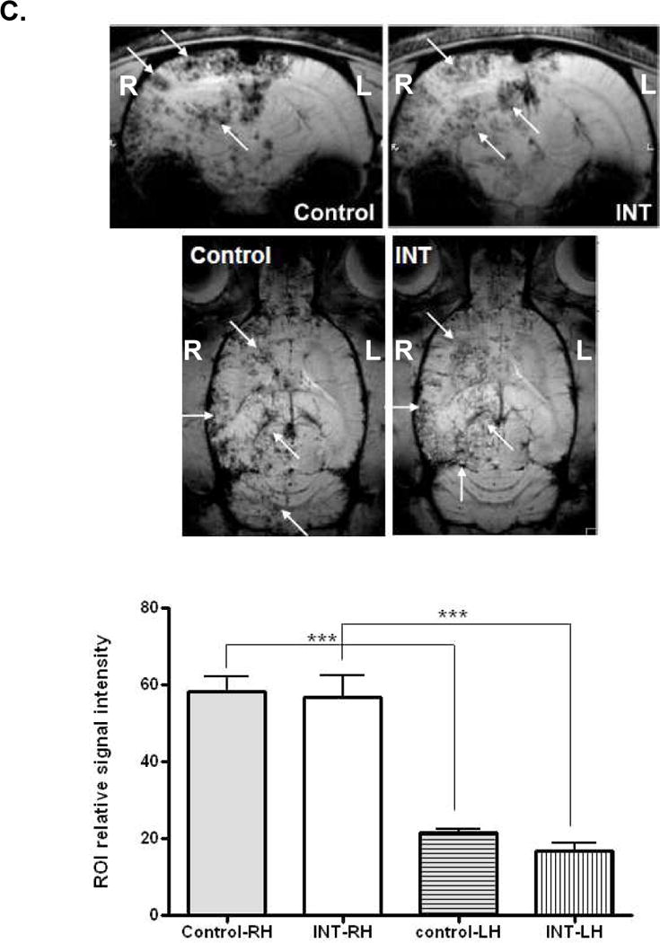
A) Immunobloting analysis of 231BR-HER2 and their no HER2 expressing matched cell line (231BR-vector) assessed for integrins αv, α5, β3 and β5. HER2 immunoblot was used to confirm the HER2 protein overexpression and anti-tubulin antibody was used as equal loading. In 231BR-HER2 cells, ELISA analysis showed abundant αVβ3 and relatively low αVβ5 integrin dimer. B) Assessment of 23BR-HER2 cell adhesion in vitro. Cells were untreated (CON) or incubated for 1 h with intetumumab (INT, 0.5 mg/mL), trastuzumab (HER, 0.5 mg/mL) or rituximab (RTN, 0.5 mg/mL). Non-adherent cell percentages are indicated as mean ± SD. * indicates p < 0.05. C) Female nude rats received intra-carotid infusion of 231BR-HER2 breast cancer cells labeled with ferumoxides-protamine sulfate and pretreated with either Intetumumab or normal human IgG (0.5 mg/ml) in vitro for 5 min prior to infusion. Coronal and horizontal T2-weighted MRI scans were performed 1 d after cell infusion. Iron is detected as signal dropout on T2-weighted images (arrows). The bar graph indicates quantification of T2* MRI signal intensity of ROI in the right hemisphere (RH) that received intra-arterial cell infusion (on the left in these MRI scans), compared to the left hemisphere (LH). *** indicates p < 0.001.
In the brain localization study, T2-weighted MRI signal hypo-intensity (black dots) indicating the presence of iron-labeled cells was observed at 1 d after infusion of both control and intetumumab treated cells (Figure 1C). There are significantly (p < 0.001) more labeled cells in the right hemisphere than left hemisphere in all animals (note MRI shows the right hemisphere on the left side). Quantification of the percentage of brain showing iron signal on MRI showed no difference due to intetumumab pretreatment (Figure 1C).
In the hematogenous metastasis study, 8 of 9 (89%) evaluable control rats developed multiple (> 3) brain metastases and only 1 of 9 rats showed no metastases on the first MRI at 49–56 wks after cell infusion (Figure 2 and Table 1). In contrast, no brain lesions were detected in 13 of 24 rats in all intetumumab groups, and only 4 rats (17%) showed multiple metastases. Individually, each intetumumab group differed from control as follows: INT_in-vitro, p=0.0071; INT_-4h+IV, p=0.0071; INT_IV, p=0.01 (Table 1). There were no differences among the three intetumumab treatment groups. Three control rats survived to 77 d and all showed multiple (> 3) brain lesions on the second MRI. Six rats in all intetumumab treatment groups combined that were negative for brain lesions on the first MRI showed metastases on the second MRI, but 7 surviving rats showed no brain metastases on the second MRI. Regardless of treatment paradigm, intetumumab reduced the incidence of multiple brain metastases on MRI.
Figure 2. MRI of the effect of intetumumab on the formation of breast cancer brain metastases.
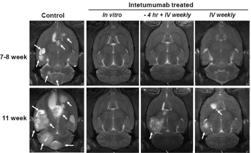
MRI was performed approximately 49–56 and 77 d after intra-carotid cell infusion. One representative rat is shown from each of four randomized treatment groups: 1) Control; 2) INT_in-vitro, cells were pretreated in vitro with intetumumab (0.5 mg/ml) prior to infusion and received no further treatment; 3) INT_-4h+IV, rats received intetumumab (10 mg/kg IV) 4 h prior to cell infusion plus weekly after infusion; 4) Rats received intetumumab (10 mg/kg IV) starting 7 d after cell infusion. The presence of brain lesions (metastases) in MRI was indicated by arrows.
Table 1.
Summary of MRI and histological analysis of breast cancer brain metastasis in nude rats
| Number of Brain Metastases§ | |||||
|---|---|---|---|---|---|
|
|
|||||
| Group | n | 0 | 1-3 | >3 | |
| 1st MRI† | Control# | 9 | 1 (11%) | 0 | 8 (89%) |
| INT_in-vitro^ | 6 | 5 (83%) | 1 (17%) | 0 | |
| INT_-4h+IV# | 10 | 4 (40%) | 3 (30%) | 3 (30%) | |
| INT IV | 8 | 4 (50%) | 3 (38%) | 1 (13%) | |
|
|
|||||
| All INT rats | 24 | 13 (54%) | 7 (29%) | 4 (17%) | |
|
| |||||
| 2nd MRI† | Contol | 3 | 0 | 0 | 3 (100%) |
| INT_in-vitro^ | 5 | 2 (40%) | 3 (60%) | 0 | |
| INT_-4h+IV | 6 | 3 (50%) | 2 (33%) | 1 (17%) | |
| INT IV | 4 | 2 (50%) | 1 (25%) | 1 (25%) | |
|
|
|||||
| All INT rats | 15 | 7 (47%) | 6 (40%) | 2 (13%) | |
|
| |||||
| Histology | Control* | 9 | 0 | 0 | 9 (100%) |
| INT_in-vitro | 8 | 3 (38%) | 2 (25%) | 3 (38%) | |
| INT_-4h+IV* | 9 | 3 (30%) | 1 (11%) | 5 (56%) | |
| INT IV | 8 | 2 (25%) | 2 (25%) | 4 (50%) | |
|
|
|||||
| All INT rats | 25 | 8 (32%) | 5 (20%) | 12 (48%) | |
For clinical relevance, we categorized the number of brain metastases into 3 groups: no tumor, 1-3 tumors, suitable for surgical removal, and >3 tumors, which would require whole brain radiation.
1st MRI was performed 49-56 d and 2nd MRI was performed 77d after cell inoculation if rats were still alive.
Two of 11 rats in the control group and 1 of 11 rats of INT_-4hr + IV group died before the first scheduled MRI.
Two rats in the INT_in-vitro group did not receive MRI due to the MR schedule.
Two of 11 rats in both the control and INT_-4hr+IV groups did not have brain histology due to post mortem degradation.
In histological analysis, all evaluable control rats developed multiple brain metastases, although the number and size were variable (median 14, mean 15 ± 9) (Figure 3A, B). Intetumumab-treated rats developed significantly fewer metastases (median 2, mean 4 ± 5, p=0.0055), with 32% of animals showing no detectable brain tumors (Table 1). Normal human IgG used to treat control rats showed no effects on altering body weight and onset of brain metastases. The number of metastases in each intetumumab group significantly (p < 0.01) differed from control with no difference found among the three intetumumab groups (Figure 3B). In those rats with brain metastases, there was no significant difference in total brain tumor volume when comparing control and intetumumab treated rats (Figure 3C). Individually, there was a total of 136 brain metastases in 9 evaluable control rats with a mean volume of 11.8 ± 20.0 mm3, while 101 metastases in 25 intetumumab rats had a mean volume of 26.0 ± 50.9 mm3 (Figure 3D).
Figure 3. Histological assessment of the effect of intetumumab on breast cancer brain metastasis.
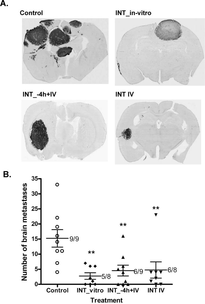
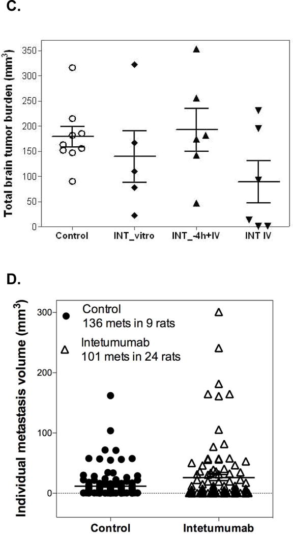
After death or sacrifice at 77 d, rat brain sections were assessed for immunohistochemistry against human mitochondria antigen as a marker of human tumor growth. A) One representative rat brain is shown from each group; B) number of brain metastases. Regardless of the route of treatment, intetumumab significantly (** p < 0.01) reduced the number of breast cancer brain metastases in nude rats. The numbers in the plot indicate the number of rats that developed brain metastasis out of total number of rats injected with cancer cells of each treatment. C) Total tumor volume of brain metastases. There was no statistical significance found in tumor volume among treatment groups. Data were presented as dot plot total brain tumor burden of individual rats with mean ± SEM. D) Tumor volume analysis from individual brain metastases in control (n=136 metastases in 9 rats) and intetumumab-treated rats (n=101 metastases in 24 animals). Data are presented as dot plot of individual brain metastasis with mean ± SEM. Intetumumab-treated rats had larger (p=0.0036) average size of individual brain metastases compared to controls by Students t-test.
In the 231BR-HER2 hematogenous metastasis model, intetumumab treatment significantly (p=0.0277) improved median survival time (52 d in control compared to 77 d in all intetumumab rats combined) (Figure 4A). All surviving animals were sacrificed on the predetermined end of study (d 77), so survival time is underestimated. No difference was found among the three intetumumab groups. Individually, only the INT_in-vitro group was statistically different from the control (p=0.0186). Survival times were negatively correlated with the number of brain metastases on the first MRI (R2=0.54) and histology (R2=0.39) with slope significantly (p < 0.0001) different from zero, but survival was not correlated with total tumor burden (R2=0.02).
Figure 4. Effect of intetumumab on survival and body weight of breast cancer brain metastases.
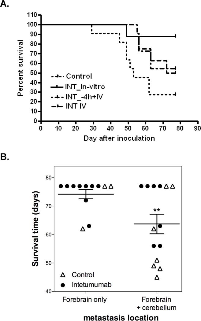
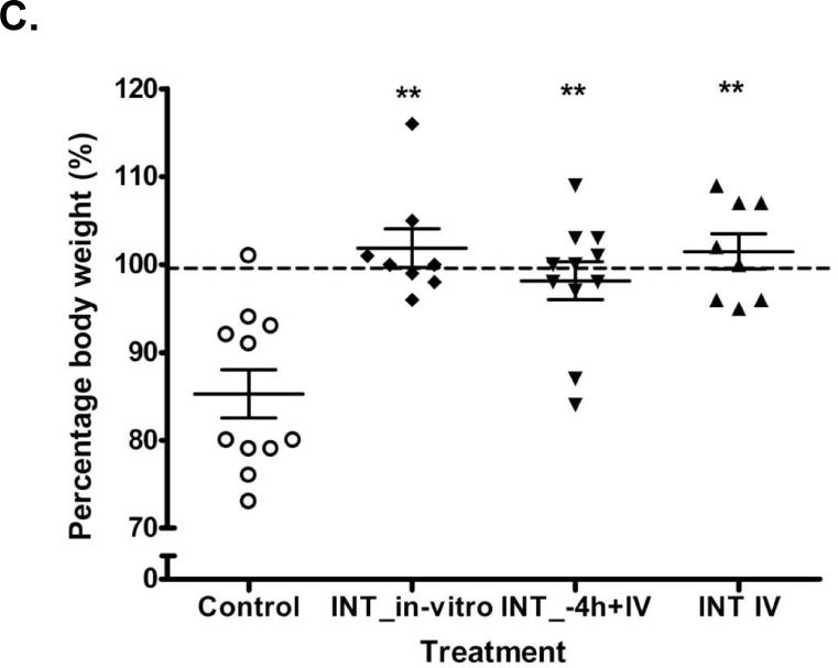
A) Kaplan-Meier survival curve: control (dotted line); INT_in-vitro (solid line); INT_-4h+IV (dashed line) and INT_IV (dotted/dashed line). The median survival time was significantly (p=0.0277) improved by intetumumab treatment. B) Brain metastasis location and survival. Data are presented as dot plot of individual rats with mean ± SEM. Lesions within the cerebellum were found more frequently in control rats and were associated with shorter survival (P=0.0058) by Students t-test. C) Plot of percentage of body weight change among the treatment groups. Intetumumab significantly prevented body weight loss compared to control rats. Data were presented as dot plot of individual rat with mean ± SEM. ** indicates p < 0.01.
All rats with histologically confirmed brain metastases had tumors within anterior brain sections (olfactory bulb through hippocampus) but only half of rats had metastases localized in the cerebellum. Twelve rats, including 3 of 9 control rats and 9 of 17 intetumumab-treated rats, had no tumor in cerebellum and survival was 74.2 ± 5.6 d. In contrast, in 14 rats (6 of 9 control and 8 of 17 intetumumab-treated) with tumor in cerebellum, survival was 63.7 ± 12.9 d (p=0.0157; Figure 4B). There was no obvious toxicity observed in rats given multiple doses of intetumumab (10 mg/kg IV). Control animals showed significant weight loss (−13.1 ± 2.3%, n=11). Intetumumab significantly (p < 0.001) prevented body weight loss independent of treatment paradigm (Figure 4C).
Two additional studies using an intracerebral xenograft model showed lack of direct anti-tumor effects of intetumumab. If 231BR-HER2 cells were pretreated with saline prior to implantation, tumor volume at 21 d was 59 ± 14, versus 87 ± 33 mm3 tumor volume when cells were pre-treated with intetumumab (p=0.24; data not shown). In other rats with untreated intracerebral xenografts, survival was 24.0 ± 5.7 d (n=13) and histological tumor volume of 75 ± 18 mm3 (n=5). Rats given a single dose of IV intetumumab 3 d after tumor implantation had a 20.3 ± 3.2 d survival (n=12), with histological tumor volume of 90 ± 19 mm3 (n=10; data not shown).
Discussion
Intetumumab and other integrin-targeting agents
In this study we demonstrate that intetumumab anti-αV integrin mAb delayed the onset and decreased the total incidence of brain metastases, and significantly improved overall survival in a hematogenous brain metastasis model. A similar result was reported in nude rats by Ning et al.[26], who found that weekly IP administration of intetumumab reduced pulmonary metastases after IV injection of lung carcinoma cells, with over half of the animals showing no pulmonary metastases. Intetumumab also decreased spontaneous metastasis of breast cancer to the lung in both an orthotropic and tail vein injection model in mice [25]. Unfortunately, brain metastases were not assessed in either study.
Other integrin-targeting agents have also shown anti-tumor and anti-metastatic characteristics [13, 15, 27]. Cilengitide, a cyclic peptide targeted to αVβ3 and αVβ5 integrins, inhibited breast cancer bone metastasis in mice [13]. The mAb etaracizumab, which specifically targets αVβ3 integrin, inhibits melanoma xenograft growth in mice, although metastasis was not evaluated [15]. Knockdown integrin αV was found to lose metastatic phenotype in human prostate cancer cells [28, 29]. Antisense αV suppresses tumor growth more strongly than antisense β3 treatment [30], suggesting that targeting a pan-αV by intetumumab would be more effective.
Intetumumab mechanisms
The αV integrins are expressed on the surface of most epithelial tumors and expression is altered in tumor progression and metastasis [8, 31]. Integrin signalling through growth factors and cytokines induces tumor cell growth and motility and blocks apoptosis [7, 32]. The absence of αVβ1 and αVβ3 mediated attachment to the ECM can trigger apoptosis [33]. Low dose intetumumab decreased breast tumor cell viability in vitro by blocking binding to vitronectin [25]. However, we found that intetumumab had no direct cytotoxic effect of breast cancer cells both in vitro and in vivo with treating 231BR-HER2 cells with intetumumab in vitro prior to direct intracerebral implantation. This effect was supported by the equivalent efficacy of delayed intetumumab treatment INT_IV and INT_-4h+IV and INT_IV groups in the hematogenous model.
Chen et al. showed that intetumumab prevents integrin binding to ECM molecules [25], which prevents downstream molecular interactions for cancer cell migration, invasion, and successful metastasis [12, 32]. Several possible integrin dimers might be targeted by intetumumab including integrin αVβ6, a key regulator in cancer cell invasion and migration [34], and integrins αVβ1 and αVβ3, which regulate cellular protrusion and mobility [35]. In addition, our results with nanoparticle labeling shows that blocking αV integrins with intetumumab prior to cell infusion did not decrease tumor cell brain localization despite the transient inhibition of cell adhesion in vitro.
The determination of a dormant or proliferative state in cancer cells is regulated by integrin signaling through the ECM [36]. Intetumumab-treated metastatic cancer cells might fail to adhere to ECM properly and enter dormancy, and will remain dormant until integrin dependant signaling shifted the cells into a proliferative state. Expression of αV integrins is also elevated in endothelial cells during angiogenesis [37, 38], and is implicated in signal transduction by vascular endothelial growth factor (VEGF) in tumor angiogenesis [39]. In contrast to a previous anti-angiogenetic report [18], we saw no obvious decrease of angiogenesis by intetumumab, as assessed the blood vessel markers VEGFR1/CD31 staining (not shown). However, blood vessel number and size is very heterogeneous among 231BR-HER2 breast cancer brain metastases [40]. We found that intetumumab increased tumor blood volume on dynamic MRI and blood vessel size not number on histology in a lung cancer brain metastasis model [24]. Based upon our results, we conclude that intetumumab reduced brain metastases in this hematogenous model mainly at the cell invasion/migration across brain vasculature and/or dormancy but not through direct cytotoxic effect to the cancer cells.
Limitations of study and clinical significance of intetumumab
The major limitations of this study are: 1) a single cell line was used, additional studies will test other brain seeking cancer cell lines (breast, lung and melanoma); 2) technical limitations of the in vivo cell trafficking with in vitro iron oxide labeling cancer cells by MRI detection and quantification; 3) The treatment regimen for maximal efficacy (dosage, timing and frequency of antibody administration) need be further defined; and 4) detailed molecular mechanism(s) of brain metastasis regulated by intetumumab remained unclear and need to be delineated.
Intetumumab has undergone Phase I and II clinical trials in treatment of different tumors and appears to be safe without major cytotoxicity [17, 19–21]. These studies demonstrated partial responses and improved overall survival in patients treated with intetumumab in combination with chemotherapy, demonstrating a therapeutic effect of blocking αv integrins in systemic cancers [17, 19–21]. Unfortunately, metastasis incidence was not reported in these clinical studies. Phase III therapeutic trials of intetumumab are planned, which will give us the opportunity to assess the impact on brain metastases.
Our results emphasize the potential prophylactic effect of intetumumab to prevent breast cancer metastases. We suggest that treatment with intetumumab may be especially important to prevent metastasis when tumor cells are released into the vasculature in breast cancer patients who undergo mastectomy. In conclusion, our studies illustrate a promising approach for the prevention of metastatic breast disease by intetumumab. These anti-metastatic effects could ultimately be extended to preventing metastasis in patients with other αV integrin-overexpressing malignancies, including lung carcinoma and melanoma.
Acknowledgments
The authors thank Sheila Taylor, Ryan Kartheiser, Murat Oztaskin and Seth Lewin for their technical assistance. This study was supported by a sponsored research agreement from Ortho Biotech Oncology R&D, DOD center of excellence grant (Pat Steeg PI), and NIH grants: NS053468, CA137488, and NS44687 to EAN.
Nonstandard abbreviations used
- INT
intetumumab
- ECM
extracellular matrix
- VEGF
vascular endothelial growth factor
Footnotes
Conflict of Interest: This study was financially sponsored in part by Ortho Biotech Oncology R&D., the manufacturer of the intetumumab antibody. DJM is an employee and stockholder of Ortho Biotech Oncology R&D. The other authors currently have no financial interest and affiliation in this agent, its developer Ortho Biotech Oncology R&D, or its parent company Johnson & Johnson.
References
- 1.Palmieri D, Smith QR, Lockman PR, Bronder J, Gril B, Chambers AF, Weil RJ, Steeg PS. Brain metastases of breast cancer. Breast Dis. 2006;26:139–147. doi: 10.3233/bd-2007-26112. [DOI] [PubMed] [Google Scholar]
- 2.Gril B, Palmieri D, Bronder JL, Herring JM, Vega-Valle E, Feigenbaum L, Liewehr DJ, Steinberg SM, Merino MJ, Rubin SD, Steeg PS. Effect of lapatinib on the outgrowth of metastatic breast cancer cells to the brain. J Natl Cancer Inst. 2008;100:1092–1103. doi: 10.1093/jnci/djn216. [DOI] [PMC free article] [PubMed] [Google Scholar]
- 3.Palmieri D, Bronder JL, Herring JM, Yoneda T, Weil RJ, Stark AM, Kurek R, Vega-Valle E, Feigenbaum L, Halverson D, Vortmeyer AO, Steinberg SM, Aldape K, Steeg PS. Her-2 overexpression increases the metastatic outgrowth of breast cancer cells in the brain. Cancer Res. 2007;67:4190–4198. doi: 10.1158/0008-5472.CAN-06-3316. [DOI] [PubMed] [Google Scholar]
- 4.Wu YJ, Muldoon LL, Dickey DT, Lewin SJ, Varallyay CG, Neuwelt EA. Cyclophosphamide enhances human tumor growth in nude rat xenografted tumor models. Neoplasia. 2009;11:187–195. doi: 10.1593/neo.81352. [DOI] [PMC free article] [PubMed] [Google Scholar]
- 5.Santarelli JG, Sarkissian V, Hou LC, Veeravagu A, Tse V. Molecular events of brain metastasis. Neurosurg Focus. 2007;22:E1. doi: 10.3171/foc.2007.22.3.2. [DOI] [PubMed] [Google Scholar]
- 6.Desgrosellier JS, Cheresh DA. Integrins in cancer: biological implications and therapeutic opportunities. Nat Rev Cancer. 2010;10:9–22. doi: 10.1038/nrc2748. [DOI] [PMC free article] [PubMed] [Google Scholar]
- 7.Guo W, Giancotti FG. Integrin signalling during tumour progression. Nat Rev Mol Cell Biol. 2004;5:816–826. doi: 10.1038/nrm1490. [DOI] [PubMed] [Google Scholar]
- 8.Hood JD, Cheresh DA. Role of integrins in cell invasion and migration. Nat Rev Cancer. 2002;2:91–100. doi: 10.1038/nrc727. [DOI] [PubMed] [Google Scholar]
- 9.Lorger M, Krueger JS, O’Neal M, Staflin K, Felding-Habermann B. Activation of tumor cell integrin alphavbeta3 controls angiogenesis and metastatic growth in the brain. Proceedings of the National Academy of Sciences of the United States of America. 2009;106:10666–10671. doi: 10.1073/pnas.0903035106. [DOI] [PMC free article] [PubMed] [Google Scholar]
- 10.Arihiro K, Kaneko M, Fujii S, Inai K, Yokosaki Y. Significance of alpha 9 beta 1 and alpha v beta 6 integrin expression in breast carcinoma. Breast Cancer. 2000;7:19–26. doi: 10.1007/BF02967183. [DOI] [PubMed] [Google Scholar]
- 11.Bandyopadhyay A, Raghavan S. Defining the role of integrin alphavbeta6 in cancer. Curr Drug Targets. 2009;10:645–652. doi: 10.2174/138945009788680374. [DOI] [PMC free article] [PubMed] [Google Scholar]
- 12.Desgrosellier JS, Barnes LA, Shields DJ, Huang M, Lau SK, Prevost N, Tarin D, Shattil SJ, Cheresh DA. An integrin alpha(v)beta(3)-c-Src oncogenic unit promotes anchorage-independence and tumor progression. Nat Med. 2009;15:1163–1169. doi: 10.1038/nm.2009. [DOI] [PMC free article] [PubMed] [Google Scholar]
- 13.Bauerle T, Komljenovic D, Merz M, Berger MR, Goodman SL, Semmler W. Cilengitide inhibits progression of experimental breast cancer bone metastases as imaged noninvasively using VCT, MRI and DCE-MRI in a longitudinal in vivo study. International journal of cancer Journal international du cancer. 2011;128:2453–2462. doi: 10.1002/ijc.25563. [DOI] [PubMed] [Google Scholar]
- 14.Delbaldo C, Raymond E, Vera K, Hammershaimb L, Kaucic K, Lozahic S, Marty M, Faivre S. Phase I and pharmacokinetic study of etaracizumab (Abegrin), a humanized monoclonal antibody against alphavbeta3 integrin receptor, in patients with advanced solid tumors. Invest New Drugs. 2008;26:35–43. doi: 10.1007/s10637-007-9077-0. [DOI] [PubMed] [Google Scholar]
- 15.Mulgrew K, Kinneer K, Yao XT, Ward BK, Damschroder MM, Walsh B, Mao SY, Gao C, Kiener PA, Coats S, Kinch MS, Tice DA. Direct targeting of alphavbeta3 integrin on tumor cells with a monoclonal antibody, Abegrin. Molecular cancer therapeutics. 2006;5:3122–3129. doi: 10.1158/1535-7163.MCT-06-0356. [DOI] [PubMed] [Google Scholar]
- 16.Reardon DA, Nabors LB, Stupp R, Mikkelsen T. Cilengitide: an integrin-targeting arginine-glycine-aspartic acid peptide with promising activity for glioblastoma multiforme. Expert Opin Investig Drugs. 2008;17:1225–1235. doi: 10.1517/13543784.17.8.1225. [DOI] [PMC free article] [PubMed] [Google Scholar]
- 17.Mullamitha SA, Ton NC, Parker GJ, Jackson A, Julyan PJ, Roberts C, Buonaccorsi GA, Watson Y, Davies K, Cheung S, Hope L, Valle JW, Radford JA, Lawrance J, Saunders MP, Munteanu MC, Nakada MT, Nemeth JA, Davis HM, Jiao Q, Prabhakar U, Lang Z, Corringham RE, Beckman RA, Jayson GC. Phase I evaluation of a fully human anti-alphav integrin monoclonal antibody (CNTO 95) in patients with advanced solid tumors. Clinical cancer research : an official journal of the American Association for Cancer Research. 2007;13:2128–2135. doi: 10.1158/1078-0432.CCR-06-2779. [DOI] [PubMed] [Google Scholar]
- 18.Trikha M, Zhou Z, Nemeth JA, Chen Q, Sharp C, Emmell E, Giles-Komar J, Nakada MT. CNTO 95, a fully human monoclonal antibody that inhibits alphav integrins, has antitumor and antiangiogenic activity in vivo. International journal of cancer Journal international du cancer. 2004;110:326–335. doi: 10.1002/ijc.20116. [DOI] [PubMed] [Google Scholar]
- 19.O’Day S, Pavlick A, Loquai C, Lawson D, Gutzmer R, Richards J, Schadendorf D, Thompson JA, Gonzalez R, Trefzer U, Mohr P, Ottensmeier C, Chao D, Zhong B, de Boer CJ, Uhlar C, Marshall D, Gore ME, Lang Z, Hait W, Ho P. A randomised, phase II study of intetumumab, an anti-alphav-integrin mAb, alone and with dacarbazine in stage IV melanoma. British journal of cancer. 2011;105:346–352. doi: 10.1038/bjc.2011.183. [DOI] [PMC free article] [PubMed] [Google Scholar]
- 20.O’Day SJ, Pavlick AC, Albertini MR, Hamid O, Schalch H, Lang Z, Ling J, Mata M, Reddy M, Foster B. Clinical and pharmacologic evaluation of two dose levels of intetumumab (CNTO 95) in patients with melanoma or angiosarcoma. Investigational new drugs. 2011 doi: 10.1007/s10637-011-9639-z. [DOI] [PubMed] [Google Scholar]
- 21.Chu FM, Picus J, Fracasso PM, Dreicer R, Lang Z, Foster B. A phase 1, multicenter, open-label study of the safety of two dose levels of a human monoclonal antibody to human alpha(v) integrins, intetumumab, in combination with docetaxel and prednisone in patients with castrate-resistant metastatic prostate cancer. Investigational new drugs. 2011;29:674–679. doi: 10.1007/s10637-010-9388-4. [DOI] [PubMed] [Google Scholar]
- 22.Wu YJ, Muldoon LL, Neuwelt EA. The chemoprotective agent N-acetylcysteine blocks cisplatin-induced apoptosis through caspase signaling pathway. The Journal of pharmacology and experimental therapeutics. 2005;312:424–431. doi: 10.1124/jpet.104.075119. [DOI] [PubMed] [Google Scholar]
- 23.Arbab AS, Yocum GT, Wilson LB, Parwana A, Jordan EK, Kalish H, Frank JA. Comparison of transfection agents in forming complexes with ferumoxides, cell labeling efficiency, and cellular viability. Mol Imaging. 2004;3:24–32. doi: 10.1162/15353500200403190. [DOI] [PubMed] [Google Scholar]
- 24.Muldoon LL, Gahramanov S, Li X, Marshall DJ, Kraemer DF, Neuwelt EA. Dynamic magnetic resonance imaging assessment of vascular targeting agent effects in rat intracerebral tumor models. Neuro-Oncol. 2011;13:51–60. doi: 10.1093/neuonc/noq150. [DOI] [PMC free article] [PubMed] [Google Scholar]
- 25.Chen Q, Manning CD, Millar H, McCabe FL, Ferrante C, Sharp C, Shahied-Arruda L, Doshi P, Nakada MT, Anderson GM. CNTO 95, a fully human anti alphav integrin antibody, inhibits cell signaling, migration, invasion, and spontaneous metastasis of human breast cancer cells. Clin & Experimental Metastasis. 2008;25:139–148. doi: 10.1007/s10585-007-9132-4. [DOI] [PubMed] [Google Scholar]
- 26.Ning S, Tian J, Marshall DJ, Knox SJ. Anti-{alpha}v integrin monoclonal antibody intetumumab enhances the efficacy of radiation therapy and reduces metastasis of human cancer xenografts in nude rats. Cancer research. 2010;70:7591–7509. doi: 10.1158/0008-5472.CAN-10-1639. [DOI] [PubMed] [Google Scholar]
- 27.MacDonald TJ, Taga T, Shimada H, Tabrizi P, Zlokovic BV, Cheresh DA, Laug WE. Preferential susceptibility of brain tumors to the antiangiogenic effects of an alpha(v) integrin antagonist. Neurosurgery. 2001;48:151–157. doi: 10.1097/00006123-200101000-00026. [DOI] [PubMed] [Google Scholar]
- 28.van den Hoogen C, van der Horst G, Cheung H, Buijs JT, Pelger RC, van der Pluijm G. Integrin alphav expression is required for the acquisition of a metastatic stem/progenitor cell phenotype in human prostate cancer. Am J Pathol. 2011;179:2559–2568. doi: 10.1016/j.ajpath.2011.07.011. [DOI] [PMC free article] [PubMed] [Google Scholar]
- 29.van der Horst G, van den Hoogen C, Buijs JT, Cheung H, Bloys H, Pelger RC, Lorenzon G, Heckmann B, Feyen J, Pujuguet P, Blanque R, Clement-Lacroix P, van der Pluijm G. Targeting of alpha(v)-integrins in stem/progenitor cells and supportive microenvironment impairs bone metastasis in human prostate cancer. Neoplasia. 2011;13:516–525. doi: 10.1593/neo.11122. [DOI] [PMC free article] [PubMed] [Google Scholar]
- 30.Li J, Tan H, Dong X, Xu Z, Shi C, Han X, Jiang H, Krissansen GW, Sun X. Antisense integrin alphaV and beta3 gene therapy suppresses subcutaneously implanted hepatocellular carcinomas. Dig Liver Dis. 2007;39:557–565. doi: 10.1016/j.dld.2007.01.025. [DOI] [PubMed] [Google Scholar]
- 31.Ramsay AG, Marshall JF, Hart IR. Integrin trafficking and its role in cancer metastasis. Cancer metastasis reviews. 2007;26:567–578. doi: 10.1007/s10555-007-9078-7. [DOI] [PubMed] [Google Scholar]
- 32.Mitra SK, Schlaepfer DD. Integrin-regulated FAK-Src signaling in normal and cancer cells. Curr Opin Cell Biol. 2006;18:516–523. doi: 10.1016/j.ceb.2006.08.011. [DOI] [PubMed] [Google Scholar]
- 33.Stupack DG, Cheresh DA. A Bit-role for integrins in apoptosis. Nat Cell Biol. 2004;6:388–389. doi: 10.1038/ncb0504-388. [DOI] [PubMed] [Google Scholar]
- 34.Marsh D, Dickinson S, Neill GW, Marshall JF, Hart IR, Thomas GJ. Alpha v beta 6 Integrin promotes the invasion of morphoeic basal cell carcinoma through stromal modulation. Cancer research. 2008;68:3295–3303. doi: 10.1158/0008-5472.CAN-08-0174. [DOI] [PubMed] [Google Scholar]
- 35.Karadag A, Ogbureke KU, Fedarko NS, Fisher LW. Bone sialoprotein, matrix metalloproteinase 2, and alpha(v)beta3 integrin in osteotropic cancer cell invasion. Journal of the National Cancer Institute. 2004;96:956–965. doi: 10.1093/jnci/djh169. [DOI] [PubMed] [Google Scholar]
- 36.Barkan D, Kleinman H, Simmons JL, Asmussen H, Kamaraju AK, Hoenorhoff MJ, Liu ZY, Costes SV, Cho EH, Lockett S, Khanna C, Chambers AF, Green JE. Inhibition of metastatic outgrowth from single dormant tumor cells by targeting the cytoskeleton. Cancer research. 2008;68:6241–6250. doi: 10.1158/0008-5472.CAN-07-6849. [DOI] [PMC free article] [PubMed] [Google Scholar]
- 37.Avraamides CJ, Garmy-Susini B, Varner JA. Integrins in angiogenesis and lymphangiogenesis. Nat Rev Cancer. 2008;8:604–617. doi: 10.1038/nrc2353. [DOI] [PMC free article] [PubMed] [Google Scholar]
- 38.Stupack DG, Cheresh DA. Integrins and angiogenesis. Curr Top Dev Biol. 2004;64:207–238. doi: 10.1016/S0070-2153(04)64009-9. [DOI] [PubMed] [Google Scholar]
- 39.Eliceiri BP, Cheresh DA. Role of alpha v integrins during angiogenesis. Cancer J. 2000;6(Suppl 3):S245–S249. [PubMed] [Google Scholar]
- 40.Lockman PR, Mittapalli RK, Taskar KS, Rudraraju V, Gril B, Bohn KA, Adkins CE, Roberts A, Thorsheim HR, Gaasch JA, Huang S, Palmieri D, Steeg PS, Smith QR. Heterogeneous Blood-Tumor Barrier Permeability Determines Drug Efficacy in Mouse Brain Metastases of Breast Cancer. Clinical cancer research : an official journal of the American Association for Cancer Research. 2010;16:5664–5678. doi: 10.1158/1078-0432.CCR-10-1564. [DOI] [PMC free article] [PubMed] [Google Scholar]