



Abstract
Hepatitis E virus (HEV) is one of the most common causes of acute hepatitis in tropical and temperate climates. Tropical genotypes 1 and 2 are associated with food-borne and waterborne transmission. Zoonotic reservoirs (mainly pigs, wild boar, and deer) are considered for genotypes 3 and 4, which exist in temperate climates. In view of the association of several zoonotic viruses with bats, we analyzed 3,869 bat specimens from 85 different species and from five continents for hepevirus RNA. HEVs were detected in African, Central American, and European bats, forming a novel phylogenetic clade in the family Hepeviridae. Bat hepeviruses were highly diversified and comparable to human HEV in sequence variation. No evidence for the transmission of bat hepeviruses to humans was found in over 90,000 human blood donations and individual patient sera. Full-genome analysis of one representative virus confirmed formal classification within the family Hepeviridae. Sequence- and distance-based taxonomic evaluations suggested that bat hepeviruses constitute a distinct genus within the family Hepeviridae and that at least three other genera comprising human, rodent, and avian hepeviruses can be designated. This may imply that hepeviruses invaded mammalian hosts nonrecently and underwent speciation according to their host restrictions. Human HEV-related viruses in farmed and peridomestic animals might represent secondary acquisitions of human viruses, rather than animal precursors causally involved in the evolution of human HEV.
INTRODUCTION
A third of the world's human population may have been infected with hepatitis E virus (HEV), the prototype of the family Hepeviridae (59). HEVs are small, nonenveloped viruses with an approximately 7,200-nucleotide (nt) positive-sense, single-stranded RNA genome. Human HEV is classified into four genotypes (41). Following food-borne and waterborne fecal-oral infection, clinical symptoms range from asymptomatic to severe hepatitis (57). Hepatitis B virus and HEV constitute the most common causes of acute hepatitis in developing countries (57). In industrialized countries, hepatitis E has long been considered rare, but there is now a growing body of evidence suggesting that particularly HEV genotypes 3 and 4 constitute major etiologies of acute viral hepatitis (13). Moreover, HEV was found to be present in blood donors at rates between 0.07% in Chinese blood donors and 19.5% in Japanese donors with elevated liver enzyme levels (27, 63) as well as in plasma fractionation pools at rates of up to 10% (5). HEV infection can become chronic in immunocompromised patients and has been associated with high rates of mortality in pregnant women (6, 14, 36). Along with a growing awareness of the clinical relevance of HEV, candidate vaccines have been developed and tested in phase II and III clinical trials (64, 72, 75).
HEV differs from all other human hepatitis viruses in that it has been linked to animal reservoirs, beginning with its isolation from swine in 1997 (43). In particular, HEV genotypes 3 and 4 are associated foremost with food-borne zoonotic transmission from deer, domestic pigs, and wild boar (55, 58, 68). Antibodies against human HEV have been detected in several other animal species, including cattle, sheep, and horses (55). Zoonotic infection seems to contribute to a high seroprevalence in swine handlers and veterinarians (7, 22, 42, 44). Critically, those viruses detected in livestock animals such as swine, deer, farmed rabbits, and mongooses all cluster closely with human viruses (12, 23, 50, 73). Beyond this monophyletic clade, more divergent animal hepeviruses have been described recently. Rat sera were long known to cross-react with human HEV antigens serologically (16, 55), and a distinct hepevirus lineage associated with mild hepatitis in infected animals was detected in wild Norway rats (31, 38, 60). Avian hepeviruses have been identified in farmed chickens but in no other birds so far (28). These viruses are globally widespread in poultry farms, causing a range of symptoms from asymptomatic infection to severe hepatitis and splenomegaly (40, 74). The genetically most distant animal hepevirus was already isolated from apparently healthy trout in 1988 but was identified as a member of the family Hepeviridae only in 2011 (3).
Because of the zoonotic nature of HEV infection, it is highly relevant to learn more about its genetic diversity and association with mammalian hosts. Current data suggest the existence of specific virus clades in specific mammalian hosts, but the restriction of previous studies to farmed and peridomestic animals provided little opportunity to investigate the origin of human viruses. The phylogenetic placement of viruses of several unrelated animal taxa with human viruses suggests an acquisition from humans, but data are insufficient to reject alternative hypotheses. Critically, the very recent detection of rodent viruses in a sister relationship with human HEV suggests the existence of a wider, undiscovered diversity of Hepeviridae in mammals. In several studies by us and other groups, bats have proven to be highly efficient indicators of mammalian virus diversity, which may be due to their exceptionally large social group sizes, which promote the acquisition and maintenance of viruses (8, 15, 19, 20, 69, 70). Bats have been linked to a growing number of emerging viruses, including lyssaviruses, coronaviruses, henipaviruses, and filoviruses (8). For all of these viruses, bats carry larger viral diversities than other mammals, supporting the notion that bats might act as viral reservoirs. To examine if bats may also play a specific role in the ecology and evolution of mammalian Hepeviridae, we investigated a globally representative biological sample from 85 different bat species, including over 3,000 specimens. Our results suggest the existence of a genetic clade of viruses whose patristic distance resembles that encountered in rodents and humans (including the associated livestock viruses), altogether yielding genetic criteria to define putative genera within the family Hepeviridae. We conclude that three putative genera of mammalian hepeviruses may exist.
MATERIALS AND METHODS
Bat sampling and specimen preparation.
Bat fecal and blood specimens were collected in Germany, Bulgaria, Spain, Ghana, Gabon, Papua New Guinea, Australia, Costa Rica, and Panama throughout 2002 to 2011 (Fig. 1) (sampling permits are provided in Acknowledgments). Bats were caught by using mist nets and kept in individual cloth bags until examination by trained ecologists. Fecal samples were collected directly from individual bags or from plastic sheets placed onto the ground below bat roost sites and stored after suspension in RNAlater solution (Qiagen, Hilden, Germany) at 8°C until further processing. Blood was taken by puncturing the wing or tail veins. Bats were released unharmed at the capture site the same night. Bat organs were available from 35 animals found dead in Germany and delivered to centers for bat protection and from 37 Eidolon helvum bats from a study site in Ghana (18).
Fig 1.

Sampling sites and covered bat evolutionary lineages. (A) Sampling sites and numbers of sampled species and specimens per family. (B) Bat evolutionary lineages according to data reported previously (67). Bat families for which samples were tested in this study are shown in boldface type. The names of bat species which tested positive for hepeviruses are shown in red type next to their family designations.
Viral RNA was purified from bat fecal and blood specimens by using the viral RNA minikit and from solid-organ specimens by using the RNeasy minikit (both from Qiagen).
Human specimens.
Sera were collected in 1998 from 453 otherwise healthy HIV-infected patients in Cameroon for studies of HIV and hepatitis C virus (51). The anonymized samples were extracted in pools of 10 to 40 by using the viral RNA minikit (Qiagen). Anonymized blood donor plasma samples collected from 2009 to 2010 in Germany were pooled in up to 96-member pools as described previously (61). Briefly, 96-member pools containing 100 μl of individual plasma donations were concentrated by ultracentrifugation, followed by RNA purification and elution in 65 μl. Due to the scarcity of available material, 20 RNA samples from these pools were merged before testing. The total number of analyzed individual donations was 93,146.
Screening for hepeviruses.
Screening for novel hepeviruses was done by heminested reverse transcription (RT)-PCR using broadly reactive oligonucleotides targeting viral RNA-dependent RNA polymerase (RdRp). The assay was designed to amplify all members of the family Hepeviridae available in GenBank. The assay sensitivity was determined to be on the order of 10 copies per reaction by using a quantified in vitro transcript (HEV genotype 3). For the generation of transcript controls, the screening RT-PCR amplicons were TA cloned (Invitrogen, Karlsruhe, Germany), reamplified with vector-specific primers, and in vitro transcribed by using T7 RNA polymerase (Asuragen, Austin, TX). Further broadly reactive nested RT-PCR assays were designed for additional genomic regions to enable a full-genome characterization (Table 1). Additional primers used for full-genome characterization are available upon request.
Table 1.
Oligonucleotides used for RT-PCR screening, complete genome sequencing, and virus quantification
| Virus targeted | Oligonucleotidea | Sequence (5′–3′)b | Polarity | Use |
|---|---|---|---|---|
| Hepeviridae | HEV-F4228 | ACYTTYTGTGCYYTITTTGGTCCITGGTT | + | Heminested screening RT-PCR |
| HEV-R4598 | GCCATGTTCCAGAYGGTGTTCCA | − | ||
| HEV-R4565 | CCGGGTTCRCCIGAGTGTTTCTTCCA | − | ||
| HEV-BS7like-R4602 | ACGACCATRTTCCAIACIGT | − | 5′ completion of 1st-round screening amplicons | |
| HEV-F14a | GTGGTCGATGCCATGGAGGCCCATCAGT | + | Heminested RT-PCR assay 1 for amplification of small sequence fragments along Hepeviridae genomes to permit full-genome sequencing by lineage-specific bridging primers | |
| HEV-F14b | GCCAGGGTAAGAATGGACGTCTCGCAGT | + | ||
| HEV-F14c | GCAACCCCCGATGGAGACCCATCAGT | + | ||
| HEV-F79a | GGCWGCTYTGGCWGCGGC | + | ||
| HEV-F79b | GGCTACTGCGGCGGCGGC | + | ||
| HEV-F102 | CYGYCTTGGCGAATGCTGTGGTGGT | + | ||
| HEV-R106 | GGCGGTGTTCGCTGCAGCTAGAGYWGC | − | ||
| HEV-R390a | GGGGCAGAATACCAGCGCTG | − | ||
| HEV-R390b | GGGGCTGAGTACCAGCGCTG | − | ||
| HEV-R390c | GGGGCGGTGTACCACCGCTG | − | ||
| HEV-R390d | GGGCAATCTCGCCAGCGCTG | − | ||
| HEV-F400 | GGIMGIGAYGTICAGCGITGG | + | Heminested RT-PCR assay 2 for full-genome sequencing | |
| HEV-F795 | GGGCIRTIGGITGYCAYTTYGT | + | ||
| HEV-F848 | CCIATGCCITAYGTICCITACCC | + | ||
| HEV-R1045 | GTCAKIAGICKIGARCARCARARIGC | − | ||
| HEV-R1065 | ATVCCICGIAGRTAIGTCATDAG | − | ||
| HEV-R1075 | GTIACYTTGTAYSWRATICCICGIAGRTAIGTCA | − | ||
| HEV-F5760 | CTGACGTTTTCGACCTGTCGT | + | Heminested RT-PCR 3 for full-genome sequencing | |
| HEV-F5770 | GCGTCTGTCGGTGGGTTTTC | + | ||
| HEV-F5790 | GGCCACAGTCCAACAATGTTC | + | ||
| HEV-R5780 | CTTATAGAAAACCCACCGACAGAC | − | ||
| HEV-R5810 | ATGTTGGAACATTGTTGGACTGTG | − | ||
| HEV-R5960 | GTYTCGACAGAGCGCCAICC | − | ||
| HEV-R6473 | CCIAGGTCTATRTCGTGIGG | − | ||
| HEV-R6493a | TCCTGCTCRTGCTGRTTATCATARTCCTG | − | ||
| HEV-R6493b | TCCTGGAGRTGCTGRTTATCAAARTCCTG | − | ||
| HEV-F6376a | GTCTCGGCCAATGGCGAGCC | + | Heminested RT-PCR 4 for full-genome sequencing | |
| HEV-F6376b | GTGTCTGAGAACGGTGAGCC | + | ||
| HEV-F6493a | CAGGAYTATGATAAYCAGCAYGAGCAGGA | + | ||
| HEV-F6493b | CAGGAYTTTGATAAYCAGCAYCTCCAGGA | + | ||
| HEV-R6865 | CRGTRGTRTTRTAATTRTARGGRTARCCRGC | − | ||
| M. bechsteinii bat hepevirus | HEV-NM8AC-rtF | TGGGTGGTTTTATGGTGATCTCT | + | Virus-specific quantitative real-time RT-PCR assays |
| HEV-NM8AC-rtP | FAM-AGGCCGACTTGCACGCGCA-BBQ1 | + (probe) | ||
| HEV-NM8AC-rtR | CGTCAGGCACAGCCATAGC | − | ||
| M. daubenonii bat hepevirus | HEV-NMS09-B-R-rtF | GCCCTGGAAAAGCGTATTGTT | + | |
| HEV-NMS09-B-R-rtP | FAM-TCAGCTTCCCCCTGGCTGGTTTTATG-BHQ1 | + (probe) | ||
| HEV-NMS09-B-R-rtR | TGAAGGTCAGCCTCAGTATAAAGRT | − | ||
| H. abae bat hepevirus | HEV-G19E-rtF | CCTGGTTGGTTCTATGGTGATCT | + | |
| HEV-G19E-rtP | FAM-AATCAGACCTGCATGCTCACACTATGGCT-BHQ1 | + (probe) | ||
| HEV-G19E-rtR | TCTCAAAAACCTTACAGCCATCAG | − | ||
| E. serotinus bat hepevirus | HEV-BS7-rtF | GCTGGTTTTACGGCGACTTG | + | |
| HEV-BS7-rtP | FAM-ATACCGAGGCTGATCTG-BHQ1 | + (probe) | ||
| HEV-BS7-rtR | AGGAACTGCCATTGCATGTG | − | ||
| V. caraccioli bat hepevirus | HEV-Pan-rtF | CCGGGCGATAGAAAAGCAT | + | |
| HEV-Pan-rtP | FAM-TTGTTGCACAGCTGCCACCTGGAT-BHQ1 | + (probe) | ||
| HEV-Pan-rtR | GGTGTAAGCGTGTATGTCAGACTCA | − |
Named after the position in the reference sequence under GenBank accession number NC_001434.
R is G/A, Y is C/T, S is G/C, W is A/T, M is A/C, K is G/T, H is A/C/T, B is C/G/T, N is A/T/C/G, and I is inosine. FAM, 6-carboxyfluorescein; BBQ/BHQ1, blackberry quencher/black hole quencher 1.
First-round RT-PCRs were carried out by using a touchdown protocol with reverse transcription at 50°C for 20 min and subsequent PCR at 95°C for 3 min, 10 cycles with 95°C for 15 s, a 1°C touchdown decrease of the annealing temperature down to 50°C, and extension at 68°C for 30 s, followed by another 40 cycles at an annealing temperature of 52°C. Second-round reactions used the same cycling protocol but without the RT step. Amplicons of the expected size (approximately 371 and 338 bp in the first and second rounds, respectively) were visualized on 2.0% agarose gels with ethidium bromide staining. First-round RT-PCR was performed by using the SuperScript III (SSIII) one-step RT-PCR kit (Invitrogen) with 5 μl of RNA, 400 nM (each) first-round primers, 1 μg bovine serum albumin, 0.2 mM each deoxynucleoside triphosphate (dNTP), and 2.4 mM MgSO4. Second-round 50-μl Platinum Taq reactions were carried out as recommended by the manufacturer (Invitrogen), using 1 μl of the first-round PCR product, 2.5 mM MgCl2, and 400 nM (each) second-round primers. All PCR products were extended up to the size of the first-round fragment for phylogenetic analyses using heminested RT-PCR oligonucleotides and an additional bat hepevirus-specific reverse primer (Table 1).
Full-genome characterization.
Following cDNA synthesis using the SuperScript III reverse transcription kit (Invitrogen), amplicons from generic PCR assays were bridged by long-range PCR using gene-specific primers and the Expand high-fidelity (Roche, Mannheim, Germany) and Phusion PCR (New England BioLabs, Frankfurt, Germany) kits. Additionally, Phi29-driven whole-transcriptome amplification (WTA) was done for the enrichment of viral sequences against the DNA/RNA background in specimens. Gene-specific reverse primers in the screening RT-PCR amplicon were designed with 5′-phosphate moieties, reverse transcribed with the SSIII kit (Invitrogen), digested with RNase H (Invitrogen), and finally ligated and amplified by using the Qiagen WTA kit (Qiagen). Genome ends were amplified by using the 5′/3′ rapid amplification of cDNA ends (RACE) kit (Roche).
Quantification of novel hepeviruses.
Viral RNA quantification was done by using photometrically quantified in vitro RNA transcripts as described above and specific real-time RT-PCR assays (see Table 1 for oligonucleotides). Quantification was done by using 5 μl of RNA extract, 300 nM each primer, and 180 nM probe, using the SSIII one-step kit as described above. Cycling in a Roche LightCycler480 instrument involved the following steps: 55°C for 15 min, 95°C for 3 min, and 45 cycles of 95°C for 15 s and 58°C for 30 s with measurement of fluorescence.
Serological analysis.
An indirect immunofluorescence assay was done by using human embryonic kidney 293T (HEK293T) cells transiently expressing the full-length ORF2-encoded capsid protein from a human HEV genotype 1 isolate (A. Osterman, unpublished data). Sera (n = 49) from eight different bat species were diluted 1:40, and detection was done by using a goat anti-bat immunoglobulin G polyclonal serum (Bethyl Laboratories, Montgomery, TX), followed by a cyanine 2-labeled donkey anti-goat serum (Dianova, Hamburg, Germany), as described previously (48). Human positive-control serum was used in dilutions of 1:40 and 1:80. Cyanine 2-labeled goat anti-human serum (Dianova) was applied as a secondary antibody. Nuclei were counterstained with 4′,6-diamidino-2-phenylindole (DAPI). All pictures were taken at the corresponding microscopic settings with a Motic Axiovision microscope (Zeiss).
Nucleotide sequence accession numbers.
Nucleotide sequences from all novel bat hepeviruses described in this study are available in GenBank under accession numbers JQ001744 to JQ001749 and JQ071861, with JQ001749 representing a full bat hepevirus genome. Nucleotide sequences from human hepeviruses determined in this study are available under GenBank accesssion numbers JQ034512 to JQ034522.
RESULTS
Heminested RT-PCR was used to screen a total of 3,869 fecal, liver, and blood specimens from 85 different bat species from five continents (Table 2). Hepevirus RNA was found in seven (0.18%) specimens, including five fecal samples, one blood sample, and one liver specimen. RNA-positive samples stemmed from Europe, Central America, and Western Africa (Fig. 1A). Detected sequences originated from five bat species of three different families (Hipposideridae, Vespertilionidae, and Phyllostomidae), representing all three major stem lineages in the phylogenetic tree of bats (Fig. 1B). The detection rate in feces (5 of 2,624) was not significantly higher than those in blood (1 of 1,173) and liver (1 of 72) (P = 0.40 and 0.15, respectively, by two-tailed Fisher's exact test). Very high virus RNA concentrations were found in all fecal samples (median, 8.5 × 10⁸ RNA copies per gram of feces; range, 2.5 × 10⁸ to 6.1 × 10¹⁰ RNA copies per gram of feces). These concentrations were several orders of magnitude higher than that in the single positive blood specimen (1.8 × 10⁵ RNA copies per ml). Hepevirus RNA was also detected in a liver sample at a high concentration comparable to those detected in feces (5.4 × 10⁹ RNA copies per gram of tissue). Virus concentrations were analyzed in all solid organs of the animal whose liver tested positive. As shown in Fig. 2, RNA concentrations in liver tissue exceeded those in any other organ by at least 1,000-fold. The comparable virus concentrations in solid organs other than liver were likely the result of a high level of viremia. Unfortunately, no blood sample was available from this animal.
Table 2.
Sample characteristics
| Chiroptera family | Species | No. of samples |
No. of RT-PCR-positive samples (%) | Virus concna (specimen type) | Sampling site(s) (yr) | ||
|---|---|---|---|---|---|---|---|
| Feces | Blood | Liver | |||||
| Pteropodidae | Dobsonia praedatrix | 9 | Papua New Guinea (2002) | ||||
| Eidolon helvum | 438 | 100 | 37 | Ghana (2008/2009/2010) | |||
| Epomophorus sp. | 3 | Ghana (2009) | |||||
| Epomops franqueti | 100 | Gabon (2009) | |||||
| Hypsignathus monstrosus | 100 | Gabon (2009) | |||||
| Melonycteris melanops | 7 | Papua New Guinea (2002) | |||||
| Micropteropus sp. | 10 | Ghana (2009) | |||||
| Micropteropus pusillus | 100 | Gabon (2009) | |||||
| Myonycteris torquata | 100 | Gabon (2009) | |||||
| Nanonycteris sp. | 7 | Ghana (2009) | |||||
| Pteropus alecto | 3 | Australia (2006) | |||||
| Pteropus poliocephalus | 24 | Australia (2006) | |||||
| Rousettus aegyptiacus | 14 | 100 | Gabon (2009); Ghana (2008) | ||||
| Rousettus amplexicaudatus | 1 | Papua New Guinea (2002) | |||||
| Rhinolophidae | Rhinolophus blasii | 82 | Bulgaria (2008) | ||||
| Rhinolophus euryale | 243 | Bulgaria (2008) | |||||
| Rhinolophus ferrumequinum | 40 | Bulgaria (2008) | |||||
| Rhinolophus hipposideros | 146 | Bulgaria (2008), Spain (2010) | |||||
| Rhinolophus landeri | 1 | Ghana (2009) | |||||
| Rhinolphus mehelyi | 13 | Bulgaria (2008) | |||||
| Hipposideridae | Hipposideros abae | 57 | 2 (3.51) | 8.50 × 10⁸ (feces), 6.05 × 10¹⁰ (feces) | Ghana (2008/2009b) | ||
| Hipposideros cf. caffer-ruber | 166 | Ghana (2008/2009) | |||||
| Nycteridae | Nycteris sp. | 4 | Ghana (2008/2009) | ||||
| Noctilionidae | Noctilio leporinus | 3 | 11 | Panama (2010) | |||
| Emballonuridae | Coleura afra | 67 | Ghana (2008/2009) | ||||
| Peropteryx kappleri | 5 | Costa Rica (2010) | |||||
| Saccopteryx bilineata | 1 | 1 | Panama (2010/2011) | ||||
| Saccopteryx leptura | 2 | Panama (2011) | |||||
| Phyllostomidae | Anoura geoffroyi | 99 | Costa Rica (2010) | ||||
| Artibeus jamaicensis | 48 | 298 | Panama (2010/2011) | ||||
| Artibeus lituratus | 4 | 22 | Panama (2010/2011) | ||||
| Artibeus phaeotis | 3 | Panama (2011) | |||||
| Artibeus watsoni | 6 | Panama (2011) | |||||
| Carollia castanea | 11 | 18 | Costa Rica (2010), Panama (2010/2011) | ||||
| Carollia perspicillata | 207 | 13 | Costa Rica (2010), Panama (2010/2011) | ||||
| Desmodus rotundus | 1 | Panama (2011) | |||||
| Enchisthenes hartii | 3 | Costa Rica (2010) | |||||
| Glossophaga commissarisi | 3 | Costa Rica (2010) | |||||
| Glossophaga soricina | 28 | 11 | Costa Rica (2010), Panama (2010/2011) | ||||
| Lampronycteris brachyotis | 2 | Panama (2011) | |||||
| Lophostoma silviculum | 3 | 10 | Panama (2010/2011) | ||||
| Micronycteris hirsuta | 1 | Panama (2010) | |||||
| Micronycteris microtis | 4 | 7 | Panama (2010) | ||||
| Micronycteris minuta | 1 | Panama (2011) | |||||
| Mimon crenulatum | 1 | 2 | Panama (2010/2011) | ||||
| Phylloderma stenops | 2 | 5 | Panama (2010/2011) | ||||
| Phyllostomus discolor | 11 | Panama (2011) | |||||
| Phyllostomus hastatus | 4 | 11 | Panama (2010/2011) | ||||
| Platyrrhinus helleri | 1 | 1 | Panama (2010) | ||||
| Tonatia saurophila | 10 | 9 | Panama (2010/2011) | ||||
| Trachops cirrhosus | 4 | 8 | Panama (2010/2011) | ||||
| Uroderma bilobatum | 3 | 23 | Panama (2010/2011) | ||||
| Vampyressa pusilla | 3 | Panama (2011) | |||||
| Vampyrodes caraccioli | 10 | 1 (10.0) | 1.75 × 10⁵ (blood) | Panama (2011b) | |||
| Mormoopidae | Pteronotus parnellii | 36 | 38 | Costa Rica (2010), Panama (2010/2011) | |||
| Vespertilionidae | Barbastella barbastellus | 8 | Bulgaria (2008) | ||||
| Eptesicus serotinus | 2 | 3 | 1 (20.0) | 5.38 × 10⁹ (liver) | Germany (2008b/2009) | ||
| Glauconycteris beatrix | 1 | Ghana (2008) | |||||
| Myotis brandtii | 18 | Germany (2008) | |||||
| Myotis alcathoe | 2 | Bulgaria (2008) | |||||
| Myotis bechsteinii | 69 | 1 | 1 (1.43) | 2.51 × 10⁸ (feces) | Bulgaria (2008), Germany (2008b/2009) | ||
| Myotis capaccini | 1 | Bulgaria (2008) | |||||
| Myotis dasycneme | 79 | 2 | Germany (2006/2007/2008/2009) | ||||
| Myotis daubentonii | 101 | 3 | 2 (1.92) | 4.41 × 10⁸ (feces), 3.73 × 10⁹ (feces) | Bulgaria (2008), Germany (2007/2008b/2009/2010) | ||
| Myotis emarginatus | 5 | Bulgaria (2008) | |||||
| Myotis myotis | 243 | 2 | Bulgaria (2008), Germany (2008/2009) | ||||
| Myotis mystacinus | 56 | Bulgaria (2008), Germany (2008) | |||||
| Myotis nattereri | 70 | 2 | Bulgaria (2008), Germany (2008/2009) | ||||
| Myotis nigricans | 4 | 2 | Panama (2010) | ||||
| Myotis oxygnathus | 1 | Bulgaria (2008) | |||||
| Myotis schreibersii | 38 | Bulgaria (2008) | |||||
| Nyctalus leisleri | 7 | Bulgaria (2008), Germany (2008) | |||||
| Nyctalus noctula | 3 | 3 | Germany (2007/2008/2009/2011) | ||||
| Pipistrellus cf. nanus | 3 | Ghana (2008/2009) | |||||
| Pipistrellus nathusii | 17 | 5 | Germany (2006/2007/2009) | ||||
| Pipistrellus pipistrellus | 44 | 7 | Germany (2006/2008/2009/2010) | ||||
| Pipistrellus pygmaeus | 54 | 1 | Bulgaria (2008), Germany (2007/2008/2009) | ||||
| Pipistrellus sp. | 7 | 3 | Ghana (2009), Germany (2009) | ||||
| Plecotus auritus | 8 | 3 | Bulgaria (2008), Germany (2006/2008/2010) | ||||
| Plecotus austriacus | 3 | Germany (2008) | |||||
| Rhogeessa tumida | 1 | Panama (2010) | |||||
| Molossidae | Molossus molossus | 1 | Panama (2010) | ||||
| Tadarida major | 1 | Ghana (2008) | |||||
| Tadarida sp. | 1 | Ghana (2009) | |||||
| Natalidae | Natalus lanatus | 4 | Costa Rica (2010) | ||||
| Total | 85 species | 2,624 | 1,173 | 72 | 7 (0.18) | ||
Concentration per gram of feces or tissue or per milliliter of serum.
Positive sampling year per site.
Fig 2.

Solid-organ distribution of bat hepevirus BS7 in an Eptesicus serotinus bat. Virus concentrations assessed by strain-specific real-time RT-PCR using quantified in vitro-transcribed RNA controls are given for individual tissue specimens. Since this animal was brought dead to a bat shelter, no blood specimen could be taken due to coagulation.
For phylogenetic analysis, a putative RNA-dependent RNA polymerase (RdRp) gene fragment corresponding to 108 amino acids (aa) was amplified from all seven positive specimens. Distance-based and probabilistic phylogenetic analyses provided an acceptable robustness of major nodes, placing all chiropteran viruses in a separate monophyletic clade within the family Hepeviridae (Fig. 3). Long phylogenetic branches linking the bat viruses to their common ancestor suggested the existence of a high level of diversity of related viruses. The clade of bat viruses was about as diversified as all human HEVs, including related HEVs found in nonhuman hosts such as rabbits, deer, and wild boar (Table 3; see also Table S1 in the supplemental material). The rodent-specific hepeviruses described more recently in wild Norway rats (Rattus norvegicus) (31, 32, 38) had a common ancestor with human HEV and related viruses but not with bat or avian HEV. The patristic distance within the clade of bat viruses exceeded that in the rat-associated clade more than 2-fold (21.1% versus 9.3% amino acid divergence) (Table 3). The bat viruses were also considerably more diversified than all known avian viruses.
Fig 3.

Partial RdRp gene phylogeny of the family Hepeviridae, including novel bat viruses. The Bayesian phylogeny was generated with MrBayes V3.1 (30), using a general time-reversible (GTR) model with a gamma distribution (G) across sites and a proportion of invariant sites (I) (GTR+G+I) as the substitution model; otherwise default settings were used, and 4,000,000 generations were sampled every 100 steps. In agreement with tree topologies from the full-length ORF1 genes (Fig. 5C), a monophyly prior was set on the root of all mammalian hepeviruses in order to stabilize the phylogenetic reconstruction over this shorter sequence fragment. After an exclusion of 15,000 of the total 40,000 trees, the final tree was annotated and visualized with TreeAnnotator and FigTree from the BEAST package. Values at the nodes indicate the fraction of times that each node was represented within the 95% highest posterior density interval of the trees. Values below 0.7 and those overlapping with taxon names are hidden for clarity of presentation. Branches leading to novel bat viruses and the corresponding taxon names are shown in red. The scale bar indicates genetic distance. The partial RNA-dependent RNA polymerase (RdRp) alignment comprised 324 nucleotides corresponding to positions 4,282 to 4,605 in an HEV genotype 1 prototype strain (GenBank accession number AF459438).
Table 3.
Percent nucleotide (below the diagonal) and amino acid (above the diagonal) sequence identities between hepevirusesa
| Hepevirus lineage (no. of strains compared) | % identity |
|||||||||
|---|---|---|---|---|---|---|---|---|---|---|
| Bat | Avian | Rodent | HEV genotype 1 | HEV genotype 2 | HEV genotype 3 | HEV genotype 4 | Rabbit | Unassigned boar | Trout | |
| Bat (7) | 78.9–100 | 67–72.5 | 58.3–63.9 | 57.4–62 | 57.4–62 | 58.3–63.9 | 63–64.8 | 58.3–62 | 59.3–68.5 | 44–46.8 |
| 70.3–100 | ||||||||||
| Avian (4) | 94.5–100 | 59.3–62 | 60.2–62 | 59.3–60.2 | 60.2–63 | 62–63.9 | 61.1–63 | 62.3–65.7 | 45.0 | |
| 60.6–65.4 | 78.3–89.3 | |||||||||
| Rodent (5) | 90.7–96.3 | 68.5–75.0 | 68.5–72.2 | 65.7–73.1 | 71.3–73.1 | 66.7–72.2 | 67–71.3 | 39.8–43.5 | ||
| 56.5–60.8 | 53.7–61.4 | 77.2–95.4 | ||||||||
| HEV genotype 1 (4) | 96.3–99.1 | 87–88.9 | 80.6–86.1 | 77.8–83.3 | 83.3–87 | 82.4–85.8 | 41.7–43.5 | |||
| 58–63.6 | 59.6–62.7 | 63.3–66.7 | 93.8–98.8 | |||||||
| HEV genotype 2 (1) | 100 | 83.3–85.2 | 81.5 | 86.1–87 | 83–84.3 | 42.6 | ||||
| 54.6–60.5 | 57.7–61.7 | 62.0–63.9 | 76.5–77.5 | 100 | ||||||
| HEV genotype 3 (13) | 91.7–100 | 82.4–86.1 | 90.7–95.4 | 83–88 | 45.4–48.1 | |||||
| 57.4–63.9 | 57.4–64.8 | 58.6–66 | 71.9–76.9 | 72.2–76.5 | 78.1–99.4 | |||||
| HEV genotype 4 (6) | 94.4–100 | 82.4–85.2 | 84–88.9 | 44.4–45.4 | ||||||
| 56.8–64.2 | 59.6–65.1 | 61.4–65.7 | 70.1–74.4 | 69.4–72.5 | 70.7–78.4 | 82.7–97.8 | ||||
| Rabbit (3) | 96.3–97.2 | 81.1–85.2 | 43.5–44.4 | |||||||
| 56.8–63 | 57.4–63.9 | 60.2–63.6 | 70.7–76.2 | 71.6–75.3 | 77.5–83.3 | 70.4–75.9 | 86.4–88.3 | |||
| Unassigned boar (2) | 88.7 | 44.4–45.3 | ||||||||
| 55.9–63.9 | 60.8–64.8 | 59.6–64.2 | 72.7–76.2 | 73–74.4 | 71.1–78.1 | 74.5–78.7 | 71.3–72.2 | 76.5 | ||
| Trout (1) | 100 | |||||||||
| 47.4–50.5 | 46.5–50.8 | 45.4–49.1 | 44.4–46 | 50.3 | 48.5–51.5 | 48.8–50.6 | 48.5–51.2 | 47.5–51.2 | 100 | |
Evolutionary analyses were conducted with MEGA5 (66). GenBank accession numbers are as follows: JQ001744 to JQ001749 and JQ071861 for bat HEV; AM943647, EF206691, AY535004, and GU954430 for avian hepevirus; GU345042, GU345043, GQ504009, GQ504010, and JN040433 for rodent HEV; AF459438, DQ459342, D11092, and L08816 for HEV genotype 1; M74506 for HEV genotype 2; EU723512, EU375463, AB291958, AB248520, AB248521, FJ998008, FJ705359, AB089824, AB443624, AB291956, AB591734, AB189071, and AB301710 for HEV genotype 3; AB097812, AB097811, AB480825, AB220974, AB521805, and GU119960 (genotype 4a) for HEV genotype 4; FJ906895, FJ906896, and GU937805 for rabbit HEV; AB602441 and AB573435 for unassigned boar HEV; and NC_015521 for trout HEV. Boldface type indicates percent nucleotide identity, and lightface type indicates percent amino acid identity.
Several viruses from livestock and peridomestic animals are placed phylogenetically within and between the four human HEV genotypes. To look for signs of human HEV-related viruses in bats, a subset of 49 bat sera representing major bat lineages, including 20 Myotis dasycneme, 5 Hipposideros gigas, 5 Hipposideros cf. caffer-ruber (Hipposideros sp. that looks like Hipposideros caffer or H. ruber), 4 Rhinolophus alcyone, 5 Rousettus aegyptiacus, 5 Miniopterus inflatus, 5 Coleura afra, and 5 Vampyrodes caraccioli sera, was analyzed. These sera were tested for antibodies to human HEV in an immunofluorescence assay using HEK293T cells transiently expressing the HEV genotype 1 capsid gene. No reactivity was observed for any sample (representative reactions are shown in Fig. 4).
Fig 4.
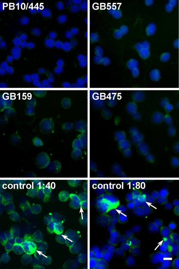
Serologic testing of bat sera for antibodies to human HEV with an HEV-specific indirect immunofluorescence assay. Slides carrying human embryonic kidney 293T cells transiently expressing the full-length ORF2 protein from a human HEV genotype 1 strain were incubated with bat sera (diluted 1:40) from eight different species. To allow the evaluation of the reaction specificity, the transfection efficiency was optimized to yield only 5 to 10% of cells expressing HEV antigen. One HEV RT-PCR-positive species (Vampyrodes caraccioli [PB10/445]) and three different RT-PCR-negative species (Hipposideros gigas [GB557], Rousettus aegyptiacus [GB159], and Miniopterus inflatus [GB475]) are shown. Notably, R. aegyptiacus specimen GB159 was chosen because we realized that it reacted nonspecifically with all cells, including those not expressing HEV antigen, and we wanted to demonstrate its clear discrimination from seropositive human sera. Detection was done by incubation with goat anti-bat immunoglobulin (Ig), followed by donkey anti-goat Ig labeled with cyanine 2. As a control, an anonymous human serum sample from a patient infected with HEV was applied in dilutions of 1:40 and 1:80 (to reduce the background signal). White arrows indicate specific serologic reactivity with HEV ORF2-expressing cells. The bar represents 25 μm. All pictures were taken with identical microscope and camera settings.
To determine, on the contrary, if descendants of bat viruses are encountered in humans, pooled plasma samples from 93,146 blood donors were tested. As a control, all samples were first screened for human HEV. HEV genotype 3 viruses were detected in 11 pools. This was in agreement with previously determined prevalences of HEV RNA in blood products (2, 27, 63), confirming the suitability of the pooled samples for HEV detection in principle. Notably, the method of blood donor pooling involved the concentration of viral particles by high-speed centrifugation, allowing a nearly quantitative recovery of viruses from pools. The sensitivity limit of the assay for individual blood donations contained in the pools could therefore be projected to be approximately 4.4 log10 copies/ml. This was compatible with previously reported viral loads in individual plasma donations ranging between 3.2 and 5.7 log10 IU/ml (4). None of the blood donor pools yielded positive results in single and heminested RT-PCR assays capable of detecting all bat-associated HEVs, ruling out the existence of human-specific viruses related to the bat-specific clade in these samples. To investigate humans with potentially closer exposures to bats and a propensity for persistent infection (11, 14, 45), an additional test of 453 anonymized sera from Cameroonian HIV-positive patients was done. No evidence of bat or human hepevirus RNA was obtained, whereas the suitability of these samples for the detection of viral RNA was proven previously (51). This may indicate that the level of immunosuppression might not have been as severe in these patients as in other cohorts of HIV-infected individuals tested for HEV (11, 14).
To compare the genome properties of the novel bat viruses with those of other members of the family Hepeviridae, the full genome of one virus, termed BS7, from a German Eptesicus serotinus bat was sequenced. As shown in Fig. 5A, the bat virus represented an independent branch among the full-length hepevirus genomes. The complete genome comprised 6,767 nucleotides (nt), excluding the poly(A) tail at the 3′ end, constituting the shortest mammalian hepevirus genome, with a size comparable to that of the avian viruses (6,654 nt in the U.S. prototype strain [GenBank accession number AY535004]). The coding regions were flanked by a 33-nt 5′-untranslated region (UTR) and a 77-nt 3′-UTR. In the coding region, at least three open reading frames (ORFs) typical of all hepeviruses were identified (Table 4). An internal putative 660-nt ORF overlapping ORF1 in the −2 reading frame was identified at positions 326 to 985 and provisionally termed ORF NX (for N-terminal unknown). Whether ORF NX corresponded to the putative ORF4 or ORF5 described for rodent hepeviruses remains unclear, since the sequence identity with these ORFs was very low (33.3% and 14.4% nucleotide identities and 30.5% and 17.5% amino acid identities with ORF4 and ORF5, respectively), and no significant similarity of the putative gene product of the internal reading frame to any described protein domain could be detected by BLAST. Similar to bat hepevirus ORF NX, rat hepevirus ORF4 is located at the N terminus of ORF1, while ORF5 is located approximately 2,000 nucleotides downstream of ORF1 (31).
Fig 5.
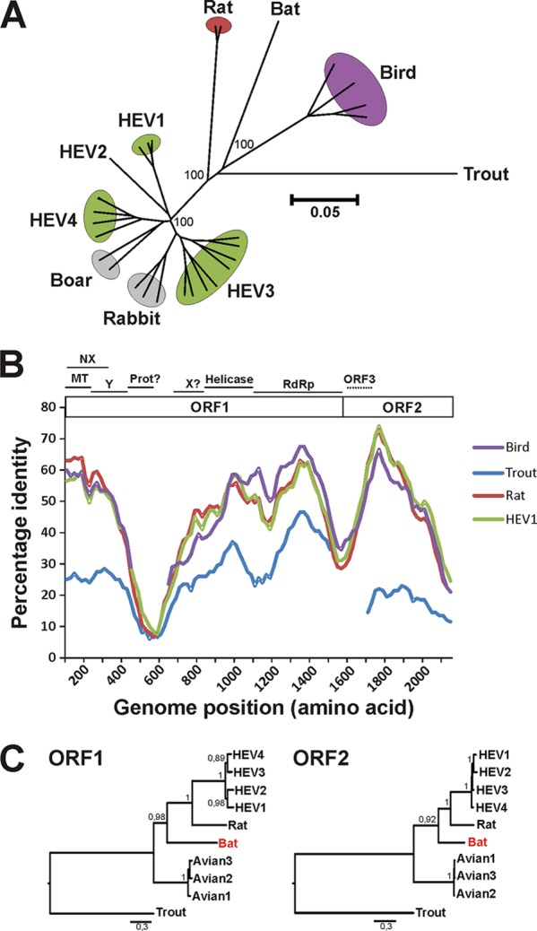
Complete genome nucleotide phylogeny, amino acid sequence identity, and ORF1/ORF2 amino acid phylogeny of bat hepevirus BS7 and prototype hepeviruses. (A) Neighbor-joining phylogeny of the complete genomes of members of the Hepeviridae using the nucleotide percentage distance substitution matrix and complete deletion option in MEGA5. Values at deep node points indicate support from 1,000 bootstrap reiterations; those at apical nodes are hidden for clarity of presentation. (B) Amino acid identity plot. The complete ORF1 and ORF2 were translated, concatenated, and compared to avian, rodent, human, and trout prototype hepeviruses. Positions containing gaps in the bat hepevirus were stripped from the alignment. The uncorrected amino acid identity was plotted with a sliding window size of 200 and a step size of 20 amino acids. For orientation, a schematic representation ORF1 and ORF2 is shown with putative nonstructural functional domains as approximated by BLAST comparisons with GenBank reference sequences depicted at the top (MT, methyltransferase; NX, putative ORF NX; Y, Y-like domain; Prot, papain-like cysteine protease; X, X domain/ADP-ribose-binding module; RdRp, RNA-dependent RNA polymerase). The protease and X domains could not be unambiguously identified and are therefore given with question marks. ORF3 is shown with a dotted line, since it is translated in a different reading frame than ORF2 and is shown only for an indication of its genomic position. (C) Bayesian phylogeny of the complete ORF1 and ORF2. Inference of Bayesian phylogenies was done by using MrBayes V3.1 with a WAG amino acid model and 4,000,000 generations sampled every 100 steps. After the exclusion of 10,000 trees as a burn-in, 15,000 final trees were annotated and visualized with TreeAnnotator and FigTree from the BEAST package. Values at the node points indicate posterior probability support (scale bar, genetic distance). GenBank accession numbers for taxa are AF459438 (HEV genotype 1), M74506 (HEV genotype 2), AB301710 (HEV genotype 3), AB220974 (HEV genotype 4), GU345042 (rat hepevirus), AM943647 (avian hepevirus genotype 1), EF206691 (avian hepevirus genotype 2), GU954430 (avian hepevirus genotype 3), and NC_015521 (trout HEV).
Table 4.
Identities of bat HEV (BS7) ORFs with HEV prototype strains
| Hepevirusa | % identity |
|||||
|---|---|---|---|---|---|---|
| ORF1 (genome positions 34–4776) |
ORF2 (genome positions 4777–6690) |
ORF3 (genome positions 4859–5272) |
||||
| Nucleotide | Amino acid | Nucleotide | Amino acid | Nucleotide | Amino acid | |
| HEV genotype 1 | 49.1–49.5 | 43.5–44.1 | 52.5–52.7 | 49.8–50.7 | 31.4–31.9 | 9.3–10.1 |
| HEV genotype 2 | 48.5 | 44.1 | 51.2 | 49.1 | 33.6 | 10.9 |
| HEV genotype 3 | 48.8–49.4 | 43.5–44.0 | 52.0–53.0 | 50.3–50.7 | 29.9–31.2 | 10.2–12.8 |
| HEV genotype 4 | 48.1–49.3 | 44.1–44.3 | 51.6–52.7 | 50.3–50.8 | 28.2–29.1 | 10–10.9 |
| Rabbit | 48.5–49.1 | 43.5–44.1 | 51–52.1 | 47–49.8 | 27.4–28.0 | 10.2–10.9 |
| Wild boar | 49–49.2 | 43.8–43.9 | 51.6–52.8 | 49.7–51.2 | 29.9–30.6 | 11.1–12.0 |
| Rodent | 48.5–48.7 | 45.1–45.3 | 51.9–52 | 48.9–49.4 | 28.9–29.6 | 7.1 |
| Avian | 51.6–52.3 | 47.5–47.6 | 47–48.8 | 42.9–43.1 | 22.7–23.5 | 6.0 |
| Trout | 38.3 | 26.5 | 33.8 | 17.3 | 28.2 | 13.2 |
GenBank accession numbers of prototype strains are as follows: D11092, L08816, AF459438, and DQ459342 for HEV genotype 1; M74506 for HEV genotype 2; EU723512, FJ705359, FJ998008, AB089824, AB248521, AB301710, AB248520, AB591734, and EU375463 for HEV genotype 3; FJ906895, FJ906896, and GU937805 for rabbit hepevirus; AB220974, GU119960, AB480825, AB097811, and AB097812 for HEV genotype 4; AB573435 and AB602441 for unassigned wild boar hepevirus; GU345042 and GU345043 for rodent hepevirus; AM943647, EF206691, AC535004, and GU954430 for avian hepevirus; and NC_015521 for trout hepevirus.
In the first ORF of BS7 (ORF1) (nt 34 to 4776), several domains could be predicted (Table 5). A putative methyltransferase domain was found to contain all four conserved amino acid residues identified previously in plus-stranded RNA viruses belonging to the alphavirus-like supergroup (62) at ORF1 amino acid positions 65 (H), 115 (D), 118 (R), and 229 (Y). The beginning of a putative Y domain, as typical for hepeviruses, was found at around aa 216 (VVTY). The level of conservation decreased downstream, and the end of the domain could not be identified. The genome region encoding the papain-like protease in HEV was highly divergent in hepeviruses from different hosts, including bats, and the amino acid residues around a putative catalytic cysteine (TCFL) defined previously by Koonin et al. (35) were found in neither the bat hepevirus nor the previously described rat, bird, or fish hepeviruses.
Table 5.
Genomic features of bat hepevirus BS7a
| Bat hepevirus genomic region and domain | Start site | End site | Length | Description |
|---|---|---|---|---|
| 5′-UTR | ||||
| Full region | nt 1 | nt 33 | 33 nt | |
| ORF1 | ||||
| Full ORF | nt 34 | nt 4776 | 4,743 nt | pI2 = 7.5 |
| Methyltransferase | Start and end could not be defined | |||
| 4 conserved motifs | aa 65 (H), 115 (D), 118 (R), and 229 (Y) | All four conserved amino acid residues exist in BS7 | ||
| Y domain | aa 216 (VVTY) | NI | Similarities in BLAST to corresponding regions in human HEV but unidentifiable end | |
| Papain-like protease | Not conserved | |||
| Proline-rich hinge region | Not conserved | |||
| X domain | aa 684 (PDGSK) | Similarities in BLAST to corresponding regions in human HEV but unidentifiable end | ||
| Helicase | Start and end could not be defined | |||
| NTP-binding site in helicase (GVPGSGKS) | aa 874 | aa 881 | 8 aa | Conserved with serine replaced by alanine |
| DEAP motif | aa 927 | aa 930 | 4 aa | 100% identity |
| RdRp | NI | NI | Start and end could not be defined | |
| RdRp motifs | ||||
| I (KDCNKFTT) | aa 1277 | aa 1284 | 8 aa | 100% identity (KDCNKFTT) |
| II (ISAWSKTFCALFGPWFRA) | aa 1297 | aa 1314 | 18 aa | 83% identity (ISAWPKTLCALFGPWFRA) |
| III (FYGDAFDDTVF) | aa 1328 | aa 1338 | 11 aa | 36% identity (FYGDLYTEADL) |
| IV (NDFSEFDSTQNN) | aa 1354 | aa 1365 | 12 aa | 100% identity (NDFSEFDSTQNN) |
| V (KHSGEPGTLLWNTVW) | aa 1410 | aa 1424 | 15 aa | 93% identity (KHSGEPGTLLFNTVW) |
| VI (AAFKGDDSIVL) | aa 1441 | aa 1451 | 11 aa | 64% identity (HVYKGDDSVVL) |
| VII (LYAGVVVAPG) | aa 1481 | aa 1490 | 10 aa | 40% identity (VFSHYIVAPG) |
| VIII (ALPDVVRFAGRLT) | aa 1493 | aa 1505 | 13 aa | 39% identity (VVKDLLRTWGRMT) |
| Conserved tripeptide GDD | aa 1445 | aa 1447 | 3 aa | 100% identity |
| ORF2 | ||||
| Full ORF | nt 4777 | nt 6690 | 1,914 nt | pI = 8.0 |
| cis-reactive element overlapping ORF2 start codon (TGAATAACA) | nt 4778 | nt 4786 | 9 nt | Conserved nucleotides in avian and human strains were also conserved in BS7 |
| Signal sequence | Not conserved at the N terminus but followed by a hydrophobic domain at aa 16-24 (FAYLLLLFL) | |||
| Arginine-rich domain | NI | aa 69 | Conserved C terminus (PLT) but deletions and fewer R residues (7 compared to 12) than human hepeviruses | |
| S domain | aa 87 | aa 277 | 191 aa | Conserved |
| M domain | aa 278 | aa 412 | 135 aa | Divergent N terminus, conserved C terminus |
| P domain | aa 413 | aa 561 | 149 aa | Partially conserved |
| C-terminal domain | Not conserved | |||
| Termination codon ORF2 | nt 6688 | nt 6690 | 3 nt | TAA typical of genotype 3 |
| ORF3 | ||||
| Full ORF | nt 4859 | nt 5272 | 414 nt | pI = 8.1 |
| 3′-UTR | ||||
| Full region | nt 6691 | nt 6767 | 77 nt |
nt, nucleotide; pI, isoelectric point (determined online at www.isoelectric.ovh.org/); aa, amino acids; NI, not identifiable.
The putative proline-rich hinge region of the highly variable sequence had a maximum proline density of 12 proline residues within 68 amino acid positions (P/68 aa).
The X domain of unknown function in human HEV (35) could not be unambiguously identified in BS7, since only the start and not the end of this putative region could be mapped in comparison to human HEV (Table 5). The putative helicase contained the nucleoside triphosphate (NTP)-binding site GVPGSGKS (aa 874 to 881) and DEAP motif (aa 927 to 930) described previously (65), with serine being replaced by alanine at aa 878. The putative RdRp contained the conserved tripeptide GDD. Out of the eight RdRp motifs described previously, two (motifs I [KDCNKFTT] and IV [NDFSEFDSTQNN]) were completely conserved in BS7. The other motifs could be identified but were less conserved (Table 5) (35).
The second ORF, coding for the putative 638-aa capsid protein, was found at nt 4777 to 6690 (Tables 4 and 5). The conserved sequence TGAATAACA within a cis-active element overlapping the ORF2 start codon in human and avian HEVs (26) was present in the bat virus genome at the homologous position. The first half of the polypeptide had a basic charge, with a predicted isoelectric point (pI) of 10.97, suggesting a potential involvement in the encapsidation process (54). In contrast to rodent hepeviruses (32), the ORF2 domains of BS7 seemed to be less conserved (Table 5) (71). Several hydrophobic residues occurred after the ORF2 start codon (FAYLLLLFL [ORF2 aa residues 16 to 24]), which characterize the N-terminal signal sequence in other mammalian hepeviruses. ORF2 was most conserved in the shell (S) domain, including tyrosine at position 288, which was described previously to be crucial for capsid formation in HEV genotype 3 (71). The middle (M) and protruding (P) domains were less conserved.
A putative ORF3 was detected in an alternative reading frame overlapping the capsid-encoding ORF2 (Table 4). In HEV, this protein encodes a phosphoprotein that is not essential for replication in hepatoma cells (21) and that might be involved in virion release, transcription, or interactions with cellular factors (34, 47, 49). The putative ORF3 product in the bat hepevirus showed little similarity to proteins encoded by ORF3 in other hepeviruses (Table 4) and did not yield significantly similar sequences in a BLAST search. It did not overlap the 3′ terminus of ORF1, as in HEV genotypes 1 to 3 (31), and its size was between that of trout hepevirus (678 nt) and that of the human or rodent hepeviruses (372 and 309 nt, respectively) (Table 5) (3, 31). The low level of similarity of ORF3 between viruses from different hosts such as bats, rodents, birds, and humans is in contrast to its marked conservation in viruses from identical hosts (Table 6) and may represent viral adaptation to particular hosts.
Table 6.
Percent nucleotide (below the diagonal) and amino acid (above the diagonal) sequence identities between hepevirus ORF3sa
| Hepevirus host or lineage (no. of compared strains) | % identity |
|||
|---|---|---|---|---|
| Bat | Avian | Rodent | HEV genotypes 1-4 | |
| Bat (1) | 9.3–12.8 | 9.8 | 12.3–16.7 | |
| Avian (4) | 88.4–96.5 | 24.3 | 25.6–31.2 | |
| 26.4–28.0 | 93.9–96.9 | |||
| Rodent (2) | 94.1 | 20.8–32.4 | ||
| 25.2–25.5 | 36.5–38.3 | 97.4 | ||
| HEV genotypes 1–4 (22) | 72.1–100 | |||
| 32.5–35.1 | 33.3–38.6 | 37–42.5 | 80.4–99.2 | |
Evolutionary analyses were conducted with MEGA5 (66). GenBank accession numbers of strains are as follows: D11092, L08816, AF459438, DQ459342, M74506, EU723512, FJ705359, FJ998008, AB089824, AB248521, AB301710, AB248520, AB591734, EU375463, FJ906895, FJ906896, GU937805, AB220974, GU119960, AB480825, AB097811, AB097812, AB573435, and AB602441 for HEV genotypes 1 to 4; GU345042 and GU345043 for rodent hepevirus; and AM943647, EF206691, AC535004, and GU954430 for avian hepevirus. Boldface type indicates percent nucleotide identity, and lightface type indicates percent amino acid identity.
The amino acid identity plot (Fig. 5B) indicated that the bat hepevirus was closely related to avian hepeviruses in some parts of ORF1 and to primate hepeviruses in ORF2 (Fig. 5B). However, multiple-change-point analysis with Dual Brothers (46) and bootscan analysis with Simplot V3.5 (39) yielded no evidence of recombination, and the complete ORF1 and ORF2 sequences clustered reliably with other mammalian hepeviruses (Fig. 5C).
According to the International Committee on Taxonomy of Viruses (ICTV), the mammalian hepeviruses known at the time of assessment constituted a single genus (41). The avian viruses were suggested previously to form another independent genus (40). To test how the bat hepeviruses fit these proposals and to develop a working criterion for the tentative classification of partially sequenced viruses, we evaluated distance-based classification criteria. First, an amino-acid-based criterion for members of the Hepeviridae was calculated by using the comparably small 108-amino-acid RdRp fragment used for this study. The fragment overlaps largely with the amplicon of another widely used RT-PCR assay (32, 60) and might therefore enable the expansion of our taxonomic attempts to previous and future studies. The distribution of pairwise distances indicated several potential taxonomic ranks (Fig. 6A). Amino acid distance values of up to 9.3% but less than 11.1% were seen within the established HEV genotypes. Distances between 11.1% and 22.2% separated established HEV genotypes 1 to 4. The rabbit viruses would thus belong to HEV genotype 3, while the unclassified wild boar viruses would correspond to a distinct genotype. The latter might therefore constitute a fifth HEV genotype. The recently described rat hepeviruses were even more divergent, with up to 34.3% substitutions relative to other mammalian HEV isolates (Table 3).
Fig 6.

Distribution of Hepeviridae partial RdRp and full ORF1 and ORF2 pairwise amino acid distances. Uncorrected pairwise amino acid distances were calculated between members of the family Hepeviridae in the same 108-amino-acid RNA-dependent RNA polymerase (RdRp) alignment as that used for Fig. 3 (A) and in the complete ORF1 (B) and ORF2 (C). The y axis indicates the number of pairwise identity scores within each range represented on the x axis. The bold line indicates a distance cutoff that separates intratypic and intertypic distances within HEV genotypes 1 to 4. The dotted lines indicate a range of possible sequence cutoffs between sequence distances within and between the four suggested hepevirus genera. Distances within NM bat hepeviruses are indicated in light gray. Distances between BS7 bat virus and NM viruses are shown in gray. Distances between PAN926, G19E36, and other bat viruses are shown in black.
The range of pairwise distances between all bat hepeviruses (up to 19.4%) suggested that they formed a taxonomic entity of the same rank as human HEV or rodent or avian hepeviruses.
Sequence distances between the closely related Myotis bat viruses from Germany (strains NMS098B and NMS09125R from M. daubentonii and NM98AC156 from M. bechsteinii) corresponded to distances observed within genotypes (Fig. 6A). Distances between PAN926 from Panama (V. caraccioli), G19E36 from Ghana (Hipposideros abae), and all other bat hepeviruses indicated that each of these viruses could be classified as a distinct genotype if the above-described criteria are applied. The classification of bat hepevirus BS7 from E. serotinus remained questionable because its sequence distances to the Myotis bat viruses fell on the border of inter- and intragenotypic distances characteristic for HEV genotypes 1 to 4.
The distance criterion within our partial RdRp gene fragment failed to discriminate bat and avian hepeviruses into two different tentative genera, which did not match the closer phylogenetic relatedness of mammalian viruses in larger parts of the ORF1-encoded polyprotein and the ORF2-encoded capsid protein (Fig. 5B and C). For better resolution, the distribution of pairwise amino acid distances was also plotted over those complete ORF1 and ORF2 sequences represented in Fig. 3. There was a clear separation between genetic distances within and between the four major clades: human HEV-like, rodent, avian, and bat hepeviruses. The distances within and between these groups were <22% and >46% in ORF1 and <18% and >42% in ORF2 (Fig. 6B and C), supporting the existence of four putative genera in the family Hepeviridae. Notably, these four putative genera were also well supported in the partial RdRp gene phylogenetic tree (Fig. 3).
DISCUSSION
In this study, we have described novel hepeviruses from a globally representative sample of bat specimens. The genomic characterization of a bat hepevirus clearly supported their classification as members of the recently established family Hepeviridae and indicated that they may be the most divergent mammalian hepeviruses described so far.
While a full genomic characterization is generally required for exact conclusions on taxonomy, the RdRp-based criterion established here may represent a useful tool for the typing of partially sequenced field specimens, similar to, e.g., the VP1 gene in enteroviruses and the RdRp gene in coronaviruses (20, 53). Our approach might help to resolve the classification of rodent hepeviruses, for which a fifth genotype has been proposed (38), as well as that of the rabbit hepeviruses, which seem to belong to HEV genotype 3 (23, 24). On the other hand, classification based on the RdRp gene may not always be sufficient to assign novel genera, and criteria based on full-ORF data may be required. The sequence of ORF3, which was found to be highly distinct between hepeviruses from different hosts, might be useful as an additional marker for classification. Our cumulative classification efforts suggested the existence of at least four putative genera in the family Hepeviridae. One genus would comprise human HEV genotypes and closely related animal viruses, while the other three would include viruses from chiropteran, rodent (rat), and avian (chicken) hosts. The trout hepevirus might correspond to a separate taxonomic unit of a higher rank, e.g., a subfamily (3).
The rate of hepevirus detection in bats was similarly low compared to that in other mammals, including humans (1, 9, 16, 24, 38, 58, 60). The highest anti-HEV seroprevalence and RNA detection rates described so far were for farmed animals, e.g., rabbits, chickens, and piglets (23, 33, 40). The accumulation of very large groups of susceptible individuals is uncommon in wild animals, with bats probably constituting the only mammals beside humans that form social groups exceeding several hundred thousand individuals in one place (52). Our study was performed on samples from very large social groups, including a breeding roost of Eidolon helvum bats in Ghana with more than 300,000 individuals (19) as well as vespertilionid bats hibernating in different roosts in Germany and Bulgaria, all exceeding 10,000 members (20, 25). While we have not observed any particularly high prevalence of viruses in these samples, the diversity of bat viruses found in our whole sample was comparable to that of the extensively studied human hepeviruses and much higher than that of the known avian or rodent viruses. We can thus be confident that our study provides a fair representation of the wider bat hepevirus diversity. Despite this, all chiropteran hepeviruses were monophyletic. In contrast to other viruses for which bats are assumed to constitute relevant mammalian reservoirs, such as mammalian coronaviruses and paramyxoviruses (19, 56), bat hepeviruses were not interspersed with human or other mammalian viruses. Bats therefore do not seem to constitute reservoirs of mammalian hepeviruses in general. The clear distinction of the four proposed hepevirus genera implicated rather that hepevirus evolution might have involved a nonrecent invasion of ancestral mammalian hosts. The marked exception to this idea is human hepeviruses, which can be found in both humans and peridomestic animals. It may be plausible that the intensification of farming activities during human history led to transmissions of some but not all human viruses to farmed animals, which would then have maintained these viruses and formed a potential source of zoonotic reinfection. This hypothesis is coherent with the observation that HEV genotypes 1 and 2 have so far been detected only in humans and not in any other animals.
Based on our data, it would now be possible to search for hepeviruses in more distant relatives of farmed or peridomestic animals. This search should include lagomorphs other than rabbits, wild (nonlivestock) ungulates, as well as carnivores other than mongooses. If, indeed, hepeviruses very distinct from human HEV could be found in these taxa, the idea of an acquisition of human hepeviruses by farmed animals would be strongly supported. HEV in wild primates should also be studied to confirm a possibly more general association of human-related viruses with primates.
As with other newly described viruses in animals, we have to address potential zoonotic risks. Human infection has been associated with the consumption of pig, wild boar, and deer meat in industrialized countries (10, 37, 58, 68). Even though bats are consumed by humans in parts of the world (45), we consider it unlikely that bat hepeviruses would easily transmit to humans. As discussed above, the viruses carried by pig, boar, and deer are closely related to human viruses. Neither avian nor rodent hepeviruses were transmissible to primates experimentally (29, 60), and the latter are even more closely related to human viruses than the bat viruses. Despite reports on the serologic reactivity of human and rodent sera with hepevirus antigens from the opposed species (16, 17), proof of zoonotic transmission would require direct virus detection. In our study, we did not find any bat hepevirus RNA in a very broad range of human specimens.
Our data underline the importance of investigating targeted and balanced samples when studying viral host associations. While the surveillance of pathogens in livestock and peridomestic animals is important for epidemiology, ecologically valid conclusions regarding host associations can be very hard to reach using such samples. It is essential to cover large geographic and phylogenetic samples from the spectrum of potential viral hosts (19).
Supplementary Material
ACKNOWLEDGMENTS
We are indebted to our recently deceased friend and colleague Elisabeth K. V. Kalko (Ulm University, Germany, and Smithsonian Tropical Research Institute, Panama) for advancing field work and reflections on bat ecology.
We thank Sebastian Brünink, Monika Eschbach-Bludau, and Tobias Bleicker at the Institute of Virology, Bonn, for technical assistance. We are grateful to Mirjam Knörnschild (Ulm University), Milen Rashkov, Justin Robarge, and Lyubomir Zhelyazkov (Strandja Natural Park); Uwe Hermanns (NABU Rostock NGO); Matthias Göttsche (University Kiel); Andreas Kiefer of the Nature Conservation Project Mayen; Elena Tilova; Liubomir Ilankov (Green Balkans NGO); Augustina Annan (KCCR); Antje Seebens and Anne Ipsen (Noctalis); the volunteers at the Bonn Consortium for Bat Conservation (BAFF); Manfred Braun (Struktur und Genehmigungsbehörde Nord, Koblenz); Henning Vierhaus (ABU Soest); Lena Grosche, Frauke Meier, and Myriam Götz (Echolot GbR); and Gael D. Maganga, Mathieu Bourgarel, Xavier Pourrut, Dieudonné Nkoghe, Peggy Motsch, André Délicat, and Philippe Engandja (CIRMF) for field work and technical assistance.
This study was funded by the European Union FP7 projects EMPERIE (grant agreement number 223498), EVA (grant agreement number 228292), and ANTIGONE (grant agreement number 278976); the German Federal Ministry of Education and Research (BMBF) (project code 01KIO701); the German Research Foundation (DFG) (grant agreement numbers DR 772/3-1 and KA 1241/18-1); the DAAD (D/00/39390); and the Australian Government Endeavor Programme (to S.M.K.).
The sampling and capture of wild animals as well as sample transfers were done under wildlife permits and ethics clearances from Costa Rica (ethics permit PI-ACCVC-005-2010 [Karen Daniela Sibaja Morales] and export permit 25939), Panama (research permit STRI STRI2563 [PI VC]-IACUC 100316-1001-18; research permit ANAM SE/A-68-11; ethics permit IACUC 100316-1001-18; and export permits SEX/A-30-11, SEX/A-55-11, SEX/A-81-10, and SEX-A-26-10), Ghana (research permit 2008-2010 [A04957] and ethics permit CHRPE49/09/CITES; export permits were not needed for any species, including Eidolon [export permit state agreement between Ghana and Hamburg {BNI}]), Australia (research permits S11828 and S11762; ethics permits TRIM 01/1118 [2], TRIM 06/3569, and University of Queensland/Animal Ethics Committee SIB600/05/DEST; and export permit DE201-12), Papua New Guinea (ethics permit PNG/NatMus/2002 and export permit conducted by the Papua New Guinea National Museum), Gabon (ethics permit 00021/MEFEPA/SG/DGEF/DFC), Germany (ethics permit LANU 314/5327.74.1.6), and Bulgaria (ethics permit 192/26.03.2009).
Footnotes
Published ahead of print 13 June 2012
Supplemental material for this article may be found at http://jvi.asm.org/.
REFERENCES
- 1. Adjei AA, et al. 2009. Hepatitis E virus infection is highly prevalent among pregnant women in Accra, Ghana. Virol. J. 6:108. [DOI] [PMC free article] [PubMed] [Google Scholar]
- 2. Adlhoch C, Kaiser M, Pauli G, Koch J, Meisel H. 2009. Indigenous hepatitis E virus infection of a plasma donor in Germany. Vox Sang. 97:303–308 [DOI] [PubMed] [Google Scholar]
- 3. Batts W, Yun S, Hedrick R, Winton J. 2011. A novel member of the family Hepeviridae from cutthroat trout (Oncorhynchus clarkii). Virus Res. 158:116–123 [DOI] [PubMed] [Google Scholar]
- 4. Baylis SA, Gartner T, Nick S, Ovemyr J, Blumel J. 2012. Occurrence of hepatitis E virus RNA in plasma donations from Sweden, Germany and the United States. Vox Sang. 103:89–90 [DOI] [PubMed] [Google Scholar]
- 5. Baylis SA, Koc O, Nick S, Blumel J. 2012. Widespread distribution of hepatitis E virus in plasma fractionation pools. Vox Sang. 102:182–183 [DOI] [PubMed] [Google Scholar]
- 6. Bose PD, et al. 2011. High viral load and deregulation of the progesterone receptor signaling pathway: association with hepatitis E-related poor pregnancy outcome. J. Hepatol. 54:1107–1113 [DOI] [PubMed] [Google Scholar]
- 7. Bouwknegt M, et al. 2008. Bayesian estimation of hepatitis E virus seroprevalence for populations with different exposure levels to swine in The Netherlands. Epidemiol. Infect. 136:567–576 [DOI] [PMC free article] [PubMed] [Google Scholar]
- 8. Calisher CH, Childs JE, Field HE, Holmes KV, Schountz T. 2006. Bats: important reservoir hosts of emerging viruses. Clin. Microbiol. Rev. 19:531–545 [DOI] [PMC free article] [PubMed] [Google Scholar]
- 9. Caron M, Kazanji M. 2008. Hepatitis E virus is highly prevalent among pregnant women in Gabon, central Africa, with different patterns between rural and urban areas. Virol. J. 5:158. [DOI] [PMC free article] [PubMed] [Google Scholar]
- 10. Colson P, et al. 2010. Pig liver sausage as a source of hepatitis E virus transmission to humans. J. Infect. Dis. 202:825–834 [DOI] [PubMed] [Google Scholar]
- 11. Colson P, Dhiver C, Poizot-Martin I, Tamalet C, Gerolami R. 2011. Acute and chronic hepatitis E in patients infected with human immunodeficiency virus. J. Viral Hepat. 18:227–228 [DOI] [PubMed] [Google Scholar]
- 12. Cossaboom CM, Cordoba L, Dryman BA, Meng XJ. 2011. Hepatitis E virus in rabbits, Virginia, USA. Emerg. Infect. Dis. 17:2047–2049 [DOI] [PMC free article] [PubMed] [Google Scholar]
- 13. Dalton HR, Bendall R, Ijaz S, Banks M. 2008. Hepatitis E: an emerging infection in developed countries. Lancet Infect. Dis. 8:698–709 [DOI] [PubMed] [Google Scholar]
- 14. Dalton HR, Bendall RP, Keane FE, Tedder RS, Ijaz S. 2009. Persistent carriage of hepatitis E virus in patients with HIV infection. N. Engl. J. Med. 361:1025–1027 [DOI] [PubMed] [Google Scholar]
- 15. Dobson AP. 2005. Virology. What links bats to emerging infectious diseases? Science 310:628–629 [DOI] [PubMed] [Google Scholar]
- 16. Dong C, et al. 2011. Restricted enzooticity of hepatitis E virus genotypes 1 to 4 in the United States. J. Clin. Microbiol. 49:4164–4172 [DOI] [PMC free article] [PubMed] [Google Scholar]
- 17. Dremsek P, et al. 2012. Seroprevalence study in forestry workers from eastern Germany using novel genotype 3- and rat hepatitis E virus-specific immunoglobulin G ELISAs. Med. Microbiol. Immunol. 201:189–200 [DOI] [PubMed] [Google Scholar]
- 18. Drexler JF, et al. 2009. Henipavirus RNA in African bats. PLoS One 4:e6367 doi:10.1371/journal.pone.0006367 [DOI] [PMC free article] [PubMed] [Google Scholar]
- 19. Drexler JF, et al. 2012. Bats host major mammalian paramyxoviruses. Nat. Commun. 3:796. [DOI] [PMC free article] [PubMed] [Google Scholar]
- 20. Drexler JF, et al. 2010. Genomic characterization of severe acute respiratory syndrome-related coronavirus in European bats and classification of coronaviruses based on partial RNA-dependent RNA polymerase gene sequences. J. Virol. 84:11336–11349 [DOI] [PMC free article] [PubMed] [Google Scholar]
- 21. Emerson SU, Nguyen H, Torian U, Purcell RH. 2006. ORF3 protein of hepatitis E virus is not required for replication, virion assembly, or infection of hepatoma cells in vitro. J. Virol. 80:10457–10464 [DOI] [PMC free article] [PubMed] [Google Scholar]
- 22. Emerson SU, Purcell RH. 2004. Running like water—the omnipresence of hepatitis E. N. Engl. J. Med. 351:2367–2368 [DOI] [PubMed] [Google Scholar]
- 23. Geng J, et al. 2011. Study on prevalence and genotype of hepatitis E virus isolated from Rex rabbits in Beijing, China. J. Viral Hepat. 18:661–667 [DOI] [PubMed] [Google Scholar]
- 24. Geng Y, et al. 2011. The serological prevalence and genetic diversity of hepatitis E virus in farmed rabbits in China. Infect. Genet. Evol. 11:476–482 [DOI] [PubMed] [Google Scholar]
- 25. Gloza-Rausch F, et al. 2008. Detection and prevalence patterns of group I coronaviruses in bats, northern Germany. Emerg. Infect. Dis. 14:626–631 [DOI] [PMC free article] [PubMed] [Google Scholar]
- 26. Graff J, et al. 2005. The open reading frame 3 gene of hepatitis E virus contains a cis-reactive element and encodes a protein required for infection of macaques. J. Virol. 79:6680–6689 [DOI] [PMC free article] [PubMed] [Google Scholar]
- 27. Guo QS, et al. 2010. Prevalence of hepatitis E virus in Chinese blood donors. J. Clin. Microbiol. 48:317–318 [DOI] [PMC free article] [PubMed] [Google Scholar]
- 28. Haqshenas G, Shivaprasad HL, Woolcock PR, Read DH, Meng XJ. 2001. Genetic identification and characterization of a novel virus related to human hepatitis E virus from chickens with hepatitis-splenomegaly syndrome in the United States. J. Gen. Virol. 82:2449–2462 [DOI] [PubMed] [Google Scholar]
- 29. Huang FF, et al. 2004. Determination and analysis of the complete genomic sequence of avian hepatitis E virus (avian HEV) and attempts to infect rhesus monkeys with avian HEV. J. Gen. Virol. 85:1609–1618 [DOI] [PubMed] [Google Scholar]
- 30. Huelsenbeck JP, Ronquist F. 2001. MRBAYES: Bayesian inference of phylogenetic trees. Bioinformatics 17:754–755 [DOI] [PubMed] [Google Scholar]
- 31. Johne R, et al. 2010. Novel hepatitis E virus genotype in Norway rats, Germany. Emerg. Infect. Dis. 16:1452–1455 [DOI] [PMC free article] [PubMed] [Google Scholar]
- 32. Johne R, et al. 2010. Detection of a novel hepatitis E-like virus in faeces of wild rats using a nested broad-spectrum RT-PCR. J. Gen. Virol. 91:750–758 [DOI] [PubMed] [Google Scholar]
- 33. Kaba M, et al. 2009. Frequent transmission of hepatitis E virus among piglets in farms in Southern France. J. Med. Virol. 81:1750–1759 [DOI] [PubMed] [Google Scholar]
- 34. Kannan H, Fan S, Patel D, Bossis I, Zhang YJ. 2009. The hepatitis E virus open reading frame 3 product interacts with microtubules and interferes with their dynamics. J. Virol. 83:6375–6382 [DOI] [PMC free article] [PubMed] [Google Scholar]
- 35. Koonin EV, et al. 1992. Computer-assisted assignment of functional domains in the nonstructural polyprotein of hepatitis E virus: delineation of an additional group of positive-strand RNA plant and animal viruses. Proc. Natl. Acad. Sci. U. S. A. 89:8259–8263 [DOI] [PMC free article] [PubMed] [Google Scholar]
- 36. Kumar A, Beniwal M, Kar P, Sharma JB, Murthy NS. 2004. Hepatitis E in pregnancy. Int. J. Gynaecol. Obstet. 85:240–244 [DOI] [PubMed] [Google Scholar]
- 37. Li TC, et al. 2005. Hepatitis E virus transmission from wild boar meat. Emerg. Infect. Dis. 11:1958–1960 [DOI] [PMC free article] [PubMed] [Google Scholar]
- 38. Li TC, et al. 2011. Characterization of self-assembled virus-like particles of rat hepatitis E virus generated by recombinant baculoviruses. J. Gen. Virol. 92:2830–2837 [DOI] [PMC free article] [PubMed] [Google Scholar]
- 39. Lole KS, et al. 1999. Full-length human immunodeficiency virus type 1 genomes from subtype C-infected seroconverters in India, with evidence of intersubtype recombination. J. Virol. 73:152–160 [DOI] [PMC free article] [PubMed] [Google Scholar]
- 40. Marek A, Bilic I, Prokofieva I, Hess M. 2010. Phylogenetic analysis of avian hepatitis E virus samples from European and Australian chicken flocks supports the existence of a different genus within the Hepeviridae comprising at least three different genotypes. Vet. Microbiol. 145:54–61 [DOI] [PubMed] [Google Scholar]
- 41. Meng XJ, et al. 2011. Family Hepeviridae, p 1021–1028 In King AMQ, Adams MJ, Carstens EB, Lefkowitz EJ. (ed), Virus taxonomy. Ninth report of the International Committee on Taxonomy of Viruses Elsevier, San Diego, CA [Google Scholar]
- 42. Meng XJ, et al. 1999. Prevalence of antibodies to the hepatitis E virus in pigs from countries where hepatitis E is common or is rare in the human population. J. Med. Virol. 59:297–302 [PubMed] [Google Scholar]
- 43. Meng XJ, et al. 1997. A novel virus in swine is closely related to the human hepatitis E virus. Proc. Natl. Acad. Sci. U. S. A. 94:9860–9865 [DOI] [PMC free article] [PubMed] [Google Scholar]
- 44. Meng XJ, et al. 2002. Prevalence of antibodies to hepatitis E virus in veterinarians working with swine and in normal blood donors in the United States and other countries. J. Clin. Microbiol. 40:117–122 [DOI] [PMC free article] [PubMed] [Google Scholar]
- 45. Mickleburgh S, Waylen K, Racey R. 2009. Bats as bushmeat: a global review. Oryx 43:217–234 [Google Scholar]
- 46. Minin VN, Dorman KS, Fang F, Suchard MA. 2005. Dual multiple change-point model leads to more accurate recombination detection. Bioinformatics 21:3034–3042 [DOI] [PubMed] [Google Scholar]
- 47. Moin SM, Chandra V, Arya R, Jameel S. 2009. The hepatitis E virus ORF3 protein stabilizes HIF-1alpha and enhances HIF-1-mediated transcriptional activity through p300/CBP. Cell. Microbiol. 11:1409–1421 [DOI] [PubMed] [Google Scholar]
- 48. Muller MA, et al. 2007. Coronavirus antibodies in African bat species. Emerg. Infect. Dis. 13:1367–1370 [DOI] [PMC free article] [PubMed] [Google Scholar]
- 49. Nagashima S, et al. 2011. A PSAP motif in the ORF3 protein of hepatitis E virus is necessary for virion release from infected cells. J. Gen. Virol. 92:269–278 [DOI] [PubMed] [Google Scholar]
- 50. Nakamura M, et al. 2006. Hepatitis E virus infection in wild mongooses of Okinawa, Japan: demonstration of anti-HEV antibodies and a full-genome nucleotide sequence. Hepatol. Res. 34:137–140 [DOI] [PubMed] [Google Scholar]
- 51. Ndjomou J, et al. 2002. Hepatitis C virus infection and genotypes among human immunodeficiency virus high-risk groups in Cameroon. J. Med. Virol. 66:179–186 [DOI] [PubMed] [Google Scholar]
- 52. Nowak RM. 1999. Walker's mammals of the world, 6th ed Johns Hopkins University Press, Baltimore, MD [Google Scholar]
- 53. Oberste MS, Maher K, Kilpatrick DR, Pallansch MA. 1999. Molecular evolution of the human enteroviruses: correlation of serotype with VP1 sequence and application to picornavirus classification. J. Virol. 73:1941–1948 [DOI] [PMC free article] [PubMed] [Google Scholar]
- 54. Panda SK, Thakral D, Rehman S. 2007. Hepatitis E virus. Rev. Med. Virol. 17:151–180 [DOI] [PubMed] [Google Scholar]
- 55. Pavio N, Meng XJ, Renou C. 2010. Zoonotic hepatitis E: animal reservoirs and emerging risks. Vet. Res. 41:46. [DOI] [PMC free article] [PubMed] [Google Scholar]
- 56. Pfefferle S, et al. 2009. Distant relatives of severe acute respiratory syndrome coronavirus and close relatives of human coronavirus 229E in bats, Ghana. Emerg. Infect. Dis. 15:1377–1384 [DOI] [PMC free article] [PubMed] [Google Scholar]
- 57. Purcell RH, Emerson SU. 2008. Hepatitis E: an emerging awareness of an old disease. J. Hepatol. 48:494–503 [DOI] [PubMed] [Google Scholar]
- 58. Purcell RH, Emerson SU. 2010. Hidden danger: the raw facts about hepatitis E virus. J. Infect. Dis. 202:819–821 [DOI] [PMC free article] [PubMed] [Google Scholar]
- 59. Purcell RH, Emerson SU. 2005. Prevention, p 635–645 In Thomas HC, Lemon S, Zuckerman AJ. (ed), Viral hepatitis, 3rd ed Blackwell Publishing, Malden, MA [Google Scholar]
- 60. Purcell RH, et al. 2011. Hepatitis E virus in rats, Los Angeles, California, USA. Emerg. Infect. Dis. 17:2216–2222 [DOI] [PMC free article] [PubMed] [Google Scholar]
- 61. Roth WK, Weber M, Seifried E. 1999. Feasibility and efficacy of routine PCR screening of blood donations for hepatitis C virus, hepatitis B virus, and HIV-1 in a blood-bank setting. Lancet 353:359–363 [DOI] [PubMed] [Google Scholar]
- 62. Rozanov MN, Koonin EV, Gorbalenya AE. 1992. Conservation of the putative methyltransferase domain: a hallmark of the ‘Sindbis-like’ supergroup of positive-strand RNA viruses. J. Gen. Virol. 73(Pt 8):2129–2134 [DOI] [PubMed] [Google Scholar]
- 63. Sakata H, et al. 2008. A nationwide survey for hepatitis E virus prevalence in Japanese blood donors with elevated alanine aminotransferase. Transfusion 48:2568–2576 [DOI] [PubMed] [Google Scholar]
- 64. Shrestha MP, et al. 2007. Safety and efficacy of a recombinant hepatitis E vaccine. N. Engl. J. Med. 356:895–903 [DOI] [PubMed] [Google Scholar]
- 65. Tam AW, et al. 1991. Hepatitis E virus (HEV): molecular cloning and sequencing of the full-length viral genome. Virology 185:120–131 [DOI] [PMC free article] [PubMed] [Google Scholar]
- 66. Tamura K, et al. 2011. MEGA5: molecular evolutionary genetics analysis using maximum likelihood, evolutionary distance, and maximum parsimony methods. Mol. Biol. Evol. 28:2731–2739 [DOI] [PMC free article] [PubMed] [Google Scholar]
- 67. Teeling EC, et al. 2005. A molecular phylogeny for bats illuminates biogeography and the fossil record. Science 307:580–584 [DOI] [PubMed] [Google Scholar]
- 68. Tei S, Kitajima N, Takahashi K, Mishiro S. 2003. Zoonotic transmission of hepatitis E virus from deer to human beings. Lancet 362:371–373 [DOI] [PubMed] [Google Scholar]
- 69. Tong S, et al. 2012. A distinct lineage of influenza A virus from bats. Proc. Natl. Acad. Sci. U. S. A. 109:4269–4274 [DOI] [PMC free article] [PubMed] [Google Scholar]
- 70. Wibbelt G, Moore MS, Schountz T, Voigt CC. 2010. Emerging diseases in Chiroptera: why bats? Biol. Lett. 6:438–440 [DOI] [PMC free article] [PubMed] [Google Scholar]
- 71. Yamashita T, et al. 2009. Biological and immunological characteristics of hepatitis E virus-like particles based on the crystal structure. Proc. Natl. Acad. Sci. U. S. A. 106:12986–12991 [DOI] [PMC free article] [PubMed] [Google Scholar]
- 72. Zhang J, et al. 2009. Randomized-controlled phase II clinical trial of a bacterially expressed recombinant hepatitis E vaccine. Vaccine 27:1869–1874 [DOI] [PubMed] [Google Scholar]
- 73. Zhao C, et al. 2009. A novel genotype of hepatitis E virus prevalent among farmed rabbits in China. J. Med. Virol. 81:1371–1379 [DOI] [PubMed] [Google Scholar]
- 74. Zhao Q, et al. 2010. Analysis of avian hepatitis E virus from chickens, China. Emerg. Infect. Dis. 16:1469–1472 [DOI] [PMC free article] [PubMed] [Google Scholar]
- 75. Zhu FC, et al. 2010. Efficacy and safety of a recombinant hepatitis E vaccine in healthy adults: a large-scale, randomised, double-blind placebo-controlled, phase 3 trial. Lancet 376:895–902 [DOI] [PubMed] [Google Scholar]
Associated Data
This section collects any data citations, data availability statements, or supplementary materials included in this article.