



Abstract
Erythrocytes are exceptionally suited for analysis of non-exocytotic release mechanisms of ATP, because these cells under physiological conditions lack vesicles. Previous studies have indicated, that Pannexin1 (Panx1) provides a key ATP permeation pathway in many cell types, including human and frog erythrocytes. Here we show that erythrocytes of Panx1-/- mice lend further support to this conclusion. However, ATP release, although attenuated, was still observed in Panx1-/- mouse erythrocytes. In contrast to Panx1+/+ cells, this release was not correlated with uptake of extracellularly applied dyes, was insensitive to Panx1 channel blockers, and was inhibited by dipyridamole and stimulated by iloprost. Thus, in erythrocytes, two independent pathways mediate the release of ATP. We also show that glyburide is a strong inhibitor of Panx1 channels.
Keywords: Pannexin, erythrocyte, ATP, probenecid, dipyridamole, glyburide
Introduction
The role of ATP as a signaling molecule is now well established [1,2]. A variety of physiological functions are regulated by ATP, including control of ciliary beat in airway epithelial cells, control of peripheral oxygen supply and trigger of immune response [3-5]. Several mechanisms mediate ATP release and include vesicular release, channel and/or transport mediated release and lytic loss of ATP from cells through compromised membranes. In many cells these mechanisms co-exist and thereby hamper mechanistic analysis, because interference with one release mechanism typically leaves another intact. Erythrocytes are exceptionally suited for mechanistic analysis of ATP release, because these cells lack vesicles and consequently exocytotic ATP release. Erythrocytes release ATP in low oxygen environment and in response to shear stress [4,6]. The physiological function of this ATP release is to control oxygen supply via endothelial production and release of NO and other relaxing factors leading to vasodilation [4,7-9].
Like in many other cell types, ATP release from human and frog erythrocytes is correlated with uptake of extracellularly applied dyes [10-13], suggesting that the release mechanism involves a large pore which allows the counter-flux of ATP and dyes. Both, dye uptake and ATP release by erythrocytes can be attenuated by inhibitors of Panx1 channels [12,13]. Furthermore, erythrocytes express Panx1 abundantly in their plasma membrane [10,11] suggesting a role of Panx1 in ATP release from these cells.
For an independent test of Panx1 involvement in ATP release by erythrocytes we used here Panx1 deficient mice. While ATP release from Panx1-/- erythrocytes was attenuated as compared to Panx1+/+ erythrocytes, a substantial residual release was still observed. This component was insensitive to Panx1 blockers but was inhibited by dipyridamole, a drug known to inhibit nucleoside transport [14]. Surprisingly, the CFTR inhibitor, glyburide, which in previous studies was shown to reduce the amount of ATP released from erythrocytes [6,15-17], is shown here to block Panx1 channels.
Materials and methods
Preparation of cells
Mice were anesthesized with isofluorane. After decapitation, mouse blood was collected in 5-10ml Krebs solution containing 5mM EGTA. The collected blood was centrifuged at low speed to remove the plasma, leukocytes and platelets. The purified erythrocytes were washed three times by low-speed centrifugation before being suspended at 20% hematocrit (2% in the experiments with dipyridamole) in Krebs as stock. To assure equal number of erythrocytes in the various samples, light transmittance was determined spectrometrically and Krebs solution was added if needed.
Preparation of oocytes and electrophysiological recording were performed as described [18]. A mouse pannexin1 construct was kindly provided by Dr. Rolf Dermietzel (University of Bochum), and Cx46 plasmid was obtained from Dr. D. L. Paul (Harvard University). The plasmid containing mouse Pannxin1 in pCS2 was linearized with Not I. In vitro transcription were performed with polymerases SP6, using the Message Machine kit (Ambion, Austin, TX). mRNAs were quantified by absorbance (260nm), and the proportion of full-length transcripts was checked by agarose gel electrophoresis. In vitro transcribed mRNAs (~40nl) were injected into Xenopus oocytes. Cells were kept in regular Ringer solution OR2 (82.5mM NaCl, 2.5mM KCl, 1mM Na2HPO4, 1mM MgCl2, 1mM CaCl2 and 5mM HEPES) with 10mg/ml antibiotics streptomycin.
Pannexin1 knock-out mouse generation
Hemizygote Panx1-knockout (KO)first (Panx1tm1a(KOMP)Wtsi) mice, in the C57Bl6 background, was generated and purchased from KOMP at UCDavis, an NIH initiative. Hemizygotes were bred till homozygosity and maintained as Panx1+/+ and Panx-/- in the animal facility at Albert Einstein College of Medicine. As previously described [19], Panx1 KOfirst were genotyped by tail PCR using 4 primers (1: 5’GAGATGGCGCAACGCAATTAAT3’; 2: 5’CTGGCTCTCATAATTCTTGCCCTG3’; 3: 5’ CTGTATCACACAACCACTTCAGAGAAGG3’; 4: 5’GAGCTGACCCCTTTCCATTCAATAG3’). The wild type Panx1 allele was targeted by primers 3 and 4 and identified as a 579 bp amplicon, while the transgene was targeted by primers 1 and 2 and identified as a 381 bp amplicon Mice lacking Panx1 are viable, fertile and do not display gross morphological defects of major organs.
Western blot
Erythrocytes derived from 2 Panx1+/+ and 3 Panx1-/- adult mice were lysed in HEPES buffered H2O. Membranes were pelleted by centrifugation at 100,000 g. After resuspension, this step was repeated until the pellet was colorless. The pellet was taken up in lysis buffer (150 mM NaCl; 10 mM Tris-base; 1% TritonX-100; protease inhibitor cocktail; pH 7.4) and total protein measured using the BCA reagents (Thermo Scientific). Samples were electrophoresed in 4-20% mini-gels.
After transfer of proteins to nitrocellulose membranes and one hour incubation in blocking phosphate buffered solution (PBS) containing 0.5% Tween-20 and 2% skinned milk, blotting was performed for two hours at room temperature (RT) using anti-Panx1 (1:1000) antibody 4515 ([11,20] and anti-beta actin (1:2000) antibodies After washes with PBS-Tween-20, membranes were incubated with goat anti-chicken or goat anti-mouse HRP conjugated secondary antibodies (1:2000; Santa Cruz Technology) for one hour at RT. Visualization of bands was performed using X-ray film and a developer.
Electrophysiology
Whole cell membrane current of single oocytes was measured using a two-electrode voltage clamp and recorded with a chart recorder. Both voltage-measuring and current-passing microelectrodes were pulled with a vertical puller (Kopf) and filled with 3M KCl. The recording chamber was perfused continuously with solution. Membrane conductance was determined using voltage pulse. Oocytes expressing mouse pannexin1 were held at -60 or -50 mV, and 5s pulses to +20 or +50 mV were applied to transiently open the channels. Glyburide and dipyridamole were obtained from Sigma-Aldrich and iloprost from Cayman. Stock solutions of glyburide and dipyridamole were in DMSO and iloprost was dissolved in aqueous solution.
ATP-release assay
ATP flux was determined by luminometry. The mouse erythrocytes were pre-incubated in Krebs solution with or without (control) drugs for 10 minutes, and aliquots of 10μl were added into 100μl stimulation solutions (150mM KGlu or 1:1 diluted with water to yield hypotonic 75 mM KGlu) for 10 minutes. After 1-minute low-speed centrifugation (~100xg), 50μl of supernatant were used with a luciferase assay (Promega, Madison, USA) to determine the ATP level. Oocytes, two days after injection of mouse Panx1 messenger RNA were pretreated in OR2 solution with and without drugs for 10 minutes and stimulated by incubation in OR2 solution (negative control), KGlu solutions (positive control), KGlu solution with drug, respectively, for 10 minutes. The supernatant was collected and assayed with luciferase/luciferin. A calibration curve was prepared with ATP supplied with the assay kit to assure that measurements were within the linear range of the assay.
Dye uptake
Erythrocytes were incubated in Krebs and hypotonic KGlu solutions containing 1mM 5,6-carboxyfluorescein for 10min, then washed repeatedly with Krebs solution before viewing under epifluorescence microscopy. Fluorescence images were captured using a Nikon inverted microcope equipped with 40X objective, 488/512 nm filter sets and Metafluor software.
Results
ATP release
Pannexin1 channels [21] can be opened by mechanical stress and by increased extracellular K+ concentration independent of the cell membrane potential [22-24]. Figure 1 shows that ATP release from Panx1+/+ mouse erythrocytes was stimulated by a hypotonic K+ solution and the release was attenuated by the Panx1 channel inhibitor probenecid [13]. In contrast to human erythrocytes, where Panx1 channel inhibitors almost completely abolish ATP release [11,25], a substantial release remained in mouse erythrocytes after probenecid treatment. This observation suggests the existence of an alternate ATP release pathway to Panx1. As indicated by Panx1-/- erythrocytes (Figure 1), this alternate pathway was also activated by hypotonic K+. However, in contrast to Panx1+/+ erythrocytes, the amount of ATP released was lower and no significant inhibition of ATP release by probenecid was observed. A probenecid-insensitive ATP release from human erythrocytes has been reported previously for iloprost-induced release [25]. Figure 2a shows that iloprost-stimulated ATP release by mouse erythrocytes to similar levels regardless whether Panx1 was present (Panx1+/+) or absent (Panx1-/-) and that probenecid had no effect on this process in either type of erythrocyte.
Figure 1.
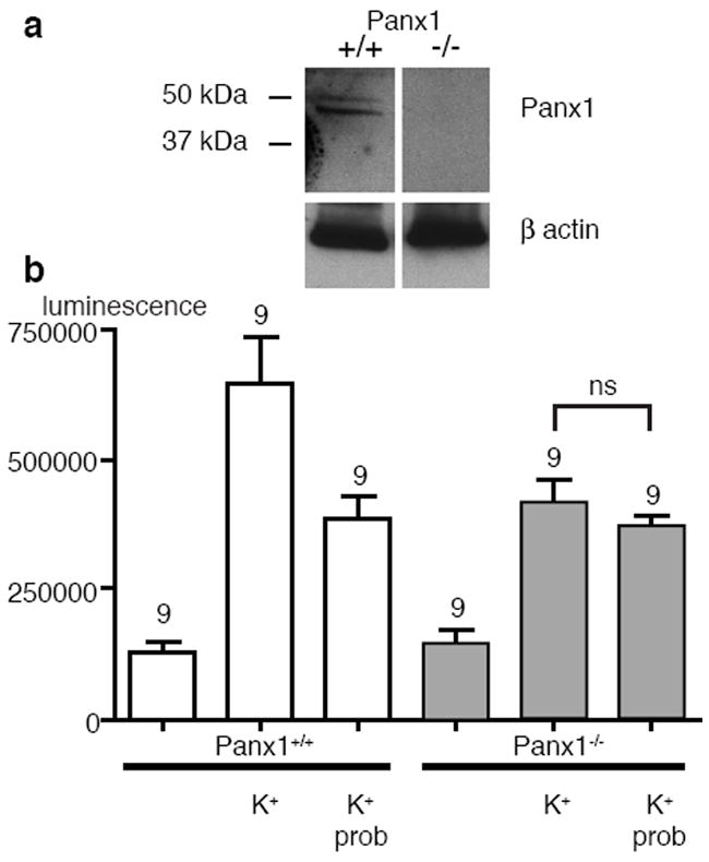
a) Example of a Western blot of erythrocyte membranes of Panx1+/+ and Panx1-/- mice. The double band characteristic for Panx1 at ~50 kD was present in Panx1+/+ erythrocytes but not in Panx1-/- cells. b) ATP release from Panx1+/+ (white bars) and Panx1-/- (grey bars) erythrocytes. ATP release, as determined with a luciferase assay, is stimulated by hypotonic K+ solution (K+) more profoundly in Panx1+/+ erythrocytes than in Panx1-/- erythrocytes. The Panx1 channel inhibitor probenecid (prob, 1 mM) attenuated ATP release in Panx1+/+ cells but not significantly (p>0.05) in Panx1-/- cells.
Figure 2.
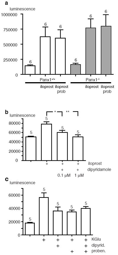
a) Iloprost (25 μM) stimulated ATP release in Panx1+/+ (white bars) and Panx1-/- (grey bars) erythrocytes to similar levels. The iloprost-induced release remained unaffected by probenecid (1 mM). b) Dipyridamole inhibited the iloprost-induced ATP release by Panx1+/+ mouse erythrocytes. c) ATP release by Panx1+/+ mouse erythrocytes induced by hypotonic K+ solution (KGlu) was inhibited by dipyridamole or by probenecid (1 mM). The combination of the drugs was not more effective than either drug alone. Note, that different hematocrits were used for part (a) (20%) as compared to parts (b and c) (2%). Significance levels (Anova with Newman-Keul’s post test): * < 0.05, **<0.01.
Effect of dipyridamole
In a screening test on Panx1 expressing ooctyes we have observed that drugs reported in the literature to affect non-vesicular ATP release also affected Panx1 currents. The Panx1 inhibiting drugs include probenecid, which hitherto had been considered to be a specific transport inhibitor, and also connexin mimetic peptides as off target effect [13,26]. One drug, however, that is well documented to inhibit ATP release and nucleoside flux, dipyridamole [4,27], did not affect Panx1 channels. We, therefore, tested whether the second, probenecid insensitive component of ATP release by erythrocytes was sensitive to dipyridamole. As shown in Figures 2b and 3a, dipyridamole attenuated iloprost-induced ATP release but did not affect Panx1 currents. The effect of dipyridamole was not restricted to iloprost-induced ATP release but also included the hypotonic K+-induced release as shown in Figure 2c. This indicates that hypotonic K+ stimulates two separate ATP release pathways, one sensitive to probenecid and one sensitive to dipyridamole. In that case, one would assume that the effects of these two drugs are additive. However, no additive effect was observed (Figure 2c). Because the data with Panx1-/- cells unequivocally demonstrate, that Panx1 cannot be the cause of iloprost-induced ATP release, the lack of additive effects of probenecid and dipyridamole on hypotonic K+-induced ATP release must have a different explanation than competition of the drugs on one single target. We, therefore, tested the effects of the two drugs on mouse Panx1 currents. Given separately, probenecid inhibited Panx1 currents while dipyridamole had no effect on the currents. When the drugs were combined, the inhibitory effect was significantly lower than that mediated by probenecid alone (Figure 3). Thus, it appears that the drugs interact. However, presently it is unclear where this interaction takes place. It could be a direct interaction of probenecid with dipyridamole in solution, thereby reducing the effective concentrations at their respective targets, or there could be interference at the targets. For example, dipyridamole, while by itself not interfering with Panx1 currents could compete at the probenecid binding site of Panx1 and consequently attenuate the inhibitory action of probenecid.
Figure 3.
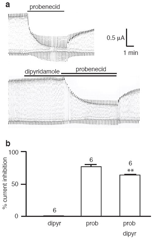
a) Panx1 channel currents in oocytes expressing mouse Panx1 exogenously were inhibited by probenecid (1 mM) but remained unaffected by dipyridamole (10 μM). The inhibition by probenecid was less pronounced when dipyridamole was also present. Currents were induced by voltage pulses from -60 to + 20 mV. The traces were selected from a continuous record from the same oocyte, The interval between the traces was 30 minutes (for full recovery from the probenecid exposure). b) Quantitative analysis of current inhibition by probenecid (1 mM) in the absence and presence of dipyridamole. Means ± SE are shown, * p<0.05.
Uptake of extracellular tracer molecules
A hallmark of non-vesicular (presumably Panx1-mediated) ATP release is the correlated uptake of extracellularly applied tracer molecules. It has been demonstrated in many cell types, including erythrocytes [10-13], that dyes with an exclusion limit >1 kD are taken up by cells under conditions similar to those inducing ATP release. As shown in Figure 4, mouse Panx1+/+ erythrocytes exhibited uptake of carboxyfluorescein when stimulated with hypotonic K+. In contrast, Panx1-/- erythrocytes did not take up the dye under the same experimental conditions. Similarly, no dye uptake was observed, when ATP release was stimulated with iloprost in Panx1-/- cells. These data show, that the Panx1 mediated, probenecid sensitive ATP release pathway allows the concomitant uptake of extracellular dyes. In contrast, the iloprost-induced/dipyridamole sensitive ATP release pathway does not allow the entry of extracellular molecules in a similar size range as ATP. Carboxyfluorescein has an abaxial diameter of about 9 Å as compared to 12 Å for ATP and thus should be small enough for passage through a non-selective, ATP permeable channel. Aside from its effect on ATP release, dipyridamole is known as a nucleoside transport inhibitor [14]. The absence of dye flux in the iloprost-induced ATP release pathway suggests a permeation pathway distinct from that provided by Panx1. The latter is in the form of a large, nonselective pore that below the exclusion limit of 1500 Daltons [26] does not discriminate by size or charge among molecules for transit through the channel. The dipyramidole sensitive pathway consequently is either an ATP selective channel or a transporter (or shares key features with a transporter).
Figure 4.
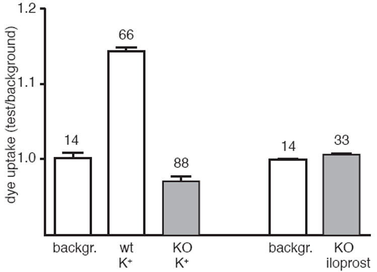
Hypotonic K+ solution (K+) stimulated the uptake of extracellularly applied carboxyfluorescein in Panx1+/+ (wt) erythroctes but not in Panx1-/- (KO) erythrocytes. Iloprost (25 μM) stimulation of Panx1-/- erythrocytes did not result in uptake of the dye.
Glyburide (glibenclamide) inhibits the Panx1 channel
ATP release by erythrocytes has been reported to involve the cystic fibrosis transmembrane conductance regulator (CFTR). Originally, it had been considered that CFTR provides the permeation pathway for ATP [28], however, the lack of ATP permeability of the CFTR channel excludes this possibility [29]. Nevertheless, the CFTR inhibitor glyburide (glibenclamide) does interfere with the release of ATP [6] and of epoxyeicosatrienoic acids [15]. Here we tested whether glyburide may have additional targets in the erythrocyte cell membrane. Specifically, because Panx1 channels evidently provide a permeation pathway for ATP, we tested whether glyburide inhibits ATP flux through Panx1 directly. Figure 5 shows, that glyburide attenuated Panx1 currents in oocytes expressing the protein exogenously with an EC50 of 45 μM. Furthermore, glyburide impaired ATP release from Panx1 expressing oocytes in the same concentration range where current inhibition was observed (Figure 5d).
Figure 5.
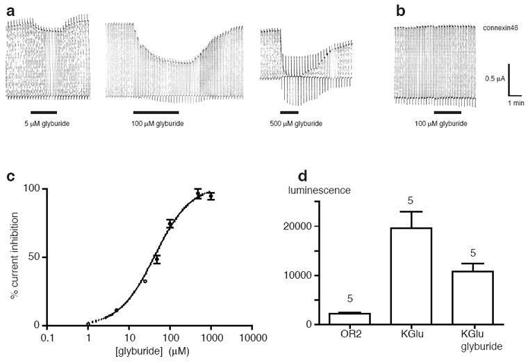
a) Membrane currents in oocytes expressing mouse Panx1 induced by voltage pulses from -50 to + 50 mV. Glyburide (glibenclamide) attenuated the currents in a dose-dependent fashion. n=3-4. b) Connexin 46 currents in oocytes were not affected by glyburide. c) Dose-response relationship of Panx1 current inhibition by glyburide. d) ATP release by Panx1 expressing ooctes was stimulated by 150 mM potassium gluconate solution (KGlu) and inhibited by glyburide (100 μM).
Discussion
It appears that erythrocytes release ATP via two independent pathways, one involving the channel formed by Panx1 and another permeation mechanism that is either a highly selective channel or a type of transporter. The contributions of these release mechanisms may vary in different species as the pannexin1 blockers have a more pronounced effect in human erythrocytes [11,25] than observed here in mouse erythrocytes. Low oxygen, the prime physiological stimulus for ATP release by erythrocytes, seems to exclusively activate the Panx1 mediated release [10,11,25]. The prostaglandin metabolite, iloprost, exclusively activates the alternate, probenecid insensitive and dipyridamole sensitive, pathway. Stimulation of ATP release by mouse erythrocytes with hypotonic K+ solution, however, activated both permeation pathways. It remains to be determined, which physiological role the non-Panx1 pathway exerts. In erythrocytes, the iloprost inducible, dipyridamole sensitive pathway may be the only alternative to Panx1 channels for ATP release. However, the repertoire of most other cells for different ATP release mechanisms is larger. Exocytotic ATP release is well established and may be the exclusive release mechanism at nerve terminals, where ATP is co-packaged and co-released with different transmitter substances. In many cell types, including astrocytes and airway epithelial cells, exocytotic release of ATP co-exists with the release through Panx1 channels and maybe other permeation pathways.
Glyburide, also known as glibenclamide, binds with high affinity to the SUR subunit of ATP regulated potassium channels [30,31]. This effect underlies the therapeutic benefit of glyburide in type 2 diabetes. At higher (micromolar) concentrations, glyburide also inhibits members of the ATP binding cassette (ABC) transporter family, including CFTR [32,33]. Here we show, that glyburide in the micromolar concentration range also inhibits Panx1 currents as well as Panx1 mediated ATP release. Consequently, effects of glyburide on ATP release cannot be used as evidence for an involvement of CFTR in the ATP release mechanism.
Recently it has been found that glyburide also affects innate immunity by inhibiting the inflammasome in various settings [34-37]. Typically, this is taken as evidence, that a key step in inflammasome activation is a potassium efflux [38-40]. However, the concentrations of glyburide required to inhibit activation of caspase and release of cytokines from various types of cells is 3 orders of magnitude higher than that for inhibition of potassium channels. Thus an alternate target for the drug is likely to mediate the inhibitory effect on the inflammasome. With the identification of Panx1 as a target of glyburide, this issue has to be reevaluated, in particular because Panx1 is an elemental part of the inflammasome activation mechanism [24,41,42]. This point is further strengthened by the observation that the inhibition of Panx1 channel currents and of Panx1 mediated ATP release occurred in the same concentration range as needed for inhibition of inflammasome activation.
Highlights.
>ATP release by erythrocytes is attenuated in cells of Pannexin1 deficient mice. >The Panx1-/- cells reveal a second Panx1 independent pathway for ATP release. >The alternate pathway is activated by iloprost and inhibited by dipyridamole. >The alternate release mechanism does not allow for the uptake of extracellular dyes. >Glibenclamide inhibits Panx1 in the same concentration range as it inhibits CFTR channels.
Acknowledgments
We thank Dr. Peter Larsson for reading the manuscript. Work by ES is supported by NIH NS052245. We are thankful to Dr. Bryen Jordan and Miss Dana Savariego (Dept. Neuroscience Albert Einstein College of Medicine) for making available and their assistance with the use of the ultracentrifuge.
Footnotes
Publisher's Disclaimer: This is a PDF file of an unedited manuscript that has been accepted for publication. As a service to our customers we are providing this early version of the manuscript. The manuscript will undergo copyediting, typesetting, and review of the resulting proof before it is published in its final citable form. Please note that during the production process errors may be discovered which could affect the content, and all legal disclaimers that apply to the journal pertain.
References
- 1.Burnstock G. Overview Purinergic mechanisms. Ann N Y Acad Sci. 1990;603:1–17. doi: 10.1111/j.1749-6632.1990.tb37657.x. discussion 18. [DOI] [PubMed] [Google Scholar]
- 2.Burnstock G, Knight GE. Cellular distribution and functions of P2 receptor subtypes in different systems. Int Rev Cytol. 2004;240:31–304. doi: 10.1016/S0074-7696(04)40002-3. [DOI] [PubMed] [Google Scholar]
- 3.Korngreen A, Priel Z. Purinergic stimulation of rabbit ciliated airway epithelia: control by multiple calcium sources. J Physiol. 1996;497(Pt 1):53–66. doi: 10.1113/jphysiol.1996.sp021749. [DOI] [PMC free article] [PubMed] [Google Scholar]
- 4.Bergfeld GR, Forrester T. Release of ATP from human erythrocytes in response to a brief period of hypoxia and hypercapnia. Cardiovasc Res. 1992;26:40–7. doi: 10.1093/cvr/26.1.40. [DOI] [PubMed] [Google Scholar]
- 5.Mariathasan S, et al. Differential activation of the inflammasome by caspase-1 adaptors ASC and Ipaf. Nature. 2004;430:213–8. doi: 10.1038/nature02664. [DOI] [PubMed] [Google Scholar]
- 6.Sprague RS, Ellsworth ML, Stephenson AH, Kleinhenz ME, Lonigro AJ. Deformation-induced ATP release from red blood cells requires CFTR activity. Am J Physiol. 1998;275:H1726–32. doi: 10.1152/ajpheart.1998.275.5.H1726. [DOI] [PubMed] [Google Scholar]
- 7.Ellsworth ML, Forrester T, Ellis CG, Dietrich HH. The erythrocyte as a regulator of vascular tone. Am J Physiol. 1995;269:H2155–61. doi: 10.1152/ajpheart.1995.269.6.H2155. [DOI] [PubMed] [Google Scholar]
- 8.Dietrich HH, Horiuchi T, Xiang C, Hongo K, Falck JR, Dacey RG., Jr Mechanism of ATP-induced local and conducted vasomotor responses in isolated rat cerebral penetrating arterioles. J Vasc Res. 2009;46:253–64. doi: 10.1159/000167273. [DOI] [PMC free article] [PubMed] [Google Scholar]
- 9.Ellsworth ML, Ellis CG, Goldman D, Stephenson AH, Dietrich HH, Sprague RS. Erythrocytes: oxygen sensors and modulators of vascular tone. Physiology (Bethesda) 2009;24:107–16. doi: 10.1152/physiol.00038.2008. [DOI] [PMC free article] [PubMed] [Google Scholar]
- 10.Dahl G, Locovei S. Pannexin: To Gap or not to Gap, is that a question? IUBMB Life. 2006;58:409–419. doi: 10.1080/15216540600794526. [DOI] [PubMed] [Google Scholar]
- 11.Locovei S, Bao L, Dahl G. Pannexin 1 in erythrocytes: Function without a gap. Proc Natl Acad Sci U S A. 2006;103:7655–7659. doi: 10.1073/pnas.0601037103. [DOI] [PMC free article] [PubMed] [Google Scholar]
- 12.Qiu F, Dahl G. A permeant regulating its permeation pore: inhibition of pannexin 1 channels by ATP. Am J Physiol Cell Physiol. 2009;296:C250–5. doi: 10.1152/ajpcell.00433.2008. [DOI] [PMC free article] [PubMed] [Google Scholar]
- 13.Silverman W, Locovei S, Dahl G. Probenecid, a gout remedy, inhibits pannexin 1 channels. Am J Physiol Cell Physiol. 2008;295:C761–7. doi: 10.1152/ajpcell.00227.2008. [DOI] [PMC free article] [PubMed] [Google Scholar]
- 14.Plagemann PG, Wohlhueter RM, Woffendin C. Nucleoside and nucleobase transport in animal cells. Biochim Biophys Acta. 1988;947:405–43. doi: 10.1016/0304-4157(88)90002-0. [DOI] [PubMed] [Google Scholar]
- 15.Jiang H, Zhu AG, Mamczur M, Falck JR, Lerea KM, McGiff JC. Stimulation of rat erythrocyte P2X7 receptor induces the release of epoxyeicosatrienoic acids. Br J Pharmacol. 2007;151:1033–40. doi: 10.1038/sj.bjp.0707311. [DOI] [PMC free article] [PubMed] [Google Scholar]
- 16.Ballerini P, Di Iorio P, Ciccarelli R, Nargi E, D’Alimonte I, Traversa U, Rathbone MP, Caciagli F. Glial cells express multiple ATP binding cassette proteins which are involved in ATP release. Neuroreport. 2002;13:1789–92. doi: 10.1097/00001756-200210070-00019. [DOI] [PubMed] [Google Scholar]
- 17.Forsyth AM, Wan J, Owrutsky PD, Abkarian M, Stone HA. Multiscale approach to link red blood cell dynamics, shear viscosity, and ATP release. Proc Natl Acad Sci U S A. 2011;108:10986–91. doi: 10.1073/pnas.1101315108. [DOI] [PMC free article] [PubMed] [Google Scholar]
- 18.Dahl G, Pfahnl A. Mutagenesis to study channel structure. Methods Mol Biol. 2001;154:251–68. doi: 10.1385/1-59259-043-8:251. [DOI] [PubMed] [Google Scholar]
- 19.Santiago MF, V J, Patel NK, Lutz SE, Caille D, Charollais A, Meda P, Scemes E. Targeting Pannexin1 improves seizure outcome. Plos One. 2011 doi: 10.1371/journal.pone.0025178. in press. [DOI] [PMC free article] [PubMed] [Google Scholar]
- 20.Boassa D, Ambrosi C, Qiu F, Dahl G, Gaietta G, Sosinsky G. Pannexin1 channels contain a glycosylation site that targets the hexamer to the plasma membrane. J Biol Chem. 2007;282:31733–43. doi: 10.1074/jbc.M702422200. [DOI] [PubMed] [Google Scholar]
- 21.Sosinsky GE, et al. Pannexin channels are not gap junction hemichannels. Channels (Austin) 2011;5:193–197. doi: 10.4161/chan.5.3.15765. [DOI] [PMC free article] [PubMed] [Google Scholar]
- 22.Bao L, Locovei S, Dahl G. Pannexin membrane channels are mechanosensitive conduits for ATP. FEBS Lett. 2004;572:65–8. doi: 10.1016/j.febslet.2004.07.009. [DOI] [PubMed] [Google Scholar]
- 23.Locovei S, Wang J, Dahl G. Activation of pannexin 1 channels by ATP through P2Y receptors and by cytoplasmic calcium. FEBS Lett. 2006;580:239–44. doi: 10.1016/j.febslet.2005.12.004. [DOI] [PubMed] [Google Scholar]
- 24.Silverman WR, de Rivero Vaccari JP, Locovei S, Qiu F, Carlsson SK, Scemes E, Keane RW, Dahl G. The pannexin 1 channel activates the inflammasome in neurons and astrocytes. J Biol Chem. 2009;284:18143–51. doi: 10.1074/jbc.M109.004804. [DOI] [PMC free article] [PubMed] [Google Scholar]
- 25.Sridharan M, Adderley SP, Bowles EA, Egan TM, Stephenson AH, Ellsworth ML, Sprague RS. Pannexin 1 is the conduit for low oxygen tension-induced ATP release from human erythrocytes. Am J Physiol Heart Circ Physiol. 2010;299:H1146–52. doi: 10.1152/ajpheart.00301.2010. [DOI] [PMC free article] [PubMed] [Google Scholar]
- 26.Wang J, Ma M, Locovei S, Keane RW, Dahl G. Modulation of membrane channel currents by gap junction protein mimetic peptides: size matters. Am J Physiol Cell Physiol. 2007;293:C1112–9. doi: 10.1152/ajpcell.00097.2007. [DOI] [PubMed] [Google Scholar]
- 27.Kirkpatrick KA, Burnstock G. Evidence that the inhibition of ATP release from sympathetic nerves by adenosine is a physiological mechanism. Gen Pharmacol. 1992;23:1045–50. doi: 10.1016/0306-3623(92)90284-q. [DOI] [PubMed] [Google Scholar]
- 28.Reisin IL, Prat AG, Abraham EH, Amara JF, Gregory RJ, Ausiello DA, Cantiello HF. The cystic fibrosis transmembrane conductance regulator is a dual ATP and chloride channel. J Biol Chem. 1994;269:20584–91. [PubMed] [Google Scholar]
- 29.Grygorczyk R, Tabcharani JA, Hanrahan JW. CFTR channels expressed in CHO cells do not have detectable ATP conductance. J Membr Biol. 1996;151:139–48. doi: 10.1007/s002329900065. [DOI] [PubMed] [Google Scholar]
- 30.Sturgess NC, Ashford ML, Cook DL, Hales CN. The sulphonylurea receptor may be an ATP-sensitive potassium channel. Lancet. 1985;2:474–5. doi: 10.1016/s0140-6736(85)90403-9. [DOI] [PubMed] [Google Scholar]
- 31.Mikhailov MV, Mikhailova EA, Ashcroft SJ. Molecular structure of the glibenclamide binding site of the beta-cell K(ATP) channel. FEBS Lett. 2001;499:154–60. doi: 10.1016/s0014-5793(01)02538-8. [DOI] [PubMed] [Google Scholar]
- 32.Sheppard DN, Welsh MJ. Inhibition of the cystic fibrosis transmembrane conductance regulator by ATP-sensitive K+ channel regulators. Ann N Y Acad Sci. 1993;707:275–84. doi: 10.1111/j.1749-6632.1993.tb38058.x. [DOI] [PubMed] [Google Scholar]
- 33.Schwiebert EM, Egan ME, Hwang TH, Fulmer SB, Allen SS, Cutting GR, Guggino WB. CFTR regulates outwardly rectifying chloride channels through an autocrine mechanism involving ATP. Cell. 1995;81:1063–73. doi: 10.1016/s0092-8674(05)80011-x. [DOI] [PubMed] [Google Scholar]
- 34.Hamon Y, Luciani MF, Becq F, Verrier B, Rubartelli A, Chimini G. Interleukin-1beta secretion is impaired by inhibitors of the Atp binding cassette transporter, ABC1. Blood. 1997;90:2911–5. [PubMed] [Google Scholar]
- 35.Muhl H, Hofler S, Pfeilschifter J. Inhibition of lipopolysaccharide/ATP-induced release of interleukin-18 by KN-62 and glyburide. Eur J Pharmacol. 2003;482:325–8. doi: 10.1016/j.ejphar.2003.09.062. [DOI] [PubMed] [Google Scholar]
- 36.Marty V, Medina C, Combe C, Parnet P, Amedee T. ATP binding cassette transporter ABC1 is required for the release of interleukin-1beta by P2X7-stimulated and lipopolysaccharide-primed mouse Schwann cells. Glia. 2005;49:511–9. doi: 10.1002/glia.20138. [DOI] [PubMed] [Google Scholar]
- 37.Lamkanfi M, et al. Glyburide inhibits the Cryopyrin/Nalp3 inflammasome. J Cell Biol. 2009;187:61–70. doi: 10.1083/jcb.200903124. [DOI] [PMC free article] [PubMed] [Google Scholar]
- 38.Muruve DA, Petrilli V, Zaiss AK, White LR, Clark SA, Ross PJ, Parks RJ, Tschopp J. The inflammasome recognizes cytosolic microbial and host DNA and triggers an innate immune response. Nature. 2008;452:103–7. doi: 10.1038/nature06664. [DOI] [PubMed] [Google Scholar]
- 39.Gross O, et al. Syk kinase signalling couples to the Nlrp3 inflammasome for anti-fungal host defence. Nature. 2009;459:433–6. doi: 10.1038/nature07965. [DOI] [PubMed] [Google Scholar]
- 40.Schroder K, Zhou R, Tschopp J. The NLRP3 inflammasome: a sensor for metabolic danger? Science. 2010;327:296–300. doi: 10.1126/science.1184003. [DOI] [PubMed] [Google Scholar]
- 41.Pelegrin P, Surprenant A. Pannexin-1 mediates large pore formation and interleukin-1beta release by the ATP-gated P2X(7) receptor. Embo J. 2006 doi: 10.1038/sj.emboj.7601378. [DOI] [PMC free article] [PubMed] [Google Scholar]
- 42.Kanneganti TD, Lamkanfi M, Kim YG, Chen G, Park JH, Franchi L, Vandenabeele P, Nunez G. Pannexin-1-mediated recognition of bacterial molecules activates the cryopyrin inflammasome independent of Toll-like receptor signaling. Immunity. 2007;26:433–43. doi: 10.1016/j.immuni.2007.03.008. [DOI] [PubMed] [Google Scholar]