



Abstract
Recapitulating the elegant structures formed during development is an extreme synthetic and biological challenge. Great progress has been made in developing materials to support transplanted cells, yet the complexity of tissues is far beyond that found in even the most advanced scaffolds. Self-assembly is a motif used in development and a route for the production of complex materials. Self-assembly of peptides, proteins and other molecules at the nanoscale is promising, but in addition, intriguing ideas are emerging for self-assembly of micron-scale structures. In this brief review, very recent advances in the assembly of micron-scale cell aggregates and microgels will be described and discussed.
Keywords: Modular scaffold, microgel, tissue engineering, regenerative medicine, hydrogel, self-assembly
Introduction
The development of biomaterials for regenerative medicine is at a crossroads. The goal of producing functional tissues is challenged by the complexity of organ architectures. It is difficult enough to vascularize tissue constructs, let alone to recapitulate the complex arrangement of cells and extracellular matrix (ECM) found in functional organs. Tremendous recent successes achieved using cell-seeded decellularized tissues confirm the important role of a scaffold or structural framework to which cells may adhere [1–3]. On the opposite end of the spectrum, scaffold-less approaches using cell aggregates or cell sheets lead to questions about the importance and necessity of a scaffold.
Despite the tremendous advances using biologically-derived scaffolds, the development of synthetically-derived scaffolds is attractive from practical, regulatory and scientific perspectives. However, will it be possible to engineer cells and materials to recapitulate the complex structures of ECM as it exists in native tissues? Of the many approaches that are currently being investigated, one promising route for the production of such materials is through bottom-up assembly.
In 1959, Feynman described the applicability of (mostly) top-down approaches to nano-manufacturing in his famous lecture, “There s plenty of room at the bottom” [4]. A top-down approach uses tools to sculpt an object – tools such as knives, saws, brushes, etchants, molds, etc. This holds true even when carried out at the nanoscale, as in photolithography. Later, Drexler described molecular engineering for nano-manufacturing, thus formalizing the bottom-up approach [5]. In regenerative medicine, production of a scaffold in a mold is an example of a top-down strategy, even if the scaffold is made porous by including leachable microparticles, or if structures are introduced post-hoc using optical pattering techniques. A bottom-up approach relies on the self-assembly or directed-assembly of a scaffold from smaller components or modules , potentially with different modules designed to carry out distinct tasks (Fig. 1).
Figure 1.

Modular approach to the production of scaffolds. Nano- or microscale modules consisting of cells, cell aggregates, particles or gels are allowed to assemble. The assembly process may be guided by specific interactions or may be random. Following crosslinking in some manner, a three-dimensional construct is available for in vitro expansion of cells or for in vivo transplantation.
A common characteristic of the current bottom-up strategies for scaffold fabrication is the assembly of molecules into nano- or microscale structures, which then assemble into macroscopic objects. Self-assembling molecules that form nanoscale structures that then form gels are excellent examples of bottom-up assembly. Self-assembly of materials from amphiphilic molecules (proteins, peptides, polymers, etc.) have been recently reviewed and will not be further described here [6]. The focus of the current review will be on the assembly of micron-scale objects into three-dimensional structures.
Modular scaffolds have been investigated by a small but growing number of groups. Previous reviews describe the synthesis of modular scaffolds up to mid-2009 [7, 8]. The current contribution will focus on work subsequent to these reviews.
Assembly of cells and cell aggregates
An exciting development is the DNA-directed self-assembly of multiple cell types. Bertozzi and colleagues have previously developed alkyne derivatives that undergo [3+2] cycloadditions in the absence copper [9–11]. Strained alkynes react readily with an azide, with no detectable cross-reactions with moieties found in cells and tissues. Saccharides such as N-azidoacetylmannosamine added to cell culture medium are taken up by cells and used to produce the extracellular glycocalyx. In a recent study, cell surfaces decorated with azide sugars were reacted with single-stranded oligonucleotides modified with a cyclooctyne derivative. Two batches of Jurkat cells were separately labeled with complementary oligonucleotides. Upon mixing in a 1:1 ratio, the cells assembled into large aggregates. To control aggregation, the ratio of the two types of cells was adjusted to 1:50. In this case, the less common cell type existed as single cells surrounded by rosettes of the other cell type, termed microtissues (Fig. 2). DNA-directed cell assembly was then used to produce a paracrine signaling network between IL-3 expressing CHO cells and an IL-3 dependent untransformed hematopoietic progenitor cell line (FL5.12). FL5.12 cells that were assembled into microtissues with the CHO cells and embedded in agarose gels were able to proliferate even after IL-3 removal. Single FL5.12 cells did not proliferate under these conditions [12].
Figure 2.
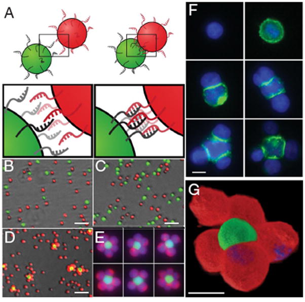
(A) Complementary oligonucleotides were covalently coupled to the surfaces of different cells by click chemistry. (B–E) Two non-adherent cell types were mixed, and did not aggregate if their surfaces were modified with: (B) no oligonucleotides, (C) non-complementary oligonulceotides. However, specific aggregation was observed if the cell surfaces were modified with complementary oligonucleotides (D&E) . (F) Aggregation of DAPI stained cells (blue), with the central cell modified with fluorescein-conjugated oligonucleotides (green). (G) 3D reconstruction of an aggregate of Texas Red-labeled (red) and fluorescein-labeled cells (green). From Gartner ZJ, Bertozzi CR. “Programmed assembly of 3-dimensional microtissues with defined cellular connectivity”, Proceedings of the National Academies of Science, USA, 106:4606–10. Copyright 2009 to the authors, permission for reuse not required.
Formation of microtissues may also be performed on larger scales by co-culturing pre-formed cell aggregates. Forgacs and colleagues have demonstrated that spheroids may be formed by cutting cylinders of a cell slurry followed by overnight culture in solution. The spheroids that form are of uniform size and are used as bioink for cell printing [13]. Spheroids of different cell types may be printed into tissue-like structures using a custom 3D printer. This highly promising technique has been very recently reviewed [14]. Morgan and colleagues have demonstrated that if a poorly adhesive surface contained small posts, the cells surrounded the posts, eventually forming a multilayer of cell aggregate with holes after removal from the substrate [15]. More recently, these honeycomb-like structures were formed from ovarian theca cells. Inside the holes, ovarian granulosa cells were added, creating a structure similar to that found in the ovary. Such techniques may allow in vitro maturation of oocytes, potentially greatly improving in vitro fertilization [16]. The same group also demonstrated the formation of toroids on poorly-adhesive posts. As the cells contracted around tapered posts, they actually pushed themselves up and off. The toroids proved to be useful building blocks to assemble highly porous tissue-like structures [17]. The group also studied the kinetics of assembly of spheroids in linear troughs. Linear arrays of spheroids tended to fuse into rods and contract over time. The kinetics of fusion and contraction could be manipulated by the length of time the spheroids were allowed to mature, or by blocking cell-cell adhesion [18, 19].
Arrays of spheroids may also be produced. Morgan and colleagues used poorly adhesive multi-wells made from agarose and seeded them with cells to form spheroids. An alginate solution was then poured over the multi-wells and gelled. When removed from the agarose, the alginate gel was decorated with spheroid-laden posts [20]. In a similar spheroid-patterning strategy, Fukada and colleagues filled microwells in a microfluidics device with HepG2 liver cells (the other surfaces of the device were coated with albumin, such that the HepG2 spheroids could only form in the microwells). Then, a solution of polylysine was flowed over the surface, adsorbing to the albumin coating and rendering the surface cell adhesive. Fibroblasts were seeded on the substrate and these cells surrounded the spheroids but did not coat them [21].
Assembly of micron-scale hydrogels
Sefton and colleagues introduced an intriguing approach to modular scaffold formation in 2006 [22]. The approach essentially inverts the architecture of the native vasculature. Cylinders (modules) are produced by cutting a tube collagen gel that is loaded with cells. The modules are then coated with endothelial cells. The modules may be packed into a chamber and subjected to blood flow. Although the thrombogenicity of the construct is low in flowing blood, adhesion of leukocytes was present [23]. This may have resulted from the flow geometry, as a endothelial-lined microfluidic chamber with similar geometry also led to greater adhesion of leukocytes [24]. Collagen/Matrigel modules were formed with embedded rat pup cardiomyocytes. The modules were seeded with endothelial cells and embedded in an alginate gel. The modules contracted in sync with electrical stimulation. However, the endothelial coating did attenuate the response [25]. The modules may be used as vehicles for cell transplantation in vivo. Human endothelial-coated collagen modules were implanted in athymic rats. The modules formed hollow channels at their interfaces and the human endothelial cells formed primitive vessels in the channels [26]. Transplanting modules coated with allogeneic rat endothelial cells into immune compromised rats resulted in the outgrowth of the transplanted cells to form functioning blood vessels that persisted for at least 60 days [27]. The modules were also implanted in immune-compromised mice by injecting through a needle. Endothelial apoptosis was noted to be present, which was decreased by coating the collagen modules with fibronectin. The fibronectin-coated modules yielded greater numbers of mature vessels lined with the transplanted endothelial cells [28]. Synthetic materials are also being developed as mechanically stronger substitutes for collagen in the cylinders [29]. A video of the methods for modular scaffold fabrication has been published [30]. The Whitesides lab has also introduced micromolding methods to produce similar, yet smaller, cylinders [31, 32].
Khademhossenei and colleagues have intensively investigated the assembly of cell-laden microgels to produce scaffolds (Fig. 3). The microgels have been assembled primarily by manipulating the hydrophilicity of surfaces and interfaces. For example, a PDMS stamp was used to pattern a hydrophobic silane onto a glass slide. Cell-laden microgels were added to the glass substrate in PBS. After pouring off the excess solution, the buffer solution remained in the hydrophilic regions of the slide, trapping some of the microgels in the remaining liquid [33]. A mathematical model of microgel assembly in oil-like media has been developed [34].
Figure 3.
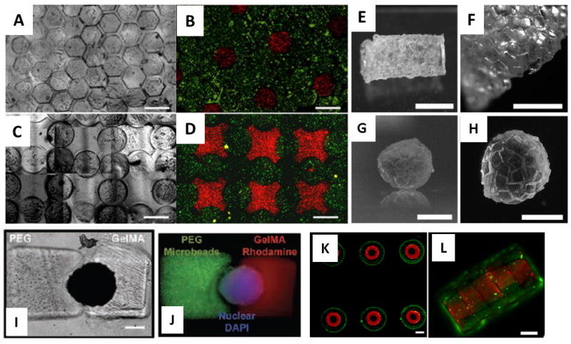
(A) Assembly of hexagonal PEG microgels floating on a perfluorinated solvent. (B) Fluorescence image of liver cells (red) in some of the hexagonal microgels and fibroblasts (green) in the rest. (C) Assembly of lock-and-key microgels. (D) Fluorescence image of liver cells (red) in the lock microgels and fibroblasts (green) in the key microgels. From: Zamanian B, Masaeli M, Nichol JW, Khabiry M, Hancock MJ, Bae H, et al. “Interface-directed self-assembly of cell-laden microgels”, Small, 2010, 6:937–44. Copyright Wiley-VCH Verlag GmbH & Co. KGaA. Reproduced with permission. (E–H) Micromasonry , using surface tension of a polymerizable solution of PEG to assemble microgels, followed by photopolymerization. (E&F) Tube formed around a PDMS cylinder. (G&H) Sphere formed in a non-aqueous solvent. From: Fernandez JG, Khademhosseini A. “Micro-Masonry: Construction of 3D Structures by Microscale Self-Assembly”, Advanced Materials. 2010;22:2538–41. Copyright Wiley-VCH Verlag GmbH & Co. KGaA. Reproduced with permission. (I&J) Photomasks were used to sequentially polymerize PEG (left) and gelatin (right) around a cell cluster (center). Qi H, Du Y, Wang L, Kaji H, Bae H, Khademhosseini A. “Patterned differentiation of individual embryoid bodies in spatially organized 3D hybrid microgels”, Adv Mater. 2010;22:5276–81. Copyright Wiley-VCH Verlag GmbH & Co. KGaA. Reproduced with permission. (K) Concentric rings formed by sequential photopolymerizations using different masks. (L) The rings stack to form tubes in mineral oil. Du Y, Ghodousi M, Qi H, Haas N, Xiao W, Khademhosseini A. “Sequential assembly of cell-laden hydrogel constructs to engineer vascular-like microchannels”, Biotechnol Bioeng. 2011; In press. Copyright John Wiley & Sons. Reproduced with permission.
Hexagonal microgels were recently produced by the photopolymerization of PEG or the calcium-induced gelation of alginate. The microgels were assembled in two-dimensional arrays and pores were generated by chelating calcium to dissolve the alginate microgels, with little impact on cell viability within the PEG microgels. Multiple layers could be stacked to produce three-dimensional structures [35]. Microgels of various shapes were also assembled while floating on the solvent perfluorodecalin. Cell-laden microgels aggregated and were crosslinked by a second photopolymerization with little loss of cell viability [36]. PEG microgel rings were also synthesized, which stacked into tubes in mineral oil. By varying the number of holes in the rings, branching structures could be introduced. Concentric rings were also produced by sequential photopolymerization with different photomasks. Endothelial cells were polymerized in the inner ring, while smooth muscle cells were polymerized in the outer ring, which could be assembled into blood vessel-like structures. [37]. The group also introduced the concept of micro-masonry . PEG microgels containing HepG2 cells were formed in various shapes (usually bricks). The microgels were suspended in a solution of PEG-methacrylate with photoinitiator and added to a PDMS template (e.g., a cylinder). When excess solution was removed, capillary forces brought the bricks together around the template. Photopolymerization was used to crosslink the solution-phase PEG-methacrylate, serving as the mortar that bound the bricks together in the shape of the template [38].
In another strategy from the same group, mouse embryoid bodies were formed and deposited into microwells on a substrate. Photopolymerizable PEG was added and photopolymerized in roughly half of the microwell. Another mask was used to photopolymerize gelatin in the other half of the microwell. Thus, half of each embryoid body was encapsulated in a block of PEG gel, and the other half was encapsulated in a block of crosslinked gelatin. Vasculogenic cells sprouted from the embryoid body into the gelatin matrix, introducing polarity into the construct [39].
Harada et al. recently used the differential affinities of cyclodextrins to selectively assemble hydrogels. Adamantane or tert-butyl groups bind strongly to β-cyclodextrin, while n-butyl groups bind strongly to α-cyclodextrin. When the various chemical groups were added to poly(acrylamide) gels, the gels self-segregated based on the specific interactions (Fig. 4A) [40]. Although the gels were in the millimeter size regime, the approach may prove to be useful for the assembly of microscale hydrogels.
Figure 4.
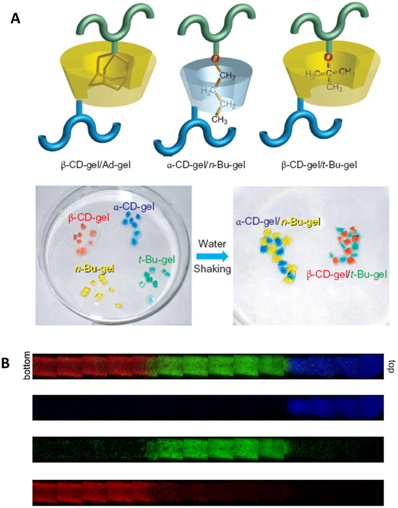
(A) Millimeter-sized gels decorated with α- or β-cyclodextrins, or n-butyl or tert-butyl groups aggregate upon shaking, but only with gels containing their appropriate binding partners. Reprinted by permission from Macmillan Publishers Ltd.: Nature Chemistry, 3:34–37. Copyright 2011. (B) PEG microspheres were formed with identical chemistries and similar size, but with different buoyancies. Upon centrifugation microspheres separated out into distinct layers based on the buoyancy differences to produce graded structures. From: Roam JL, Xu H, Nguyen PK, Elbert DL., “The Formation of Protein Concentration Gradients Mediated by Density Differences of Poly(ethylene glycol) Microspheres”, Biomaterials. 2010;8642–8650. Copyright Elsevier, used with permission.
In the author s lab, we have recently produced macroporous scaffolds by the assembly and chemical reaction of hydrogel microspheres. Microspheres with different functions (structural, drug delivery and porogenic) were synthesized from reactive PEG derivatives and proteins. The method that was developed for microsphere fabrication relied a thermally induced phase separation of PEG in the presence of sodium sulfate [41]. The microspheres retained reactive groups, allowing crosslinking upon mixing the microspheres and centrifugation, forming a scaffold. The crosslinking was mild enough to be performed in the presence of living cells. The porogenic microspheres dissolved with two days in culture to produce porous scaffolds without affecting cell viability [42]. The density of the crosslinks in the microspheres affected their buoyancy relatively independently of their size, allowing the formation of gradients in microspheres upon centrifugation (Fig. 4B) [43]. Microspheres used for scaffold assembly may also be produced by the precipitation photopolymerization of PEG-diacrylate [44].
A number of groups are investigating aggregates consisting of cells and drug-delivering hydrogel microspheres. The overall hypothesis is that growth factor diffusion into spheroids is a limiting factor in the differentiation of stem cells. Hyaluronic acid microspheres containing perlecan for controlled release of BMP-2 were added to mesenchymal stem cells in a micromass to promote chondrogenesis [45]. Others have developed chondroitin sulfate, agarose and gelatin microspheres were developed to release growth factors to affect cell differentiation. The microspheres were incorporated within mouse embryoid bodies, with good viability and some effects on stem cell differentiation [46, 47]. Gelatin/chitosan microspheres have been similarly used to promote the differentiation of adipose derived stem cells [48].
These strategies are intermediate between scaffold-based and scaffold-free approaches, and evolved from techniques using non-hydrogel microspheres made from degradable poly(esters) [49–54].
Conclusions
The self-assembly strategies discussed here may evolve to allow the formation of spatially organized tissues. The ability to control the architecture of 3D cell/material hybrid structures may prove to be critical for the development of highly advanced scaffolds for cell transplantation that are better mimics of natural ECM.
Acknowledgments
This work was supported by NIH R01HL085364.
Footnotes
Publisher's Disclaimer: This is a PDF file of an unedited manuscript that has been accepted for publication. As a service to our customers we are providing this early version of the manuscript. The manuscript will undergo copyediting, typesetting, and review of the resulting proof before it is published in its final citable form. Please note that during the production process errors may be discovered which could affect the content, and all legal disclaimers that apply to the journal pertain.
References
- 1.Petersen TH, Calle EA, Zhao L, Lee EJ, Gui L, Raredon MB, et al. Tissue-Engineered Lungs for in Vivo Implantation. Science. 2010;329:538–41. doi: 10.1126/science.1189345. [DOI] [PMC free article] [PubMed] [Google Scholar]
- 2*.Ott HC, Clippinger B, Conrad C, Schuetz C, Pomerantseva I, Ikonomou L, et al. Regeneration and orthotopic transplantation of a bioartificial lung. Nat Med. 2010;16:927–33. doi: 10.1038/nm.2193. This paper along with reference 1 demonstrates the tremendous amount of information encoded in ECM. Additionally, the architecture of the tissue seems to be of utmost importance. The bottom-up approach to scaffold formation may eventually be able to assemble such complex structures, but much more research will be required. [DOI] [PubMed] [Google Scholar]
- 3.Badylak SF, Freytes DO, Gilbert TW. Extracellular matrix as a biological scaffold material: Structure and function. Acta biomaterialia. 2009;5:1–13. doi: 10.1016/j.actbio.2008.09.013. [DOI] [PubMed] [Google Scholar]
- 4.Feynman RP. There's plenty of room at the bottom. Eng and Sci. 1961;23:22–36. [Google Scholar]
- 5.Drexler KE. Molecular engineering: An approach to the development of general capabilities for molecular manipulation. Proceedings of the National Academy of Sciences of the United States of America. 1981;78:5275–8. doi: 10.1073/pnas.78.9.5275. [DOI] [PMC free article] [PubMed] [Google Scholar]
- 6.Branco MC, Schneider JP. Self-assembling materials for therapeutic delivery. Acta Biomaterialia. 2009;5:817–31. doi: 10.1016/j.actbio.2008.09.018. [DOI] [PMC free article] [PubMed] [Google Scholar]
- 7.Nichol JW, Khademhosseini A. Modular Tissue Engineering: Engineering Biological Tissues from the Bottom Up. Soft Matter. 2009;5:1312–9. doi: 10.1039/b814285h. [DOI] [PMC free article] [PubMed] [Google Scholar]
- 8.Rivron NC, Rouwkema J, Truckenmuller R, Karperien M, De Boer J, Van Blitterswijk CA. Tissue assembly and organization: Developmental mechanisms in microfabricated tissues. Biomaterials. 2009;30:4851–8. doi: 10.1016/j.biomaterials.2009.06.037. [DOI] [PubMed] [Google Scholar]
- 9.Codelli JA, Baskin JM, Agard NJ, Bertozzi CR. Second-generation difluorinated cyclooctynes for copper-free click chemistry. Journal of the American Chemical Society. 2008;130:11486–93. doi: 10.1021/ja803086r. [DOI] [PMC free article] [PubMed] [Google Scholar]
- 10.Jewett JC, Bertozzi CR. Cu-free click cycloaddition reactions in chemical biology. Chemical Society Reviews. 2010;39:1272–9. doi: 10.1039/b901970g. [DOI] [PMC free article] [PubMed] [Google Scholar]
- 11.Jewett JC, Sletten EM, Bertozzi CR. Rapid Cu-free click chemistry with readily synthesized biarylazacyclooctynones. Journal of the American Chemical Society. 2010;132:3688–90. doi: 10.1021/ja100014q. [DOI] [PMC free article] [PubMed] [Google Scholar]
- 12*.Gartner ZJ, Bertozzi CR. Programmed assembly of 3-dimensional microtissues with defined cellular connectivity. Proceedings of the National Academy of Sciences. 2009;106:4606–10. doi: 10.1073/pnas.0900717106. The cells that are labeled with complementary DNA strands tend to aggregate severely unless present in vastly different ratios. Although clusters could be purified by flow cytometry, methods need to be developed to allow the formation of complex structures of self-limiting size. Furthermore, many biological structures are tubular rather than spherical. Anisotropic assembly seems difficult but necessary for use in tissue fabrication. [DOI] [PMC free article] [PubMed] [Google Scholar]
- 13.Jakab K, Norotte C, Damon B, Marga F, Neagu A, Besch-Williford CL, et al. Tissue engineering by self-assembly of cells printed into topologically defined structures. Tissue Eng Part A. 2008;14:413–21. doi: 10.1089/tea.2007.0173. [DOI] [PubMed] [Google Scholar]
- 14.Jakab K, Norotte C, Marga F, Murphy K, Vunjak-Novakovic G, Forgacs G. Tissue engineering by self-assembly and bio-printing of living cells. Biofabrication. 2010;2:022001. doi: 10.1088/1758-5082/2/2/022001. [DOI] [PMC free article] [PubMed] [Google Scholar]
- 15*.Dean DM, Napolitano AP, Youssef J, Morgan JR. Rods, tori, and honeycombs: the directed self-assembly of microtissues with prescribed microscale geometries. Faseb J. 2007;21:4005–12. doi: 10.1096/fj.07-8710com. The presented technique is scaffoldless. Is a scaffold necessary for cell transplantation in regenerative medicine? Can the ECM secreted by the cells be considered a scaffold ? Is the density of cells in a spheroid unnaturally high for many applications? [DOI] [PubMed] [Google Scholar]
- 16.Krotz S, Robins J, Ferruccio T-M, Moore R, Steinhoff M, Morgan J, et al. In vitro maturation of oocytes via the pre-fabricated self-assembled artificial human ovary. Journal of Assisted Reproduction and Genetics. 2010;27:743–50. doi: 10.1007/s10815-010-9468-6. [DOI] [PMC free article] [PubMed] [Google Scholar]
- 17.Livoti CM, Morgan JR. Self-assembly and tissue fusion of toroid-shaped minimal building units. Tissue Eng Part A. 2010;16:2051–61. doi: 10.1089/ten.tea.2009.0607. [DOI] [PMC free article] [PubMed] [Google Scholar]
- 18.Rago AP, Dean DM, Morgan JR. Controlling cell position in complex heterotypic 3D microtissues by tissue fusion. Biotechnol Bioeng. 2009;102:1231–41. doi: 10.1002/bit.22162. [DOI] [PubMed] [Google Scholar]
- 19.Bao B, Jiang J, Yanase T, Nishi Y, Morgan JR. Connexon-mediated cell adhesion drives microtissue self-assembly. Faseb J. 2011;25:255–64. doi: 10.1096/fj.10-155291. [DOI] [PMC free article] [PubMed] [Google Scholar]
- 20.Rago AP, Chai PR, Morgan JR. Encapsulated arrays of self-assembled microtissues: an alternative to spherical microcapsules. Tissue Eng Part A. 2009;15:387–95. doi: 10.1089/ten.tea.2008.0107. [DOI] [PubMed] [Google Scholar]
- 21.Okuyama T, Yamazoe H, Mochizuki N, Khademhosseini A, Suzuki H, Fukuda J. Preparation of arrays of cell spheroids and spheroid-monolayer cocultures within a microfluidic device. J Biosci Bioeng. 2010;110:572–6. doi: 10.1016/j.jbiosc.2010.05.013. [DOI] [PubMed] [Google Scholar]
- 22.McGuigan AP, Sefton MV. Vascularized organoid engineered by modular assembly enables blood perfusion. Proceedings of the National Academy of Sciences. 2006;103:11461–6. doi: 10.1073/pnas.0602740103. [DOI] [PMC free article] [PubMed] [Google Scholar]
- 23.McGuigan AP, Sefton MV. The thrombogenicity of human umbilical vein endothelial cell seeded collagen modules. Biomaterials. 2008;29:2453–63. doi: 10.1016/j.biomaterials.2008.02.010. [DOI] [PMC free article] [PubMed] [Google Scholar]
- 24.Khan OF, Sefton MV. Endothelial cell behaviour within a microfluidic mimic of the flow channels of a modular tissue engineered construct. Biomed Microdevices. 2011;13:69–87. doi: 10.1007/s10544-010-9472-8. [DOI] [PMC free article] [PubMed] [Google Scholar]
- 25.Leung BM, Sefton MV. A modular approach to cardiac tissue engineering. Tissue Eng Part A. 2010;16:3207–18. doi: 10.1089/ten.tea.2009.0746. [DOI] [PMC free article] [PubMed] [Google Scholar]
- 26.Gupta R, Van Rooijen N, Sefton MV. Fate of endothelialized modular constructs implanted in an omental pouch in nude rats. Tissue Eng Part A. 2009;15:2875–87. doi: 10.1089/ten.tea.2008.0494. [DOI] [PMC free article] [PubMed] [Google Scholar]
- 27.Chamberlain MD, Gupta R, Sefton MV. Chimeric vessel tissue engineering driven by endothelialized modules in immunosuppressed Sprague-Dawley rats. Tissue Eng Part A. 2011;17:151–60. doi: 10.1089/ten.tea.2010.0293. [DOI] [PMC free article] [PubMed] [Google Scholar]
- 28*.Cooper TP, Sefton MV. Fibronectin coating of collagen modules increases in vivo HUVEC survival and vessel formation in SCID mice. Acta biomaterialia. 2011;7:1072–83. doi: 10.1016/j.actbio.2010.11.008. The authors went to great lengths to reduce inflammation by minimizing surgical trauma and use of immune compromised animals. However, transplanted endothelial cell survival was still an issue, indicating the relatively harsh environment encountered by cells upon transplantation. The authors found that serum free medium was a better mimic of the in vivo environment, although platelet poor plasma may be even better. [DOI] [PMC free article] [PubMed] [Google Scholar]
- 29.Ciucurel EC, Sefton MV. A Poloxamine-Polylysine Acrylate Scaffold for Modular Tissue Engineering. Journal of biomaterials science. 2011 doi: 10.1163/092050610X541133. In press. [DOI] [PubMed] [Google Scholar]
- 30.Chamberlain MD, Sefton MV, Butler MJ, Ciucurel EC, Fitzpatrick LE, Khan OF, et al. Fabrication of Micro-tissues using Modules of Collagen Gel Containing Cells. J Vis Exp. 2010:e2177. doi: 10.3791/2177. [DOI] [PMC free article] [PubMed] [Google Scholar]
- 31.McGuigan AP, Bruzewicz DA, Glavan A, Butte MJ, Whitesides GM. Cell encapsulation in sub-mm sized gel modules using replica molding. PLoS One. 2008;3:e2258. doi: 10.1371/journal.pone.0002258. [DOI] [PMC free article] [PubMed] [Google Scholar]
- 32.Bruzewicz DA, McGuigan AP, Whitesides GM. Fabrication of a modular tissue construct in a microfluidic chip. Lab Chip. 2008;8:663–71. doi: 10.1039/b719806j. [DOI] [PubMed] [Google Scholar]
- 33.Du Y, Ghodousi M, Lo E, Vidula MK, Emiroglu O, Khademhosseini A. Surface-directed assembly of cell-laden microgels. Biotechnol Bioeng. 2010;105:655–62. doi: 10.1002/bit.22552. [DOI] [PMC free article] [PubMed] [Google Scholar]
- 34.Shi Z, Chen N, Du Y, Khademhosseini A, Alber M. Stochastic model of self-assembly of cell-laden hydrogels. Phys Rev E Stat Nonlin Soft Matter Phys. 2009;80:061901. doi: 10.1103/PhysRevE.80.061901. [DOI] [PMC free article] [PubMed] [Google Scholar]
- 35.Yanagawa F, Kaji H, Jang YH, Bae H, Yanan D, Fukuda J, et al. Directed assembly of cell-laden microgels for building porous three-dimensional tissue constructs. Journal of biomedical materials research. 2011 doi: 10.1002/jbm.a.33034. [DOI] [PMC free article] [PubMed] [Google Scholar]
- 36.Zamanian B, Masaeli M, Nichol JW, Khabiry M, Hancock MJ, Bae H, et al. Interface-directed self-assembly of cell-laden microgels. Small. 2010;6:937–44. doi: 10.1002/smll.200902326. [DOI] [PMC free article] [PubMed] [Google Scholar]
- 37*.Du Y, Ghodousi M, Qi H, Haas N, Xiao W, Khademhosseini A. Sequential assembly of cell-laden hydrogel constructs to engineer vascular-like microchannels. Biotechnol Bioeng. 2011 doi: 10.1002/bit.23102. In press. The assembly of tube-like structures, particularly with branches, is fundamental to the production of tissue architectures. Although the tubes were of appropriate sizes for some biological structures, further reduction in the size of the assembled tubules may open up new avenues. [DOI] [PMC free article] [PubMed] [Google Scholar]
- 38.Fernandez JG, Khademhosseini A. Micro-Masonry: Construction of 3D Structures by Microscale Self-Assembly. Advanced Materials. 2010;22:2538–41. doi: 10.1002/adma.200903893. [DOI] [PMC free article] [PubMed] [Google Scholar]
- 39.Qi H, Du Y, Wang L, Kaji H, Bae H, Khademhosseini A. Patterned differentiation of individual embryoid bodies in spatially organized 3D hybrid microgels. Adv Mater. 2010;22:5276–81. doi: 10.1002/adma.201002873. [DOI] [PMC free article] [PubMed] [Google Scholar]
- 40.Harada A, Kobayashi R, Takashima Y, Hashidzume A, Yamaguchi H. Macroscopic self-assembly through molecular recognition. Nat Chem. 2011;3:34–7. doi: 10.1038/nchem.893. This study shows the power of specific interactions in directing assembly. However, similar to reference 12, the introduction of anisotropy in the aggregates seems necessary to produce tissue-like structures. [DOI] [PubMed] [Google Scholar]
- 41.Nichols MD, Scott EA, Elbert DL. Factors affecting size and swelling of poly(ethylene glycol) microspheres formed in aqueous sodium sulfate solutions without surfactants. Biomaterials. 2009;30:5283–91. doi: 10.1016/j.biomaterials.2009.06.032. [DOI] [PMC free article] [PubMed] [Google Scholar]
- 42.Scott EA, Nichols MD, Willits RK, Elbert DL. Modular scaffolds assembled around living cells using poly(ethylene glycol) microspheres with macroporation via a non-cytotoxic porogen. Acta Biomaterialia. 2010;6:29–38. doi: 10.1016/j.actbio.2009.07.009. [DOI] [PMC free article] [PubMed] [Google Scholar]
- 43*.Roam JL, Xu H, Nguyen PK, Elbert DL. The Formation of Protein Concentration Gradients Mediated by Density Differences of Poly(ethylene glycol) Microspheres. Biomaterials. 2010:8642–8650. doi: 10.1016/j.biomaterials.2010.07.085. The microspheres were added on top of the cell pellet but seem to have become incorporated within the pellet. [DOI] [PMC free article] [PubMed] [Google Scholar]
- 44.Flake MM, Nguyen PK, Scott RA, Vandiver LR, Willits RK, Elbert DL. Poly(ethylene glycol) microparticles produced by precipitation polymerization in aqueous solution. Biomacromolecules. 2011 doi: 10.1021/bm1011695. In press. [DOI] [PMC free article] [PubMed] [Google Scholar]
- 45.Jha AK, Yang W, Kirn-Safran CB, Farach-Carson MC, Jia X. Perlecan domain I-conjugated, hyaluronic acid-based hydrogel particles for enhanced chondrogenic differentiation via BMP-2 release. Biomaterials. 2009;30:6964–75. doi: 10.1016/j.biomaterials.2009.09.009. [DOI] [PMC free article] [PubMed] [Google Scholar]
- 46.Lim JJ, Hammoudi TM, Bratt-Leal AM, Hamilton SK, Kepple KL, Bloodworth NC, et al. Development of nano- and microscale chondroitin sulfate particles for controlled growth factor delivery. Acta biomaterialia. 2011;7:986–95. doi: 10.1016/j.actbio.2010.10.009. [DOI] [PMC free article] [PubMed] [Google Scholar]
- 47*.Bratt-Leal AM, Carpenedo RL, Ungrin MD, Zandstra PW, McDevitt TC. Incorporation of biomaterials in multicellular aggregates modulates pluripotent stem cell differentiation. Biomaterials. 2011;32:48–56. doi: 10.1016/j.biomaterials.2010.08.113. The particles incorporated in the cell clusters are not in contact with each other, but an increase in the volume ratio of particles to cells would lead to an interpenetrating network of cells and materials. The density of cells in spheroids may be unnaturally high, and simple dilution of the cells with materials may have interesting effects even without drug delivery. [DOI] [PMC free article] [PubMed] [Google Scholar]
- 48.Han Y, Wei Y, Wang S, Song Y. Cartilage regeneration using adipose-derived stem cells and the controlled-released hybrid microspheres. Joint Bone Spine. 2010;77:27–31. doi: 10.1016/j.jbspin.2009.05.013. [DOI] [PubMed] [Google Scholar]
- 49.Mahoney MJ, Saltzman WM. Transplantation of brain cells assembled around a programmable synthetic microenvironment. Nat Biotech. 2001;19:934–9. doi: 10.1038/nbt1001-934. [DOI] [PubMed] [Google Scholar]
- 50.Ferreira L, Squier T, Park H, Choe H, Kohane DS, Langer R. Human embryoid bodies containing nano- and microparticulate delivery vehicles. Advanced Materials. 2008;20:2285. [Google Scholar]
- 51.Solorio LD, Fu AS, Hernandez-Irizarry R, Alsberg E. Chondrogenic differentiation of human mesenchymal stem cell aggregates via controlled release of TGF-beta1 from incorporated polymer microspheres. Journal of biomedical materials research. 2010;92:1139–44. doi: 10.1002/jbm.a.32440. [DOI] [PMC free article] [PubMed] [Google Scholar]
- 52.Bouffi C, Thomas O, Bony C, Giteau A, Venier-Julienne MC, Jorgensen C, et al. The role of pharmacologically active microcarriers releasing TGF-beta3 in cartilage formation in vivo by mesenchymal stem cells. Biomaterials. 2010;31:6485–93. doi: 10.1016/j.biomaterials.2010.05.013. [DOI] [PubMed] [Google Scholar]
- 53.Carpenedo RL, Bratt-Leal AM, Marklein RA, Seaman SA, Bowen NJ, McDonald JF, et al. Homogeneous and organized differentiation within embryoid bodies induced by microsphere-mediated delivery of small molecules. Biomaterials. 2009;30:2507–15. doi: 10.1016/j.biomaterials.2009.01.007. [DOI] [PMC free article] [PubMed] [Google Scholar]
- 54.Carpenedo RL, Seaman SA, McDevitt TC. Microsphere size effects on embryoid body incorporation and embryonic stem cell differentiation. Journal of biomedical materials research. 2010;94:466–75. doi: 10.1002/jbm.a.32710. [DOI] [PubMed] [Google Scholar]