



Abstract
Inactivation of the tumor suppressor adenomatous polyposis coli, with the resultant activation of β-catenin, is the initiating event in the development of a majority of colorectal cancers. Krüppel-like factor 5 (KLF5), a proproliferative transcription factor, is highly expressed in the proliferating intestinal crypt epithelial cells. To determine whether KLF5 contributes to intestinal adenoma formation, we examined tumor burdens in ApcMin/+ mice and ApcMin/+/Klf5+/− mice. Compared with ApcMin/+ mice, ApcMin/+/Klf5+/− mice had a 96% reduction in the number of intestinal adenomas. Reduced tumorigenicity in the ApcMin/+/Klf5+/− mice correlated with reduced levels and nuclear localization of β-catenin as well as reduced expression of two β-catenin targets, cyclin D1 and c-Myc. In vitro studies revealed a physical interaction between KLF5 and β-catenin that enhanced the nuclear localization and transcriptional activity of β-catenin. Thus, KLF5 is necessary for the tumor-initiating activity of β-catenin during intestinal adenoma formation in ApcMin/+ mice, and reduced expression of KLF5 offsets the tumor-initiating activity of the ApcMin mutation by reducing the nuclear localization and activity of β-catenin.
Introduction
Homeostasis of intestinal epithelial cells requires stringent regulation of proliferation, differentiation, migration, and apoptosis to ensure continued tissue renewal. Intestinal tumors occur when these processes become dysregulated, resulting in hyperproliferation at the cost of differentiation (1). A gene proved critical in the initiation of colorectal cancer (CRC) is adenomatous polyposis coli (APC). Germline loss-of-function mutations in APC were identified as the genetic basis for familial adenomatous polyposis (2). Subsequently, APC has been shown to be inactivated in >80% of sporadic CRC (2). The association between loss of APC function and CRC formation has been confirmed in experimental mouse models, with the most widely used model being the ApcMin/+ mouse (Min for multiple intestinal neoplasia; refs. 3, 4). Mice heterozygous for the ApcMin mutation exhibit the intestinal manifestations observed in familial adenomatous polyposis patients—they develop numerous adenomas but predominantly in the small intestine rather than the colon. One of the primary contributions of APC to intestinal tumorigenesis is its regulation of β-catenin (5). β-catenin is the key mediator of Wnt signaling, which is critical for maintaining proliferation of the intestinal crypt epithelium (6). In the absence of Wnt signaling, APC allows for destabilization of β-catenin through phosphorylation events (7), and the loss of APC results in accumulation and constitutive activation of β-catenin (8). Mutations that affect stabilization of β-catenin have been identified in CRC cell lines and tumors (9); thus, it seems that any mutation event that constitutively activates Wnt signaling via β-catenin may lead to tumor initiation in the gut (10).
Krüppel-like factor 5 (KLF5) is a zinc-finger transcription factor that is highly expressed in proliferating epithelial cells of the intestinal crypts. KLF5 belongs to the KLF family of transcription factors that regulate diverse cellular functions, including proliferation, differentiation, apoptosis, and embryogenesis (11, 12). KLF5 has been shown to drive proliferation in cultured cells and in mouse intestinal tissues (13–15). In addition, KLF5 has been reported to be a target of Wnt signaling (16). Because KLF5 is growth promoting and has been implicated as an oncogene in various types of solid tumors (17–20), we sought to determine whether KLF5 contributes to the tumorigenic effects of the ApcMin mutation by investigating mice that are heterozygous for mutations in each gene.
Materials and Methods
Mice
Founder C57BL/6J male mice heterozygous for the ApcMin allele (ApcMin/+) were purchased from the Jackson Laboratory. Founder C57BL/6J mice heterozygous for the Klf5 alleles (Klf5+/−) were previously developed (15). ApcMin/+ males were mated with Klf5+/− females to obtain mice wild-type (WT) for both alleles, mice heterozygous for either the Klf5 allele (Klf5+/−) or the ApcMin allele (ApcMin/+), or mice heterozygous for both the ApcMin and Klf5 alleles (ApcMin/+/Klf5+/−).
Tumor assessment
Mice were sacrificed at age 16 to 18 wk or at age 28 wk by CO2 asphyxiation. The entire small intestine was dissected longitudinally. Intestinal tissues were examined under a dissecting microscope in a blinded fashion for the presence of adenomas. Intestinal adenomas were counted and grouped by size as follows: <1, 1 to 2, 2 to 3, and >3 mm.
Cell culture
Cell culture medium and fetal bovine serum (FBS) were purchased from Invitrogen. COS-1 cells were acquired from American Type Culture Collection and maintained in DMEM and 10% FBS with 100 U/mL penicillin and 100 μg/mL streptomycin.
Plasmid constructs and antibodies
Information regarding plasmids and antibodies used can be found in Supplementary Materials and Methods.
Immunohistochemistry
Immunohistochemical staining using a chromogenic substrate was performed as described previously (19). For immunofluorescence staining, tissue sections were deparaffinized, rehydrated, subjected to antigen retrieval using Rodent Decloaker (Biocare Medical), and incubated with primary antibody overnight at 4°C. Slides were then incubated with Alexa Fluor secondary antibodies (Invitrogen) and counterstained with Hoechst 33258 to visualize nuclei. Fluorescent staining was viewed with a Zeiss Axioskop 2 plus microscope or a Zeiss LSM 510 confocal microscope (Carl Zeiss MicroImaging, Inc.). Images were analyzed for intensities of nuclear KLF5 staining using Metamorph Imaging software (Molecular Devices).
Immunocytochemistry
Cells were grown in 4-well chamber slides cells, fixed in 4% ultrapure formaldehyde (Polysciences), and blocked in 3% bovine serum. Cells were stained overnight at 4°C with primary antibodies followed by staining with Alexa Fluor secondary antibodies (Invitrogen).
Reverse transcription-quantitative PCR
Total RNA was isolated from mouse distal jejunum, which was inverted and scraped to isolate mucosal tissue. First-strand cDNA was prepared using the SuperScript III First-Strand Synthesis System for reverse transcription-PCR (RT-PCR; Invitrogen). Primers used are listed in Supplementary Materials and Methods. PCR reactions were amplified and detected with the iCycler iQ real-time PCR Detection System (Bio-Rad Laboratories), using iQ SYBR Green Supermix (Bio-Rad Laboratories). Relative fold changes were calculated based on the comparative CT (−ΔΔCT) method (21).
Luciferase reporter assays
COS-1 cells at 80% to 90% confluence were transfected with pCI-neo, pCI-neo-β-catenin-S33Y-FLAG, pCI-neo-KLF5, or in combination using Lipofectamine 2000 (Invitrogen). Cells were additionally transfected with firefly luciferase reporter constructs: pGL2, pTOPFLASH or pA3-cyclin D1, and a Renilla luciferase reporter to normalize for transfection efficiencies. Luciferase activities were determined at 24 h posttransfection using the Dual Luciferase Assay System kit (Promega).
For small interfering RNA (siRNA) experiments, validated Stealth Select siRNAs for mouse KLF5 (1126 or 1127) were obtained from Invitrogen. HCT 116 or DLD-1 cells were transfected using siRNAMax (Invitrogen) with either nonspecific siRNA or siRNAs specific for KLF5, 1126, or 1127 (Invitrogen) and the firefly luciferase reporter, pTOPFLASH. Cells were assayed for luciferase activity 48 h after transfection.
Western blot analysis
Equal amounts of protein lysates were subjected to standard Western blotting procedures. Lysates from COS-1 cells were prepared using 1 × Cell Extraction Buffer (Biosource/Invitrogen). Whole cell lysates from mouse jejunum were prepared from 1-cm pieces of small intestine isolated 10 cm proximal to the cecum using Cell Disruption Buffer (Applied Biosystems/Ambion). Before lysates preparation, tissues were screened for absence of polyps.
Northern blot analysis
Total RNA was prepared from cells using Trizol reagent (Invitrogen), per manufacturer's instructions. Northern blots were carried out as previously described (13).
Coimmunoprecipitation
In COS-1 cells, nuclear extracts were prepared using the Nuclear Complex Co-IP kit (Active Motif) and immunoprecipitations were performed according to the manufacturer's protocol. For immunoprecipitation from DLD-1 lysates, cells were pretreated with 2 mmol/L dithiobis[succinimidylpropionate] to cross-link interacting proteins (22) before lysate preparation.
Subcellular fractionation
Fractionation of crude cell lysates was carried out as described previously (23).
Statistical analysis
Numerical values in text and figures are expressed as the mean ± SE. Data were evaluated for normality and homogeneity of variance by Kolmogorov-Smirnov and Levene's tests, respectively. When assumptions of normality and equal variance were met, multigroup data were analyzed statistically by ANOVA with multiple pair-wise comparisons between means performed using Tukey's tests. Data failing to meet the assumptions required for analysis by parametric statistical tests were evaluated using distribution-free methods. Differences between means were considered to be statistically significant when P value are <0.05. Probability values of greater than P values of 0.001 are reported as exact P values.
Results
Haploinsufficiency of Klf5 inhibits intestinal adenoma formation in ApcMin/+ mice
To investigate the role of KLF5 in the well-established ApcMin/+ mouse model of intestinal tumorigenesis, we crossed ApcMin/+ mice with Klf5+/− mice, and compared adenoma formation between ApcMin/+ mice and ApcMin/+/Klf5+/− double heterozygous mice. Mice were screened at age 16 to 18 weeks when intestinal adenomas were clearly visible or at age 28 weeks, nearing the end of the approximate life span of the ApcMin/+ mice. As seen in Fig. 1A, at age 16 to 18 weeks, only 41% of ApcMin/+/Klf5+/− mice developed adenomas in the small intestine (n = 22), compared with 100% of ApcMin/+ mice (n = 22). Furthermore, the number of adenomas per mouse was reduced by 96% in ApcMin/+/Klf5+/− mice, which developed an average of 1.5 ± 0.8 adenomas per mouse, compared with 37.7 ± 4.5 in ApcMin/+ mice (Fig. 1B). Average numbers of colonic tumors in the ApcMin/+/Klf5+/− mice were not significantly different compared with the ApcMin/+ mice (0.36 ± 0.14 versus 0.50 ± 0.16 adenomas per mouse, respectively). The reduction in tumor incidence in the double heterozygous ApcMin/+/Klf5+/− mice compared with the ApcMin/+ mice was maintained as the mice aged. At 28 weeks, ApcMin/+/Klf5+/− mice had an average of 0.73 ± 0.38 small intestinal adenomas per mouse (n = 11) compared with 37.3 ± 1.8 in the ApcMin/+ mice (n = 11; Fig. 1B). These results indicate that tumor formation was not merely delayed in the ApcMin/+/Klf5+/− mice but that haploinsufficiency of Klf5 prevents tumor initiation.
Figure 1.
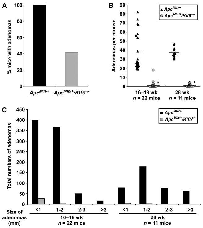
Characterization of tumor incidence and tumor burden in the ApcMin/+ and ApcMin/+/Klf5+/− mice. Mice at age 16 to 18 wk or age 28 wk were examined macroscopically for adenomas in the small intestine. A, graph of percentage of mice with small intestinal adenomas at age 16 to 18 wk (n = 22). B, scatter plot of the number of adenomas per mouse in the small intestine; ▲, ApcMin/+;  , ApcMin/+/Klf5+/−. Black horizontal lines, average number of adenomas for each genotype (*, P < 0.001; 16–18 wk, n = 22; 28 wk, n = 11; Wilcoxon-Mann-Whitney test). C, graph of total numbers of adenomas from mice screened in B, separated by size of adenomas.
In addition to tumor numbers, tumor sizes were compared between the two genotypes. As seen in Fig. 1C, at 16 to 18 weeks, the mode for adenoma size was <1 mm for both ApcMin/+ and ApcMin/+/Klf5+/− mice. By 28 weeks, the mode for ApcMin/+ mice was increased to 1 to 2 mm, indicating growth of the tumors, yet the mode for ApcMin/+/Klf5+/− mice remained at <1 mm. These data suggest that Klf5 haploinsufficiency may affect tumor growth as well as tumor initiation.
KLF5 and β-catenin levels are reduced in the ApcMin/+/Klf5+/− mice
To confirm that KLF5 protein was reduced in the small intestine of ApcMin/+/Klf5+/− mice, KLF5 levels were examined by immunofluorescence (Fig. 2A). KLF5 was restricted to the nuclei of epithelial cells in proliferating crypts of the small intestine. In Klf5+/− mice, KLF5 levels were significantly reduced to approximately half the levels in WT mice (P = 0.015) as determined by quantification of immunofluorescence intensities, correlating with haploinsufficiency of Klf5 (Fig. 2B). Similarly, KLF5 levels in ApcMin/+/Klf5+/− mice were reduced to approximately half those in ApcMin/+ mice (P = 0.016). In the ApcMin/+ mice, KLF5 showed intense staining in the transit-amplifying cells in the crypts of normal-appearing tissue (Fig. 2A), but intensities were not significantly increased compared with WT levels.
Figure 2.

Levels and localization of KLF5 and β-catenin in the small intestine of WT, Klf5+/−, ApcMin/+, and ApcMin/+/Klf5+/− mice. A, KLF5 levels in the small intestine of 16-wk-old mice were determined by immunofluorescence staining of KLF5 in normal-appearing tissues. B, quantification of KLF5 immunofluorescence intensities. Columns, mean value of four mice per group; bars, SE. Average intensities were determined from at least 50 cells per mouse. Results are shown as KLF5 fluorescence intensity per cell relative to intensities in WT mice (*, P = 0.015; **, P = 0.016; Tukey's test). C, immunohistochemical staining of β-catenin in small intestinal tissues from 16-wk-old mice. Bottom row, enlargements of the corresponding panels above. Arrows, nuclear staining. D, Western blot analysis of protein lysates from normal-appearing jejunum of 16-wk-old mice.
Because β-catenin is the key effector of APC mutations in intestinal cancers, we examined whether β-catenin levels and/or localization were influenced by Klf5 haploinsufficiency. Immunohistochemical staining of small intestinal tissues showed strong nuclear and cytoplasmic staining of β-catenin at the base of the crypts in the ApcMin/+ mice, whereas nuclear staining was less pronounced in the WT, Klf5+/−, and ApcMin/+/Klf5+/− mice (Fig. 2C). By Western blot analysis, β-catenin levels were higher in intestinal tissues from ApcMin/+ mice compared with WT or Klf5+/− mice (Fig. 2D), confirming previous findings that mutations in the Apc gene increase the stability and accumulation of β-catenin (7). Importantly, this effect was abrogated in ApcMin/+/Klf5+/− mice, where β-catenin levels were comparable with those in the WT and Klf5+/− mice (Fig. 2C and D). Similarly, when comparing levels of β-catenin in nuclear extracts from intestinal tissues by Western blot analysis, β-catenin was reduced in ApcMin/+Klf5+/− mice compared with ApcMin/+ mice (Supplementary Fig. S1), agreeing with results in Fig. 2C indicating reduced nuclear localization of β-catenin in the crypts of ApcMin/+/Klf5+/− mice. Thus, haploinsufficiency of Klf5 may “offset” the effect of the ApcMin mutation on the β-catenin protein.
KLF5 interacts with β-catenin
To investigate the mechanisms by which KLF5 affects β-catenin protein levels and localization, KLF5 and β-catenin were coexpressed in COS-1 cells. Protein levels of β-catenin were higher in cells expressing both ectopic β-catenin and KLF5 compared with cells expressing β-catenin alone (Supplementary Fig. S2A). This affect was not due to higher transcript levels, as both endogenous and exogenous β-catenin mRNA levels were unchanged in the presence of ectopic KLF5 (Supplementary Fig. S2B). Another potential mechanism by which KLF5 could affect β-catenin protein levels is through physical interaction and stabilization. Therefore, immunoprecipitation experiments were conducted in cells expressing KLF5-HA and β-catenin-FLAG to determine whether the two proteins interact. β-Catenin-FLAG coprecipitated with KLF5-HA, and likewise, KLF5-HA immunoprecipitated with β-catenin-FLAG (Fig. 3A). Neither protein was precipitated with the IgG antibody control. To verify interaction of endogenous β-catenin and KLF5 proteins, KLF5 was immunoprecipitated from DLD-1 human colon cancer cell lysates. β-catenin coprecipitated with KLF5 but was not precipitated in the preimmune serum negative control (Fig. 3B).
Figure 3.

Interaction of KLF5 and β-catenin. A, COS-1 cells were transfected as indicated, and lysates were subjected to immunoprecipitation and Western blotting with tagged antibodies to HA (for KLF5) and FLAG (for β-catenin). B, lysates from DLD-1 cells were immunoprecipitated (IP) with either a polyclonal anti-KLF5 antibody or preimmune serum as a negative control and examined for coprecipitation of β-catenin. C, COS-1 cells were transfected as indicated and stained for immunofluorescence with antibodies derived against FLAG (red) and HA (green) to determine colocalization. D, COS-1 cells were transfected with β-catenin-FLAG alone and stained for localization of endogenous KLF5 (green) and FLAG-tagged β-catenin (red). White arrow, cell expressing ectopic β-catenin; orange arrow, cell not expressing ectopic β-catenin.
To further evaluate the interaction between β-catenin and KLF5, the two proteins were examined for colocalization by immunofluorescence staining. Expression of KLF5-HA alone showed intense, diffuse staining of KLF5 throughout the nucleus, whereas β-catenin-FLAG-alone transfected cells showed both cytoplasmic and nuclear staining of β-catenin (Fig. 3C). When KLF5 and β-catenin were coexpressed, KLF5 was redistributed within the nucleus in a pattern that corresponded to β-catenin localization. In addition, there was an increase in nuclear staining for β-catenin in the cotransfected cell (Fig. 3C). Redistribution of KLF5 was also seen for endogenous KLF5 in cells transfected with β-catenin-FLAG alone. Although KLF5 exhibited diffuse nuclear staining in cells lacking ectopic β-catenin (Fig. 3D, orange arrow), KLF5 was redirected to distinct intranuclear structures in cells expressing β-catenin–FLAG (Fig. 3D, white arrow). This altered localization of KLF5 correlated with the expression pattern of ectopic β-catenin.
KLF5 facilitates nuclear localization of β-catenin
To investigate in-depth the ability of KLF5 to promote nuclear localization of β-catenin, cells transfected with either β-catenin alone or β-catenin and KLF5 were analyzed for subcellular distribution of β-catenin by immunofluorescence and protein fractionation. In examining a field of cells stained for β-catenin and KLF5, β-catenin exhibited more intense nuclear localization in the presence of ectopic KLF5 than when expressed alone (Fig. 4A). β-catenin localization was also analyzed by subcellular fractionation, with protein lysates separated into nuclear, cytoplasmic, and membrane fractions (Fig. 4B). Amounts of β-catenin localized to the membrane fraction were approximately equal in the two sets of transfections. However, a significant proportion of β-catenin was redistributed from the cytoplasm to the nucleus upon coexpression with KLF5. Thus, cells with ectopic expression of both β-catenin and KLF5 had higher levels of nuclear β-catenin.
Figure 4.
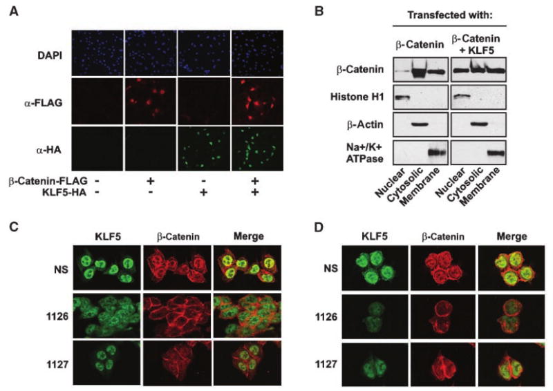
Alterations in KLF5 expression affect β-catenin nuclear localization. A, COS-1 cells were transfected as indicated and examined by immunofluorescence for nuclear β-catenin (red); KLF5 (green). Cells were counterstained with Hoechst 33528 (blue) to visualize nuclei. B, COS-1 cells were transfected with either β-catenin or β-catenin and KLF5 and fractionated into nuclear, cytosolic, and membranous components. Lysates were blotted for histone H1 (nuclear), β-actin (cytosolic), and Na+/K+ATPase (membranous) to confirm fractionation of compartments. C, HCT116 colon cancer cells were transfected with nonspecific siRNA or two distinct KLF5 siRNAs (1126 or 1127). Seventy-two hours after transfection, cells were stained by immunofluorescence for expression and localization of KLF5 (green) and β-catenin (red). D, repeat of experiment in C using DLD-1 colon cancer cells.
To determine whether loss of KLF5 would have a reciprocal effect on β-catenin localization, HCT116 human colon cancer cells were transfected with KLF5-specific siRNA and examined for sub-cellular localization of β-catenin by immunofluorescence (Fig. 4C). In cells transfected with nonspecific siRNA, β-catenin exhibited visible nuclear staining. However, this localization was in large part disrupted in cells transfected with two different KLF5-specific siRNAs, where β-catenin was localized predominantly to the cell membranes and cytoplasm. A similar effect was seen in another colon cancer cell line, DLD-1 (Fig. 4D), Taken together, these data indicate that KLF5 facilitates localization of β-catenin to the nucleus.
KLF5 modulates transcriptional activity of β-catenin
Because increased expression of KLF5 enhances nuclear localization of β-catenin, KLF5 may also promote the transcriptional activity of β-catenin. To test this possibility, KLF5 and β-catenin were coexpressed in COS-1 cells with pTOPFLASH, a luciferase reporter construct that measures TCF/β-catenin activity (8). Compared with empty vector, cells transfected with β-catenin alone showed a 3.6-fold increase in TOPFLASH luciferase activity (P = 0.003), whereas ectopic expression of KLF5 alone had no effect (Fig. 5A). Cells transfected with both β-catenin and KLF5 showed a 6.2-fold increase in luciferase activity, an effect that was significantly greater than with β-catenin alone (P = 0.006). As a transcriptional target for both KLF5 and β-catenin (24, 25), Ccnd1 promoter activity was also measured (Fig. 5A). Transfection with β-catenin alone did not significantly increase Ccnd1 luciferase activity compared with cells transfected with empty vector. In cells transfected with KLF5 alone, luciferase activity was increased by 3.2-fold over vector control (P < 0.001). Coexpression of β-catenin and KLF5 resulted in a 7.6-fold increase in activity, which was significantly greater than with β-catenin (P < 0.001) or KLF5 alone (P < 0.001). Moreover, when β-catenin and KLF5 were coexpressed, they interacted to produce a synergistic effect on Ccnd1 promoter luciferase activity (P < 0.001). Thus, coexpression of KLF5 with β-catenin enhances the transcription of β-catenin targets.
Figure 5.
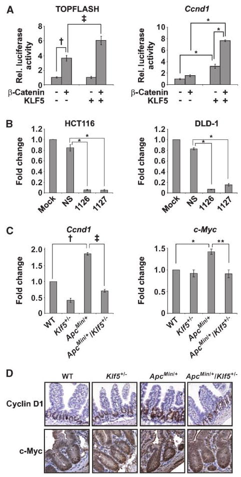
Changes in KLF5 expression affect activation of β-catenin transcriptional targets. A, luciferase assays in COS-1 cells transfected with expression constructs as indicated and measured for activity of TOPFLASH and Ccnd1 promoters. Rel. luciferase activity, relative luciferase activity. Coexpression of β-catenin and KLF5 produced significant synergistic effects for Ccnd1 (F [1,20] = 74.0; P < 0.001). Comparison of means (Tukey's procedure): for TOPFLASH: †, P = 0.003; ‡, P = 0.006; n = 3 per group; for Ccnd1: *, P < 0.001; n = 6 per group. B, luciferase assays in HCT116 and DLD-1 colon cancer cells transfected with indicated siRNAs. (*, P < 0.001; n = 3 per group). C, quantitative RT-PCR analysis of Ccnd1 and c-Myc mRNA levels using RNA from distal jejunum. Results were normalized to β-actin levels. For Ccnd1: †, P = 0.009; ‡, P < 0.001 0.0; n = 3; for c-Myc: *, P = 0.002; **, P = 0.009; n = 3. D, immunohistochemical staining of cyclin D1 and c-Myc in jejunum from mice at age 16 wk.
To determine if loss of KLF5 would have a corresponding negative effect on TCF/β-catenin transcriptional activity, HCT 116 and DLD-1 cells were transfected with a nonspecific siRNA or two different siRNAs specific for KLF5 and the TOPFLASH luciferase reporter (Fig. 5B). Transfection with either KLF5-specific siRNA dramatically reduced TOPFLASH activity in both cell lines compared with transfection with nonspecific siRNA (P < 0.001), correlating with loss of β-catenin from the nucleus in Fig. 4C and D.
Expression of β-catenin targets is reduced in ApcMin/+/Klf5+/− mice
To address whether reduced KLF5 expression affects transcriptional activity of β-catenin in vivo, RNA was isolated from the jejunum of WT, Klf5+/−, ApcMin/+, and ApcMin/+/Klf5+/− mice, and expression of two transcriptional targets of β-catenin that promote proliferation were examined by reverse transcriptase-quantitative PCR. Transcript levels of Ccnd1, the gene encoding cyclin D1, were increased 1.9-fold in the ApcMin/+ mice compared with WT (P = 0.009; Fig. 5C). In contrast, levels of Ccnd1 in the ApcMin/+/Klf5+/− mice were similar to WT controls (Fig. 5C). As a transcriptional target of KLF5 (24, 25), Ccnd1 transcript levels were significantly reduced in the Klf5+/− mice compared with WT (0.41-fold; P = 0.03), suggesting that this target gene is strongly influenced by KLF5 levels. In examining c-Myc transcript levels, c-Myc was increased 1.4-fold (P = 0.002) in ApcMin/+ mice compared with WT mice. This change was offset in the ApcMin/+/Klf5+/− mice, with levels comparable with WT (P < 0.001). No significant change in c-Myc levels was observed in the Klf5+/− mice. Target gene expression was further confirmed by immunohistochemical staining of the protein products (Fig. 5D). In concordance with mRNA levels, cyclin D1 staining was more intense in small intestinal tissues of ApcMin/+ mice compared with WT mice (Fig. 5D), whereas no induction of cyclin D1 was observed in the ApcMin/+/Klf5+/− mice. Likewise, c-Myc expression was increased in the ApcMin/+ mice, but this effect was abrogated in the ApcMin/+/Klf5+/− animals, which exhibited c-Myc levels similar to WT (Fig. 5D). Therefore, KLF5 is necessary for the downstream activation of β-catenin target genes in the ApcMin/+ mouse.
KLF5 and β-catenin colocalize in intestinal adenomas
Because KLF5 seems to contribute to the tumorigenic effects of β-catenin in the ApcMin/+ setting, immunohistochemical analyses were performed to determine the distribution of KLF5 and β-catenin in intestinal tumors derived from ApcMin/+ mice. As seen in Fig. 6A, there was intense nuclear staining of KLF5 in epithelial cells in the dysplastic epithelium adjacent to the adenomas as well as cells throughout the adenomas. β-catenin exhibited strong cytoplasmic staining throughout the adenomas, and showed a high degree of nuclear staining, particularly in cells at the leading edges of the adenomas. To examine colocalization of KLF5 and β-catenin in ApcMin/+ adenomas, tissues were stained by immunofluorescence (Fig. 6B). Both KLF5 and β-catenin showed nuclear staining in a majority of the cells in the adenomas, with considerable colocalization of the two proteins in individual cells. Thus, KLF5 and β-catenin are coexpressed in ApcMin/+ adenomas, and can work together to drive growth-promoting transcriptional activity in those tissues.
Figure 6.

Expression of KLF5 and β-catenin in intestinal adenomas of ApcMin/+ mice. Adenomas isolated from ApcMin/+ mice at age 16 wk were stained for localization of KLF5 and β-catenin. A, representative immunohistochemical staining of KLF5 and β-catenin in ApcMin/+ adenomas. B, ApcMin/+ adenomas were stained for KLF5 (green) and β-catenin (red) by immunofluorescence to determine colocalization. Nuclei are also shown to indicate total cells.
When comparing β-catenin levels in the adenomas from ApcMin/+ and ApcMin/+/Klf5+/− mice by immunofluorescence, no significant differences were observed in β-catenin intensities in small-size adenomas (<1 mm) from the two genotypes (Supplementary Fig. S3). Both sets of adenomas exhibited nuclear localization of β-catenin. The only subjective difference seemed to be a more diffuse cytoplasmic localization of β-catenin in the ApcMin/+ mice compared with a more intense localization to cell junctions in the ApcMin/+/Klf5+/− mice. These results reinforce the notion that haploinsufficiency of Klf5 affects adenoma formation at the earliest stages; and that these effects can be bypassed at low frequency to result in high β-catenin levels and subsequent establishment of adenomas.
Discussion
Mutational inactivation of the APC tumor suppressor gene and subsequent activation of the Wnt cascade is the rate-limiting event in CRC (26). In cases of CRC in which APC is not mutated, other mutations disrupting the normal regulation of β-catenin occur in a mutually exclusive manner (27, 28), highlighting the importance of dysregulation of β-catenin in the transformation of epithelial cells. Here, we show that KLF5 is a critical component for maintaining increased levels and activities of β-catenin in the ApcMin/+ mouse model of intestinal tumorigenesis. With heterozygous deletion of Klf5, levels of β-catenin in the ApcMin/+ setting are reduced to WT levels, resulting in reduced β-catenin activity as reflected in reduced expression of transcriptional targets including Ccnd1 and c-Myc. Other genetic crosses with ApcMin/+ mice have been reported to have significant effects on intestinal tumorigenesis, including ApcMin/+/MyD88−/− (80% reduction in number of adenomas; ref. 29) and ApcMin/+/cyclin D1−/− (84% reduction; ref. 30). However, the ApcMin/+/Klf5+/− mouse is the first example of haploinsufficiency of a gene rescuing the tumorigenic effects of the ApcMin mutation.
The effect of KLF5 on β-catenin seems to be through physical interaction. We showed here that the two proteins are coimmunoprecipitated and that they colocalize within the cell. In addition, we showed that overexpression of KLF5 enhances nuclear localization of β-catenin, and that reducing KFL5 expression, both in vitro and in vivo, significantly reduces concentrations of β-catenin in the nucleus. Because KLF5 contains a putative nuclear localization signal with homology to the nuclear localization signal confirmed in the related protein KLF4 (31), KLF5 may bind and assist β-catenin in localizing to the nucleus. Enhanced levels of nuclear β-catenin would therefore increase its availability to bind and activate transcriptional partners, such as TCF-4. This premise is supported by increased nuclear localization of β-catenin and increased TOPFLASH luciferase activity in the presence of ectopically expressed KLF5 (Figs. 4A, B and 5A).
KLF5 has been reported to promote proliferation of intestinal epithelial cells, both in vitro and in vivo (13, 14). KLF5 is a target of the extracellular signal-regulated kinase (ERK)1/2 mitogen-activated protein kinase pathway (32, 33), a signaling pathway critical for the G1-S phase cell cycle transition (34). As a downstream target of ERK1/2 signaling, KLF5 is activated in response to a number of mitogenic and stress stimuli (14, 35, 36) and thus may play a role in enhancing proliferation in intestinal tissues in situations of damage or stress. In support of this point, our laboratory has shown that KLF5 contributes to colonic hyperplasia in mice infected with the bacterial pathogen, Citrobacter rodentium (15).
In human cancer, KLF5 has been reported as a downstream target of oncogenic HRas in vitro and shown to mediate pro-proliferative and transforming activities of HRas (24). In addition, KLF5 is highly expressed in human primary colorectal cancers exhibiting KRas mutations (19). Thus, in addition to its potential involvement with the APC/β-catenin pathway, KLF5 may contribute to colorectal carcinogenesis through mutational activation of KRas. In a study of 247 gastric carcinoma patients, high KLF5 expression is found in 63% of early-stage gastric cancer patients (18). Furthermore, KLF5 expression has been reported as a prognostic factor for overall survival in patients with sporadic breast cancer, with higher KLF5 expression correlating with shorter disease-free survival and poorer overall survival (20). In contrast to these studies, a number of reports examining KLF5 status in various types of tumors show that KLF5 is down-regulated by several mechanisms, implicating KLF5 as a tumor suppressor (37–39). This apparent discrepancy may be attributable to the timing of changes in KLF5 expression, where increased expression early in tumor formation is growth-promoting, and loss of expression in later stages of cancer aids tumor cell survival. Studies that examine KLF5 expression by stage support this possibility, reflecting high KLF5 expression in early stages of cancer progression and lower KLF5 expression in later stages (18). What is clear from the results presented here is that reduced levels of KLF5 in the ApcMin/+ context inhibit tumor initiation, implying that the activity of KLF5 contributes to early events in adenoma formation.
It has been argued that there may be an “optimal” level of Wnt signaling for colorectal tumorigenesis to occur, given that different functional mutations in the APC gene have varying abilities to drive formation of colorectal polyps (40). Thus, modulation of β-catenin activity, especially in patients with hereditary APC mutations, may be therapeutically beneficial. Given that partial reduction of KLF5 has a significant effect on β-catenin activity and adenoma formation in the ApcMin/+ intestinal tumorigenesis model, KLF5 may present an attractive target for the development of novel therapeutic agents for early stage CRC treatment or prevention.
Supplementary Material
Acknowledgments
Grant support: NIH (DK52230, DK64399, DK76742, CA84197, and CA130308).
Footnotes
Note:Supplementary data for this article are available at Cancer Research Online (http://cancerres.aacrjournals.org/).
Disclosure of Potential Conflicts of Interest: The authors have no potential conflicts of interest.
References
- 1.Pinto D, Clevers H. Wnt, stem cells and cancer in the intestine. Biol Cell. 2005;97:185–96. doi: 10.1042/BC20040094. [DOI] [PubMed] [Google Scholar]
- 2.Kinzler KW, Vogelstein B. Lessons from hereditary colorectal cancer. Cell. 1996;87:159–70. doi: 10.1016/s0092-8674(00)81333-1. [DOI] [PubMed] [Google Scholar]
- 3.Moser AR, Pitot HC, Dove WF. A dominant mutation that predisposes to multiple intestinal neoplasia in the mouse. Science. 1990;247:322–4. doi: 10.1126/science.2296722. [DOI] [PubMed] [Google Scholar]
- 4.Su LK, Kinzler KW, Vogelstein B, et al. Multiple intestinal neoplasia caused by a mutation in the murine homolog of the APC gene. Science. 1992;256:668–70. doi: 10.1126/science.1350108. [DOI] [PubMed] [Google Scholar]
- 5.Fodde R. The APC gene in colorectal cancer. Eur J Cancer. 2002;38:867–71. doi: 10.1016/s0959-8049(02)00040-0. [DOI] [PubMed] [Google Scholar]
- 6.Gregorieff A, Pinto D, Begthel H, et al. Expression pattern of Wnt signaling components in the adult intestine. Gastroenterology. 2005;129:626–38. doi: 10.1016/j.gastro.2005.06.007. [DOI] [PubMed] [Google Scholar]
- 7.Rubinfeld B, Albert I, Porfiri E, et al. Binding of GSK3β to the APC-β-catenin complex and regulation of complex assembly. Science. 1996;272:1023–6. doi: 10.1126/science.272.5264.1023. [DOI] [PubMed] [Google Scholar]
- 8.Korinek V, Barker N, Morin PJ, et al. Constitutive transcriptional activation by a β-catenin-Tcf complex in APC−/− colon carcinoma. Science. 1997;275:1784–7. doi: 10.1126/science.275.5307.1784. [DOI] [PubMed] [Google Scholar]
- 9.Sparks AB, Morin PJ, Vogelstein B, Kinzler KW. Mutational analysis of the APC/β-catenin/Tcf pathway in colorectal cancer. Cancer Res. 1998;58:1130–4. [PubMed] [Google Scholar]
- 10.Brabletz T, Jung A, Kirchner T. Beta-catenin and the morphogenesis of colorectal cancer. Virchows Arch. 2002;441:1–11. doi: 10.1007/s00428-002-0642-9. [DOI] [PubMed] [Google Scholar]
- 11.Ghaleb AM, Nandan MO, Chanchevalap S, et al. Kruppel-like factors 4 and 5: the yin and yang regulators of cellular proliferation. Cell Res. 2005;15:92–6. doi: 10.1038/sj.cr.7290271. [DOI] [PMC free article] [PubMed] [Google Scholar]
- 12.McConnell BB, Ghaleb AM, Nandan MO, Yang VW. The diverse functions of Kruppel-like factors 4 and 5 in epithelial biology and pathobiology. Bioessays. 2007;29:549–57. doi: 10.1002/bies.20581. [DOI] [PMC free article] [PubMed] [Google Scholar]
- 13.Chanchevalap S, Nandan MO, Merlin D, Yang VW. All-trans retinoic acid inhibits proliferation of intestinal epithelial cells by inhibiting expression of the gene encoding Kruppel-like factor 5. FEBS Lett. 2004;578:99–105. doi: 10.1016/j.febslet.2004.10.079. [DOI] [PMC free article] [PubMed] [Google Scholar]
- 14.Sun R, Chen X, Yang VW. Intestinal-enriched Kruppel-like factor (Kruppel-like factor 5) is a positive regulator of cellular proliferation. J Biol Chem. 2001;276:6897–900. doi: 10.1074/jbc.C000870200. [DOI] [PMC free article] [PubMed] [Google Scholar]
- 15.McConnell BB, Klapproth JM, Sasaki M, Nandan MO, Yang VW. Kruppel-like factor 5 mediates transmissible murine colonic hyperplasia caused by Citrobacter rodentium infection. Gastroenterology. 2008;134:1007–16. doi: 10.1053/j.gastro.2008.01.013. [DOI] [PMC free article] [PubMed] [Google Scholar]
- 16.Ziemer LT, Pennica D, Levine AJ. Identification of a mouse homolog of the human BTEB2 transcription factor as a β-catenin-independent Wnt-1-responsive gene. Mol Cell Biol. 2001;21:562–74. doi: 10.1128/MCB.21.2.562-574.2001. [DOI] [PMC free article] [PubMed] [Google Scholar]
- 17.Chaib H, Cockrell EK, Rubin MA, Macoska JA. Profiling and verification of gene expression patterns in normal and malignant human prostate tissues by cDNA microarray analysis. Neoplasia. 2001;3:43–52. doi: 10.1038/sj.neo.7900126. [DOI] [PMC free article] [PubMed] [Google Scholar]
- 18.Kwak MK, Lee HJ, Hur K, et al. Expression of Kruppel-like factor 5 in human gastric carcinomas. J Cancer Res Clin Oncol. 2008;134:163–7. doi: 10.1007/s00432-007-0265-2. [DOI] [PMC free article] [PubMed] [Google Scholar]
- 19.Nandan MO, McConnell BB, Ghaleb AM, et al. Kruppel-like factor 5 mediates cellular transformation during oncogenic KRAS-induced intestinal tumorigene-sis. Gastroenterology. 2008;134:120–30. doi: 10.1053/j.gastro.2007.10.023. [DOI] [PMC free article] [PubMed] [Google Scholar]
- 20.Tong D, Czerwenka K, Heinze G, et al. Expression of KLF5 is a prognostic factor for disease-free survival and overall survival in patients with breast cancer. Clin Cancer Res. 2006;12:2442–8. doi: 10.1158/1078-0432.CCR-05-0964. [DOI] [PubMed] [Google Scholar]
- 21.Livak KJ, Schmittgen TD. Analysis of relative gene expression data using real-time quantitative PCR and the 2(-ΔΔ C(T)) Method. Methods. 2001;25:402–8. doi: 10.1006/meth.2001.1262. [DOI] [PubMed] [Google Scholar]
- 22.Zhang L, Rayner S, Katoku-Kikyo N, Romanova L, Kikyo N. Successful co-immunoprecipitation of Oct4 and Nanog using cross-linking. Biochem Biophys Res Commun. 2007;361:611–4. doi: 10.1016/j.bbrc.2007.07.089. [DOI] [PMC free article] [PubMed] [Google Scholar]
- 23.Guillemin I, Becker M, Ociepka K, Friauf E, Nothwang HG. A subcellular prefractionation protocol for minute amounts of mammalian cell cultures and tissue. Proteomics. 2005;5:35–45. doi: 10.1002/pmic.200400892. [DOI] [PubMed] [Google Scholar]
- 24.Nandan MO, Yoon HS, Zhao W, et al. Kruppel-like factor 5 mediates the transforming activity of oncogenic H-Ras. Oncogene. 2004;23:3404–13. doi: 10.1038/sj.onc.1207397. [DOI] [PMC free article] [PubMed] [Google Scholar]
- 25.Shtutman M, Zhurinsky J, Simcha I, et al. The cyclin D1 gene is a target of the β-catenin/LEF-1 pathway. Proc Natl Acad Sci U S A. 1999;96:5522–7. doi: 10.1073/pnas.96.10.5522. [DOI] [PMC free article] [PubMed] [Google Scholar]
- 26.Fodde R, Brabletz T. Wnt/β-catenin signaling in cancer stemness and malignant behavior. Curr Opin Cell Biol. 2007;19:150–8. doi: 10.1016/j.ceb.2007.02.007. [DOI] [PubMed] [Google Scholar]
- 27.Morin PJ, Sparks AB, Korinek V, et al. Activation of β-catenin-Tcf signaling in colon cancer by mutations in β-catenin or APC. Science. 1997;275:1787–90. doi: 10.1126/science.275.5307.1787. [DOI] [PubMed] [Google Scholar]
- 28.Oving IM, Clevers HC. Molecular causes of colon cancer. Eur J Clin Invest. 2002;32:448–57. doi: 10.1046/j.1365-2362.2002.01004.x. [DOI] [PubMed] [Google Scholar]
- 29.Rakoff-Nahoum S, Medzhitov R. Regulation of spontaneous intestinal tumorigenesis through the adaptor protein MyD88. Science. 2007;317:124–7. doi: 10.1126/science.1140488. [DOI] [PubMed] [Google Scholar]
- 30.Hulit J, Wang C, Li Z, et al. Cyclin D1 genetic heterozygosity regulates colonic epithelial cell differentiation and tumor number in ApcMin mice. Mol Cell Biol. 2004;24:7598–611. doi: 10.1128/MCB.24.17.7598-7611.2004. [DOI] [PMC free article] [PubMed] [Google Scholar]
- 31.Shields JM, Yang VW. Two potent nuclear localization signals in the gut-enriched Kruppel-like factor define a subfamily of closely related Kruppel proteins. J Biol Chem. 1997;272:18504–7. doi: 10.1074/jbc.272.29.18504. [DOI] [PMC free article] [PubMed] [Google Scholar]
- 32.Aizawa K, Suzuki T, Kada N, et al. Regulation of platelet-derived growth factor-A chain by Kruppel-like factor 5: new pathway of cooperative activation with nuclear factor-κB. J Biol Chem. 2004;279:70–6. doi: 10.1074/jbc.M306621200. [DOI] [PubMed] [Google Scholar]
- 33.Nagai R, Kowase K, Kurabayashi M. Transcriptional regulation of smooth muscle phenotypic modulation. Ann N Y Acad Sci. 2000;902:214–22. doi: 10.1111/j.1749-6632.2000.tb06316.x. discussion 222–213. [DOI] [PubMed] [Google Scholar]
- 34.Meloche S, Pouyssegur J. The ERK1/2 mitogen- activated protein kinase pathway as a master regulator of the G1- to S-phase transition. Oncogene. 2007;26:3227–39. doi: 10.1038/sj.onc.1210414. [DOI] [PubMed] [Google Scholar]
- 35.Chanchevalap S, Nandan MO, McConnell BB, et al. Kruppel-like factor 5 is an important mediator for lipopolysaccharide-induced proinflammatory response in intestinal epithelial cells. Nucleic Acids Res. 2006;34:1216–23. doi: 10.1093/nar/gkl014. [DOI] [PMC free article] [PubMed] [Google Scholar]
- 36.Shindo T, Manabe I, Fukushima Y, et al. Kruppel-like zinc-finger transcription factor KLF5/BTEB2 is a target for angiotensin II signaling and an essential regulator of cardiovascular remodeling. Nat Med. 2002;8:856–63. doi: 10.1038/nm738. [DOI] [PubMed] [Google Scholar]
- 37.Chen C, Bhalala HV, Vessella RL, Dong JT. KLF5 is frequently deleted and down-regulated but rarely mutated in prostate cancer. Prostate. 2003;55:81–8. doi: 10.1002/pros.10205. [DOI] [PubMed] [Google Scholar]
- 38.Chen C, Sun X, Ran Q, et al. Ubiquitin-proteasome degradation of KLF5 transcription factor in cancer and untransformed epithelial cells. Oncogene. 2005;24:3319–27. doi: 10.1038/sj.onc.1208497. [DOI] [PubMed] [Google Scholar]
- 39.Rozenblum E, Vahteristo P, Sandberg T, et al. A genomic map of a 6-Mb region at 13q21–22 implicated in cancer development: identification and characterization of candidate genes. Hum Genet. 2002;110:111–21. doi: 10.1007/s00439-001-0646-6. [DOI] [PubMed] [Google Scholar]
- 40.Segditsas S, Tomlinson I. Colorectal cancer and genetic alterations in the Wnt pathway. Oncogene. 2006;25:7531–7. doi: 10.1038/sj.onc.1210059. [DOI] [PubMed] [Google Scholar]
Associated Data
This section collects any data citations, data availability statements, or supplementary materials included in this article.