



Abstract
Escherichia coli K1 invasion of human brain microvascular endothelial cells (HBMEC) requires the reorganization of host cytoskeleton at the sites of bacterial entry. Both actin and myosin constitute the cytoskeletal architecture. We have previously shown that myosin light chain (MLC) phosphorylation by MLC kinase is regulated during E. coli invasion by an upstream kinase, p21-activated kinase 1 (PAK1), which is an effector protein of Rac and Cdc42 GTPases, but not of RhoA. Here, we report that the binding of only Rac1 to PAK1 decreases in HBMEC upon infection with E. coli K1, which resulted in increased phosphorylation of MLC. Overexpression of a constitutively active (cAc) form of Rac1 in HBMEC blocked the E. coli invasion significantly, whereas overexpression of a dominant negative form had no effect. Increased PAK1 phosphorylation was observed in HBMEC expressing cAc-Rac1 with a concomitant reduction in the phosphorylation of MLC. Immunocytochemistry studies demonstrated that the inhibition of E. coli invasion into cAc-Rac1/HBMEC is due to lack of phospho-MLC recruitment to the sites of E. coli entry. Taken together the data suggest that E. coli modulates the binding of Rac1, but not Cdc42, to PAK1 during the invasion of HBMEC.
Keywords: Rac1, PAK1, Myosin light chain, Endothelial cells, Meningitis
Abbreviations: HBMEC, human brain microvascular endothelial cells; MLC, myosin light chain; MLCK, myosin light chain kinase; OmpA, outer membrane protein A; PAK1, p21-activated kinase 21
1. Introduction
Despite advances in antimicrobial therapy and general supportive care, bacterial meningitis remains a significant cause of morbidity and mortality in neonates. According to a variety of studies, up to 80% of cases of neonatal meningitis are due to Escherichia coli K1, a Gram-negative bacterium, and incidence has been recently reported to be on the rise again, surpassing the infections by group B Streptococcus and Listeria monocytogenes [1]. Entry of E. coli into the central nervous system via human brain microvascular endothelial cells (HBMEC) is initiated subsequent to the binding of the bacterial surface protein outer membrane protein A (OmpA) to its receptor on the host cell [2,3]. E. coli K1 binding to HBMEC induces protrusions in the host cells that eventually enwrap the bacterium for the uptake, a process that requires cytoskeletal reorganization of HBMEC [4].
Myosins are actin-activated Mg-ATPases that convert the energy of ATP hydrolysis into force between actin and myosin filaments, leading to either contraction or tension. Conventional myosins that form filaments consists of two heavy chain actin-activatedATPases and two light chains, one essential and one regulatory. The phosphorylation of regulatory myosin light chain (MLC) at Ser19 (and Thr18) by Ca2+-dependent myosin light chain kinase (MLCK) has been shown to be a critical regulatory step for physiological modulation of myosin contractility [5]. Our recent studies have shown that p21-activated kinase 1 (PAK1) modulates the phosphorylation of MLC in E. coli invasion of HBMEC [6]. The phosphorylated MLC subsequently recruits to the actin accumulation points at the bacterial attachment site. PAK1 was initially identified as a Rac binding protein and was further shown to interact significantly with GTP-bound form of Rac and Cdc42 [5]. The catalytic activity of PAK1 is regulated by the binding of Rac or Cdc42 to a highly conserved motif in the N-terminus, known as the p21-binding domain (PBD) or Cdc42/Rac interactive binding (CRIB) domain [5]. The binding of Rac/Cdc42 induces a conformational change in PAK1, which in turn is thought to be necessary for autophosphorylation at several sites and enabling the phosphorylation of exogenous substrates. The activated (phosphorylated) PAK1 phosphorylates MLCK thereby inactivating it, which in turn reduces the MLC phosphorylation [7]. Thus, Rac/Cdc42 interaction with PAK1 in GTP state inhibits the phosphorylation of MLC.
Invasion of eukaryotic cells is frequently a result of bacterial manipulation of the host cell cytoskeleton at the level of these small GTPases, the activity of which is associated with particular membrane and cytoskeletal rearrangements such as lamellipodia (Rac1), filopodia (Cdc42) and stress fibers (RhoA) [8–15]. Uropathogenic E. coli internalization, mediated by fimH, utilizes Rac1 and Cdc42; and expression of active forms of these GTPases increases the invasion significantly [14]. Interestingly, Neisseria gonorrhoeae uses distinct mechanisms of internalization by members of CEACAM receptor family involving Rac1 and Cdc42-dependent and independent pathways, but not Rho [16]. In contrast, to our previous studies, in which PAK1 activity was down regulated by E. coli K1, Helicobacter pylori induce Rac1- and Cdc42-dependent activation of PAK1 [17]. Here, we report that binding of Rac1, but not Cdc42, to PAK1 is down regulated, which in turn increases the phosphorylation of MLC in E. coli invasion. Overexpression of constitutively active, but not dominant negative, Rac1 blocked the E. coli invasion as well as the recruitment of phospho-MLC at the actin condensation sites.
2. Materials and methods
2.1. Bacterial strains
The E. coli strains E44 and E91 used in this study are derivatives of the E. coli K1 strain RS218 (serotype 018:K1:H7) [2]. Strain E44 is a spontaneous rifampicin-resistant mutant and invades HBMEC. E91 is a non-invasive mutant lacking the ompA gene and was used as a negative control. The strains were grown in brain heart infusion (BHI) medium supplemented with antibiotics rifampicin (100 μg/ml) for E44 and tetracycline (12.5 μg/ml) for E91.
2.2. Plasmids and antibodies
Rac1 mutants, dominant negative (Rac1N17) with myc tag, constitutively active (Rac1Q61L) with HA tag in a pcDNA3 vector were kindly provided by Dr. G.M. Bokoch. Anti-PAK1, anti-Rac1, anti-Cdc42, and anti-phospho-MLC antibodies were obtained from Santa Cruz Biotechnology Inc. (Santa Cruz, CA). Anti-MLC antibody was purchased from Sigma Laboratories. Antiphosphotyrosine (4G10) antibody was from Up-State Biotechnology Inc. (Waltham, MA).Anti-phospho-PAK1 (Thr423)/PAK2 (Thr402) antibody and Rac1 activation assay kit were purchased from Cell Signaling (Beverly, MA) and Cytoskeleton Inc. (Denver, CO), respectively.
2.3. Cell culture, DNA transfection, and invasion assays
HBMEC were maintained at 37 °C in a humidified atmosphere of 5% CO2 in medium containing M-199/Ham F-12 (1:1) supplemented with 10% fetal bovine serum, sodium pyruvate and 2 mM glutamine and cultivated in a cell culture incubator at 5% CO2 and 37 °C. For stable transfection HBMEC were transfected with either Rac1 mutants or empty vector using lipofectamine reagent, allowed to recover for 24 h, and the transfected cells were selected using 400 μg/ml G418.
For invasion assays, HBMEC grown in 24-well cell culture plates to 95% confluence were infected with 107 cfu of E. coli strains in experimental medium (1:1 mixture of Ham F-12 and M-199 containing 5% heat inactivated fetal bovine serum) and incubated for 90 min at 37 °C in an atmosphere containing 5% CO2. The monolayers were washed three times with RPMI 1640, medium containing gentamicin (100 μg/ml) was added to the wells, and further incubated for 1 h at 37 °C. Then the cells were washed three times with RPMI 1640 and lysed with 0.5% of Triton X-100. The released bacteria were diluted with saline and enumerated by plating on blood agar. In duplicate experiments, the total cell associated bacteria were determined as described for invasion, except that the gentamicin step was omitted. Results were expressed as an average of bacterial colony forming units recovered per well from three independent determinations ± S.D. of the means.
2.4. Western blotting and immunoprecipitations
Confluent cell cultures of 100 mm dishes were incubated with either E44 or E91 for varying time points at 37 °C. The cells were then rinsed with ice-cold phosphate buffered saline (PBS) and scraped off into 500 μl of RIPA buffer (1 × PBS, 1% Nonidet P-40, 0.5% sodium deoxycholate, 0.1% SDS, 10 μg/ml PMSF, aprotinin, and 1 mM sodium orthovanadate). The lysates were clarified by centrifugation for 10 min at 10,000 × g to remove cell debris. The supernatants were collected and approximately 30 μg of total protein of cell lysates was fractionated by electrophoresis on a 4.0% stacking–12% separating SDS polyacrylamide gel. Proteins were then transferred to nitrocellulose membranes using a Bio-Rad semi-dry transfer apparatus. The blot was incubated with blocking buffer (100 mM Tris–HCl, pH 7.5; 150 mM NaCl; 0.1% Tween 20; 2% bovine serum albumin and 2% milk) for 1 h at room temperature and then probed with the appropriate antibody. The same blot was stripped and re-blocked for subsequent probing with additional antibodies.
For immunoprecipitation, HBMEC transfected with empty vector or Rac1 mutants were grown in 100 mm dishes and incubated with E. coli (1:100 cell to bacteria ratio) at 37 °C for varying time periods. Cells were washed in cold PBS before lysis in RIPA Buffer. Lysates were clarified by centrifugation at 12,000 × g for 10 min before incubation with antibody at 4 °C overnight. The lysates were then incubated with protein A-agarose beads for 1 h at 4 °C. The beads were washed three times in ice-cold lysis buffer and the bound proteins were released by adding SDS sample buffer, resolved by SDS-PAGE, and analyzed by Western blotting with various antibodies. The proteins were detected using a chemiluminescence detection kit (Super Signal Chemiluminescence Kit, Pierce Co.) followed by exposure to an X-ray film.
2.5. Immunofluorescence
HBMEC infected with either OmpA+ or OmpA– E. coli for 15–30 min, washed with RPMI 1640, and fixed with 2% paraformaldehyde in PBS for 15 min at room temperature. The cells were washed and permeabilized with 0.1% Triton X-100 in PBS containing 1% bovine serum albumin for 30 min at room temperature. Then the cells were incubated with the primary antibody (p-MLC antibody 1:100) for 1 h followed by 1:250 dilution of anti-goat secondary antibody coupled to FITC. To label actin, rhodamine phalloidin was included with the secondary antibody. Finally the slides were mounted with Vectashield containing DAPI (Vector Laboratories). Cells were viewed with a Leica (Wetzlar, Germany) DMRA microscope with Planapochromat ×40/1.25 NA and ×63/1.40 NA oil immersion objective lenses. Image acquisition was with a SkyVision-2/VDS digital CCD (12-bit, 1280 × 1024 pixel) camera in unbinned or 2 × 2-binned models into EasyFISH software, saved as 16-bit monochrome, and merged as 24-bit RGB TIFF images (Applied Spectral Imaging Inc., Carlsbad, CA).
2.6. Activated Rac pull-down assay
The assay uses the Rac/Cdc42 interactive PBD of PAK1 fused to GST, which was immobilized to glutathione beads. The PBD protein motif has been shown to bind specifically to the GTP-bound form of Rac and/or Cdc42 proteins. The assay therefore provides a simple means of quantitating Rac/Cdc42 activation in cells. Briefly, HBMEC grown in 100 mm dishes were treated with invasive E. coli, washed with ice-cold PBS twice to remove unbound bacteria, and lysed in 500 μl of RIPA buffer. Lysates were centrifuged to remove particulates, incubated with equal quantities of PBD-GST beads for 60 min at 4 °C. The PBD-GST beads were washed three times and the bound proteins were eluted with SDS sample buffer. The proteins were separated on SDS polyacrylamide gels and subjected to Western blotting using a polyclonal antibody to Rac1. The same blot was stripped and reprobed with anti-Cdc42 antibody. The intensity of the bands corresponding Rac1, Cdc42 and PBD-GST was analyzed using ImageJ software (http://rsb.info.nih.gov/ij).
2.7. PepTag assay
The PepTag assays utilize brightly colored, fluorescent peptide substrates that are highly specific for the kinases in question. The hot pink color is imparted by the addition of a dye molecule to the PepTag peptide substrate. Phosphorylation by PKC or PKA of their specific substrate alters the peptide’s net charge from +1 to –1. This change in the net charge of the substrate allows the phosphorylated and non-phosphorylated versions of the substrate to be rapidly separated on an agarose gel. The phosphorylated species migrates toward the positive electrode, while the non-phosphorylated substrate migrates toward the negative electrode. HBMEC total lysates 10–25 μg in 10 μl) were incubated with the PKC reaction mixture (25 μl) according to the manufacturer’s protocol at 30 °C for 30 min. The reactions were stopped by placing the tubes in a boiling water bath.After adding 80% glycerol (1 μl), the samples were loaded onto an agarose gel (0.8% agarose in 50 mM Tris–HCl, pH 8.0) and separated at 100 V for 15 min. The peptide bands were visualized under UV light.
3. Results
3.1. Decreased GTPase activity of Rac1 and its association with PAK1 in E. coli K1 invasion of HBMEC
Our previous studies have demonstrated that the phosphorylation of PAK1 is down regulated in E. coli invasion of HBMEC [6]. The binding of Rho family of GTPases to PAK1, especially Rac and Cdc42, increases the phosphorylation of PAK1. Therefore, it was of interest to examine which of these two GTPases plays a role in regulating the PAK1 activity. These GTPases bind to PBD domain of PAK1 solely in their active forms i.e. the GTP-bound state, resulting in stimulation of the kinase activity [5]. Therefore, Rac/Cdc42 activation assays utilizing PBD-GST fusion protein coupled to glutathione-beads were performed. In several other studies, Rac pull-down assays were carried out with the cells grown under serum-starved conditions [8,9]. Since maintaining HBMEC in serum-free conditions is a problem for bacterial infections, we used HBMEC grown under normal serum (5%) concentrations for the pull-down assays. Approximately 0.75 mg HBMEC total proteins, infected with either OmpA+ or OmpA– E. coli for varying periods, were used for Rac/Cdc42 activation assays. Immunoblotting of the PBD-bead-bound proteins with anti-Rac1 antibody showed that the association of Rac1 to PBD-beads in HBMEC infected with OmpA+ E. coli was significantly decreased between 15 and 30 min when compared to control uninfected cells (Fig. 1A). The association of Rac1 with PBD-GST beads returned to normal levels by 60 min post-infection. OmpA– E. coli infection of HBMEC, however, showed no decreased association of Rac1 with PBD-GST beads at any time point. In contrast, immunoblotting of the same blots with anti-Cdc42 antibody revealed small quantities of Cdc42 associated with PBD-GST beads when compared to Rac with no significant differences between OmpA+ and OmpA– E. coli infected cells. A small portion of total cell lysate (25 μg) was also immunoblotted with anti-β-actin antibody, which showed that equal quantities of total proteins were loaded in all lanes. In addition, the PBD-GST fusion protein (34 kDa) in the blot, released from beads after treatment with SDS-buffer along with Rac and Cdc42, was stained with Ponceau. The blot revealed the presence of similar quantities of PBD-GST, indicating that the assays were carried out with the addition of equal quantities of beads to the cell lysates. Densitometric analysis of the Rac1 and Cdc42 bands from pull-down assays from four separate experiments demonstrated that Rac1 association but not Cdc42 was decreased by at least threefold at 15–30 min post-infection (Fig. 1B). These results suggest that the GTPase activity of Rac1 decreases between 15 and 30 min post-infection with OmpA+ E. coli, which could be responsible for decreased activity of PAK1.
Fig. 1.

Assessment of Rac1 and Cdc42 GTPase activity in HBMEC infected with E. coli by PBD-GST bead pull-down assay. HBMEC grown in 100 mm dishes were treated with E. coli for varying periods as indicated, the treated cells were washed with ice-cold PBS, and lysed in 500 μl of lysis buffer. Lysates were centrifuged to remove particulates, 750 μg proteins was incubated with PBD-GST beads in the presence of protease inhibitor cocktail for 60 min at 4 °C. The beads were washed three times with ice-cold PBS; the bound proteins were eluted with SDS sample buffer, and separated by SDS polyacrylamide gel electrophoresis. The blot was stained with Ponceau for few minutes, washed and subjected to Western blotting using a polyclonal antibody against Rac1. The same blot was stripped and reprobed with anti-Cdc42 antibody. In separate experiments, total proteins (20 μg) were separated and immunoblotted with anti-β-actin antibody. (B) The densities of the bands were calculated using ImageJ software after scanning the blots, normalized to the density of PBD-GST, and graphed.
Since the interaction of activated Rac1/Cdc42 with PAK1 increases autophosphorylation at Thr423 of PAK1, we next examined the PAK1 phosphorylation as an indicator of Rac1 activity using an anti-phospho-PAK1 antibody specific to Thr423. Total cell lysates, obtained from HBMEC infected with OmpA+ or OmpA– E. coli, were immunoprecipitated with either Rac1 or Cdc42 antibodies followed by Western blotting with anti-phospho-PAK1 antibody. Rac1 immunoprecipitates revealed that the level of PAK1 phosphorylation in HBMEC infected with OmpA+ E. coli for 5 min was similar to that of uninfected control cells, which was significantly reduced between 15 and 30 min (Fig. 2A). However, the phosphorylation levels of PAK1 reappeared at 60 min post-infection similar to the levels of control. The variations of phospho-PAK1 levels are not due to unequal loading of immunocomplexes as the blot revealed equal amounts of Rac1 in all lanes. In contrast, HBMEC infected with OmpA– E. coli showed no variation in the phosphorylation pattern of PAK1 at any time point. Interestingly, Cdc42 immunoprecipitates revealed the presence of very minute amounts of phopho-PAK1. The blots were also reprobed with anti-Cdc42 antibodies to verify the presence of equal amounts of proteins.
Fig. 2. Rac1 binding to PAK1 decreases in HBMEC upon infection with OmpA+ E. coli.

A. Total cell lysates (300 μg proteins) of HBMEC, infected with OmpA+ E. coli for different periods of time as indicated, were immunoprecipitated with anti-Rac1 or anti-Cdc42 antibody, the immune complexes were subjected to Western blot analysis with anti-Rac1 antibody or anti-Cdc42 (mouse). Lower portion of the blot was immunoblotted with anti-phospho-PAK1 antibody. B. Total cell lysates of HBMEC infected with E. coli for varying periods of time were immunoprecipitated with anti-PAK1 antibody and the immune complexes were separated on 12% SDS polyacrylamide gel. The proteins were transferred to nitrocellulose and the upper portion of the blot was probed with anti-phospho-PAK1 and the lower half of the same blot with anti-Rac1 antibody. The blots were stripped and reprobed with either anti-PAK1 or anti-Cdc42 antibodies, respectively.
In addition, the cell lysates were also immunoprecipitated with anti-PAK1 antibody and the immune complexes were probed with antibodies to Rac1, Cdc42 and phospho-PAK1. As shown in Fig. 2B, Rac1 is associated with PAK1 in similar quantities in the control cells as well as at 5 min post-infection, which was decreased between 15 and 30 min followed by an increase at 60 min. No differences in Cdc42 association were observed in cell lysates of either control or HBMEC infected with E. coli. The blot when reprobed with anti-phospho-PAK1 showed decreased phosphorylation of PAK1 between 15 and 30 min post-infection. Lack of phospho-PAK1 in the cell lysates is not due to lack of PAK1 proteins as the blot when immunoblotted with anti-PAK1 showed equal quantities of PAK1. These results suggest that the invasive E. coli modulates the activity of Rac1 rather than Cdc42. Reduced binding of Rac1 with PAK1 increases the phosphorylation of MLC due to increased MLCK activity, which we determined is important for E. coli invasion of HBMEC in our previous studies [6].
3.2. Overexpression of constitutively active Rac1, but not dominant negative Rac1 inhibits E. coli invasion
To further confirm the role of Rac1 in E. coli invasion, HBMEC were separately transfected with mammalian expression vectors containing dominant negative and constitutively active Rac1. Dominant negative (DN) mutant of Rac1 (N17) preferentially binds GDP rather than GTP thereby inhibiting the activation of endogenous Rac1 GTPase by sequestering and titrating out the guanidine exchange factors [17]. Constitutively active (cAc) Rac1 (Q61L) is defective in the intrinsic GTPase activity thus, exists in the GTP-bound form in cells. Both the transfectants were selected using G418 and overexpression of the individual protein was verified by immunoblotting of the total cell lysates with anti-Rac1-antibody. As shown in Fig. 3A, both DN-Rac1- and cAc-Rac1-transfected HBMEC showed significantly more proteins when compared to pcDNA3/HBMEC. The same blot when reprobed with anti-β-actin antibody revealed the presence of equal quantities of proteins in all lanes. These cells were used for E. coli invasion assays and the results showed that a >80% inhibition of E. coli invasion into HBMEC overexpressing cAc-Rac1 (10,560 ± 1250 cfu per well for pcDNA3/HBMEC vs. 3000 ± 590 cfu per well for cAc-Rac1/HBMEC, P < 0.001 by paired two-tailed t-test)(Fig. 3B). E. coli invasion in DN-Rac1/HBMEC was similar to the invasion of vector control transfected HBMEC. Lack of invasion of E. coli into cAc/HBMEC is not due to inefficient binding of the bacteria to the transfected cells, as the total cell associated bacteria did not differ significantly among these transfectants. OmpA– E. coli was used as a negative control and showed background levels of binding and invasion.
Fig. 3. Inhibition of E. coli K1 invasion in HBMEC transfected with cAc-Rac1.
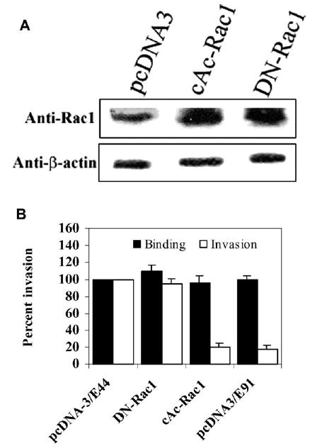
A. Total cell lysates (20 μg) of either cAc-Rac1/HBMEC or DN-Rac1/HBMEC or pcDNA3/HBMEC were separated on SDS polyacrylamide gels and were analyzed by immunoblotting with anti-Rac1 or anti-β-actin antibody. B. HBMEC overexpressing the Rac1 mutants along with pcDNA3 were incubated with OmpA+ E. coli (E44) and the invasion assays were carried out as described in Section 2. OmpA– E. coli (E91) was also used in these assays as a negative control. The invasion of E44 into these transfectants was expressed as a percent invasion of pcDNA3/HBMEC being taken as 100%. These data are from at least three separate experiments performed in triplicate. Error bars indicate the standard deviation from the mean. The inhibition of E. coli invasion into cAc-Rac1/HBMEC is significantly lower when compared to control cells. (P < 0.01 by unpaired, two-tailed t-test). Similarly, the total cell associated bacteria was represented as binding for each transfectant.
Next, we examined the phosphorylation states of PAK1 and MLC in these transfectants after infection with OmpA+ E. coli for various time periods. The cell lysates, from vector alone, DN-Rac1/HBMEC, and cAc-Rac1/HBMEC infected with OmpA+ E. coli, were immunoprecipitated with anti-phospho-PAK1 antibody and immunoblotted with the same antibody. As expected, the pcDNA3/HBMEC revealed low levels of phosphorylation of PAK1 between 10 and 30 min post-infection compared to uninfected control cells, which levels were increased to control levels by 60 min (Fig. 4A). In contrast, the phosphorylation of PAK1 was considerably less at all time points in DN-Rac1/HBMEC, suggesting that overexpression of DN-Rac1 mutant in HBMEC blocks the endogenous GTPase activity (Fig. 4B). In agreement with the phospho-PAK1 levels in these cells, phosphorylation of MLC in both pcDNA3- and DN-Rac1/HBMEC is greater from 10 to 60 min postinfection with OmpA+ E. coli. Reprobing the blot with anti-MLC antibody revealed that each lane contains equal amounts of MLC. Thus, reduced phosphorylation of PAK1 might lead to activation of MLCK, which increases the phosphorylation of MLC necessary for E. coli invasion. In contrast, overexpression of cAc-Rac1 in HBMEC increased the PAK1 activity at every time point irrespective of E. coli infection, which in turn reduced the MLC phosphorylation to lower than the basal levels (Fig. 4C). These results are not due to unequal loading of the proteins as the blots when reprobed with anti-MLC antibodies showed the presence of equal amount of proteins. Analysis of the intensities of phospho-PAK1 bands after normalized to the intensities of IgG bands demonstrated that the basal level phosphorylation of PAK1 in DN-Rac1/HBMEC was lower than the level of pcDNA3/HBMEC (Fig. 4C). In contrast, the phosphorylation levels of PAK1 were significantly greater in cAc-Rac1/HBMEC when compared to the levels of phospho-PAK1 in pcDNA3/HBMEC. Similar analysis of phospho-MLC levels in these cells revealed that the basal phosphorylation levels of MLC in DN-Rac1/HBMEC were greater than that of pcDNA3/HBMEC (Fig. 4C). Taken together these results suggest that cAc-Rac1 overexpression in HBMEC increased the basal levels of phopho-PAK1 and thus lower phosphorylation of MLC, which did not change due to infection with E. coli.
Fig. 4. Phosphorylation levels of MLC and PAK1 in HBMEC overexpressing DN-Rac1 or cAc-Rac1.

Total cell lysates of pcDNA3/HBMEC (A), DN-Rac1/HBMEC (B) or cAc-Rac1/HBMEC (C) infected with OmpA+ E. coli for varying periods as indicated were analyzed by Western blot analysis using either anti-pMLC or anti-MLC antibodies. In separate experiments, the total cell lysates were immunoprecipitated with anti-phospho-PAK1 antibody and the immune complexes were analyzed by anti-phospho-PAK1 antibody. Arrows indicate either phospho-PAK1 or phospho-MLC bands. (D). The densities of phospho-PAK1 were determined from the scanned blot using ImageJ software, normalized to the densities of IgG bands, and graphed.
3.3. Lack of phospho-MLC at the sites of E. coli entry is responsible for the inhibition of E. coli invasion in HBMEC overexpressing cAc-Rac1
Our previous studies showed that phospho-MLC recruits to the sites of bacterial entry during the invasion of E. coli into HBMEC [6]. Therefore, to examine whether overexpression of either DN-Rac1 or cAc-Rac1 has any effect on the recruitment of phospho-MLC, HBMEC transfected with the Rac1 constructs were subjected to immunocytochemistry after infecting with E. coli (Fig. 5). OmpA+ E. coli infection of HBMEC transfected with pcDNA3 induced the accumulation of actin filaments underneath several groups of bacteria (Fig. 5B), which were co-localized with phospho-MLC (Fig. 5C, D). In contrast, OmpA– E. coli infected cells demonstrated no condensation of either actin or phospho-MLC despite the presence of a number of bacteria attached to the cells (U–X). The cAc-Rac1/HBMEC showed slightly increased spreading of the cells with a few numbers of protrusions without bacterial infection (E–H). These cells when infected with OmpA+ E. coli showed very little accumulation of actin beneath the bacterial attachment (Fig. 5J). In addition, no phospho-MLC was observed at the sites of E. coli attachment (K). By contrast, the uninfected DN-Rac1/HBMEC showed cell spreading and several lamellipodia more than that observed in the non-transfected cells (Fig. 5M–P). Interestingly, increased accumulation of phospho-MLC in the nucleus besides its distribution through out the cell was observed. Infection with OmpA+ E. coli induced the condensation of actin in several places and was co-localized with phospho-MLC (Q–T). These results confirm that overexpression of cAc-Rac1 blocks the E. coli invasion by reducing the phosphorylation levels of MLC and might inhibit actin condensation. In contrast, DN-Rac1 inhibits the binding of endogenous Rac1 to PAK1, thereby decreasing its activity, which in turn induces the phosphorylation of MLC via MLCK. Lack of phospho-MLC and actin condensation is responsible for the inhibition of E. coli invasion in cAc-Rac1/HBMEC.
Fig. 5. Inhibition of phospho-MLC and actin accumulation at the E. coli entry site in cAc-Rac1/HBMEC.

HBMEC transfected with pcDNA3 were transfected with OmpA+ (A–D) or OmpA– E. coli (U–X) and HBMEC transfected with cAc-Rac1 or DN-Rac1 were infected with OmpA+ E. coli for 15 min (I–L) and (Q–T) or left uninfected (E–H) and (M–P), respectively. The cells were washed, fixed and stained with rhodamine phalloidin (B, F, J, N, R and V) and anti-pMLC antibody (C, G, K, O, S and W). The localization of phospho-MLC was achieved by incubating the anti-pMLC antibody followed by Cy2-conjugated secondary antibody and mounted with anti-fade solution containing DAPI. The bacteria were visualized using transmitted light optics equipped with a blue filter (A, E, I, M, Q and U). The cells were also visualized with dual filter mode for both red and green fluorescence (D, H, L, P, T and X). The arrows indicate the localized bacteria, actin or p-MLC in the infected monolayers. The magnification of panels containing bacteria is 60×, whereas other panels are of 40×.
3.4. Activation of protein kinase C and PI3-kinase was not affected in HBMEC transfected with cAc-Rac1
Previous studies from our laboratory showed that the invasive E. coli induces the phosphorylation of PKC-α and PI3-kinase (PI3-K) [18,19]. The activation of PKC-α has been shown to be downstream of FAK and is PI3-K dependent. To rule out the possibility that other signaling pathways are not affected in DN-Rac1/HBMEC and cAc/HBMEC, the activation of PKC-α in these transfectants was performed using a non-radioactive PepTag assay [19]. As shown in Fig. 6A, no significant differences were observed in the PKC-α activation between DN-Rac1/HBMEC and cAc-Rac1/HBMEC. The activation of PKC-α increased up to 15 min post-infection followed by a decline by 30 min in all the cell lysates, which results were similar to that of pcDNA3-transfected cells as shown in our previous studies [19].
Fig. 6.

Activation PKC-α and PI3-K in HBMEC overexpressing cAc-PAK1. A. Total cell lysates (20 μg of proteins) of DN-Rac1/HBMEC and cAc-Rac1/HBMEC infected with OmpA+ E. coli for varying periods of time were analyzed for PKC-α activity by a PepTag non-radioactive assay. B. Total cell lysates of HBMEC either non-transfected, overexpressing DN-Rac1 or cAc-Rac1 infected with OmpA+ E. coli were separated by SDS polyacrylamide gel electrophoresis, transferred to nitrocellulose, and immunoblotted with anti-phospho-Akt antibody to examine the activation of PI3K.
In addition, the cell lysates were also assessed for PI3K activation. Phosphorylation of Akt, which is a substrate to PI3K, was examined using anti-phospho Akt antibody. Non-transfected HBMEC when infected with E. coli showed increased phosphorylation of Akt between 15 and 30 min and declined by 60 min postinfection (Fig. 6B). Similar phosphorylation pattern was also observed with HBMEC overexpressing either DN-Rac1 or cAc-Rac1 showed increased. Each blot was also reprobed with anti-Akt antibody to examine the equality of protein loading in all the lanes. The results obtained with HBMEC are similar to that of the data obtained with pcDNA3/HBMEC in our previous studies [18]. These data suggest that the inhibition of E. coli invasion by overexpression of cAc-Rac1 in HBMEC might not be due to the blockade of other signaling pathways necessary for invasion.
4. Discussion
To invade, pathogens often trigger pre-existing host-signal transduction cascades involving phosphorylation of host proteins [20,21]. The small GTPases of the Rho family (Rho, Rac1 and Cdc42) are involved in the regulation of the actin cytoskeleton and act as molecular switches in various signaling processes [17,22,23]. Rho induces formation of stress fibers and focal adhesions, Rac1 is involved in lamellipodia and membrane ruffle formation, and Cdc42 regulates microspike formation [24]. Several proteins have been identified that directly interact with the activated (GTP-bound) forms of these GTPases and therefore are likely candidates to be effectors of cellular responses involving these small G proteins [25]. PAK1 is a target of the small GTPases Rac1 and Cdc42. Our previous studies have shown that E. coli K1 down-regulates the PAK1 activity during the entry of HBMEC [6]. In addition, our studies also demonstrated that recruitment of phosphorylated MLC along with actin at the bacterial attachment site in E. coli K1 invasion process. The regulation of MLC phosphorylation was shown to be dependent on PAK1. However, it is not clearly known whether Rac1 or Cdc42 is responsible for down-regulating the PAK1 activity. In this study, we show that greater quantities of Rac1 bound to PAK1 when compared to Cdc42, which reduced its activity with a consequent increase in the MLC phosphorylation levels in HBMEC. E. coli may be utilizing different GTPases at various steps of the transcellular process. Since the activation of GTPases has a spatial relationship in different signaling events, E. coli might be activating Rac1 at the cell surface and other GTPases inside the cell. Therefore, we are not excluding the role of Cdc42 in E. coli entry. For example, Rac1 and Cdc42 co-operate in cell movement, whereas Cdc42 is not necessary for the movement itself; it controls polarity signals that are required for directed migration [22]. In serum-starved Swiss 3T3 fibroblasts, Nobes and Hall demonstrated that the activities of Cdc42, Rac1 and Rho are linked to each other in hierarchical fashion, with Cdc42 rapidly activating Rac1, which in turn activates Rho [17].
Earlier, we showed that PAK1 activity is inhibited in the E. coli entry up to 15 min, which may lead to increased MLC kinase activity [6]. Interestingly, the phosphorylation pattern of PAK1 in the present study showed a decrease of between 15 and 30 min. The shift in the time frame of PAK1 activation may relate to the passage number of HBMEC, although the concept has not been affected. In agreement with PAK1 activity, the present data also showed that Rac1 association was inhibited up to 30 min with PAK1, leading to MLC phosphorylation upon infection with E. coli K1. Chen et al., have shown that PAK1 activation is important for Salmonella induced nuclear response, but not for actin mediated entry into cultured cells [9]. These responses are dependent on the interaction of Cdc42 with PAK1. However, a dominant negative Cdc42 mutant did not block invasion of Salmonella, thereby pointing out the role for Rac1. In contrast, Criss et al. showed that Rac1 selectively regulates the Salmonella entry at the apical plasma membrane, which was blocked by the overexpression of a dominant inhibitory Rac1 [26]. Of interest, our studies show that overexpression of DN-Rac1 has no effect on E. coli invasion, whereas cAc-Rac1 did. In this scenario, cAc-Rac1 increases the PAK1 activity as shown by Western blot analysis, which modifies the MLCK activity. In agreement with these results, the phosphorylation of MLC was significantly decreased in these cells although infected with invasive E. coli. Similar decrease in the MLC phosphorylation was observed in BHK-21 cells expressing cAc-Rac1 [7]. In contrast, the overexpression of DN-Rac1 might titrate out the factors required for the activation of endogenous Rac1 and therefore cannot activate PAK1. Interestingly, despite increased basal levels of MLC phosphorylation at all time points, infection with E. coli showed a further increase in MLC phosphorylation in DN-Rac1/HBMEC from 15 to 60 min. It is possible that an E. coli factor released into the cytoplasm after OmpA interaction with its receptor might further modulate the PAK1 activity.
Actin rearrangements are important for E. coli invasion of HBMEC [4]. Our previous studies utilizing dominant negative FAK or PKC-α mutants showed that actin accumulation was absent at the bacterial entry site, thus blocking the invasion [19,20]. Similarly, overexpression of cAc-Rac1 mutant in HBMEC significantly inhibited actin condensation underneath the E. coli binding sites, suggesting that a cross talk between Rac1 and actin polymerization pathways in E. coli invasion of HBMEC. Another significant difference between control and Rac1 overexpressing HBMEC is the lack of phospho-MLC at the bacterial entry site, indicating the requirement of myosin driven motor function for internalization of the bacteria.Although we have shown that OmpA interaction with HBMEC receptor is important for the actin condensation, it still remains to be elucidated how OmpA and its receptor interaction causes the down-regulation of PAK1 activity via Rac1 interaction. In several other bacteria, the effector molecules are injected into host cells by a specialized protein secretion system and translocation apparatus type III. SopE, a Salmonella typhimurium secreted protein was characterized as a guanidine exchange factor for Rho-GTPases by its ability to stimulate conformational change in Cdc42 thereby promotes guanine nucleotide release [27]. ExoT of Pseudomonas aeruginosa on the other hand acts as a GTPase activating protein for RhoA, Rac1, and Cdc42 [12]. However, no secretion system has been identified in E. coli K1 thus far. Nonetheless, OmpA interaction appears to be crucial for further expression of other virulence genes, whose products (secreted or surface expressed) might modulate the function of Rac1. Our present view is that two different signaling pathways are triggered by E. coli invasion of HBMEC, an actin rearrangement related and a myosin related pathway whose convergence is required for efficient internalization of bacteria.
In conclusion, our studies presented here along with previously published data demonstrate that E. coli K1 modulates the Rac1 binding to PAK1, thereby inactivating the enzyme. This in turn activates the MLCK, which phosphorylates MLC in greater quantities. The phospho-MLC is subsequently recruited to the sites of E. coli entry along with actin filaments for the formation of a fruitful cytoskeleton complex necessary for generating the force to pull the bacteria into the HBMEC.
Acknowledgments
We thank Gary Bokoch, The Scripps Research Institute, La Jolla for providing Rac1 mutant constructs and Sunil Sukumaran for helping with immunocytochemistry. We also thank Dr. G. McNamara, Children’s Hospital LosAngeles Research Institute Image Core, for assistance with fluorescence imaging and Dr. Martine Torres for critical reading of the manuscript. This work was supported by NIH grants AI40567 and HD 41525 (N.V.P).
References
- 1.Stoll BJ, Hansen N, Fanaroff AA, Wright LL, Carlo WA, Ehrenkranz RA, Lemons JA, Donovan EF, Stark AR, Tyson JE, Oh W, Bauer CR, Korones SB, Shankaran S, Laptook AR, Stevenson DK, Papile LA, Poole WK. Changes in pathogens causing early-onset sepsis in very-low-birth-weight infants. N Engl J Med. 2002;347:240–247. doi: 10.1056/NEJMoa012657. [DOI] [PubMed] [Google Scholar]
- 2.Prasadarao NV. Identification of Escherichia coli outer membrane protein A receptor on human brain microvascular endothelial cells. Infect Immun. 2002;70:4556–4563. doi: 10.1128/IAI.70.8.4556-4563.2002. [DOI] [PMC free article] [PubMed] [Google Scholar]
- 3.Prasadarao NV, Wass CA, Weiser JN, Stins MF, Huang SH, Kim KS. Outer membrane protein A of Escherichia coli contributes to invasion of brain microvascular endothelial cells. Infect Immun. 1996;64:146–153. doi: 10.1128/iai.64.1.146-153.1996. [DOI] [PMC free article] [PubMed] [Google Scholar]
- 4.Prasadarao NV, Wass CA, Stins MF, Shimada H, Kim KS. Outer membrane protein A-promoted actin condensation of brain microvascular endothelial cells is required for Escherichia coli invasion. Infect Immun. 1999;67:5775–5783. doi: 10.1128/iai.67.11.5775-5783.1999. [DOI] [PMC free article] [PubMed] [Google Scholar]
- 5.Bokoch GM. Biology of the p21-activated kinases. Annu Rev Biochem. 2003;72:743–781. doi: 10.1146/annurev.biochem.72.121801.161742. [DOI] [PubMed] [Google Scholar]
- 6.Rudrabhatla RS, Sukumaran SK, Prasadarao NV. Modulation of myosin light chain phosphorylation by p21 activated kinase interaction with Rac1 in E. coli invasion of human brain microvascular endothelial cells. Infect Immun. 2003;71:2787–2797. doi: 10.1128/IAI.71.5.2787-2797.2003. [DOI] [PMC free article] [PubMed] [Google Scholar]
- 7.Sanders LC, Matsumura F, Bokoch GM, de Lanerolle P. Inhibition of myosin light chain kinase by p21-activated kinase. Science. 1999;283:2083–2085. doi: 10.1126/science.283.5410.2083. [DOI] [PubMed] [Google Scholar]
- 8.Akeda Y, Kodama T, Kashimoto T, Cantarelli V, Horiguchi Y, Nagayama K, Iida T, Honda T. Dominant-negative Rho, Rac, and Cdc42 facilitate the invasion process of Vibrio parahaemolyticus into Caco-2 cells. Infect Immun. 2002;70:970–973. doi: 10.1128/IAI.70.2.970-973.2002. [DOI] [PMC free article] [PubMed] [Google Scholar]
- 9.Barz C, Abahji TN, Trulzsch K, Heesemann J. The Yersinia Ser/Thr protein kinase YpkA/YopO directly interacts with the small GTPases RhoA and Rac-1. FEBS Lett. 2000;29:139–143. doi: 10.1016/s0014-5793(00)02045-7. [DOI] [PubMed] [Google Scholar]
- 10.Chen LM, Bagrodia S, Cerione RA, Galan JE. Requirement of p21-activated kinase (PAK) for Salmonella typhimurium-induced nuclear responses. J Exp Med. 1999;189:1479–1488. doi: 10.1084/jem.189.9.1479. [DOI] [PMC free article] [PubMed] [Google Scholar]
- 11.Hauck CR, Meyer TF, Lang F, Gulbins E. CD66-mediated phagocytosis of Opa52 Neisseria gonorrhoeae requires a Src-like tyrosine kinase- and Rac1-dependent signaling pathway. EMBO J. 1998;17:443–454. doi: 10.1093/emboj/17.2.443. [DOI] [PMC free article] [PubMed] [Google Scholar]
- 12.Kazmierczak BI, Jou TS, Mostov K, Engel JN. Rho GTPase activity modulates Pseudomonas aeruginosa internalization by epithelial cells. Cell Microbiol. 2001;3:85–98. doi: 10.1046/j.1462-5822.2001.00091.x. [DOI] [PubMed] [Google Scholar]
- 13.Kazmierczak BI, Engel JN. Pseudomonas aeruginosa ExoT acts in vivo as GTPase-activating protein for RhoA, Rac1, and Cdc42. Infect Immun. 2002;70:2198–2205. doi: 10.1128/IAI.70.4.2198-2205.2002. [DOI] [PMC free article] [PubMed] [Google Scholar]
- 14.Lerm M, Selzer J, Hoffmeyer A, Rapp UR, Aktories K, Schmidt G. Deamidation of Cdc42 and Rac by Escherichia coli cytotoxic necrotizing factor 1: activation of c-Jun N-terminal kinase in HeLa cells. Infect Immun. 1999;67:496–503. doi: 10.1128/iai.67.2.496-503.1999. [DOI] [PMC free article] [PubMed] [Google Scholar]
- 15.Martinez JJ, Hultgren SJ. Requirement of Rho-family GTPases in the invasion of Type 1-piliated uropathogenic Escherichia coli. Cell Microbiol. 2002;4:19–28. doi: 10.1046/j.1462-5822.2002.00166.x. [DOI] [PubMed] [Google Scholar]
- 16.Billker O, Popp A, Brinkmann V, Wenig G, Schneider J, Caron E, Meyer TF. Distinct mechanisms of internalization of Neisseria gonorrhoeae by members of the CEACAM receptor family involving Rac1- and Cdc42-dependent and -independent pathways. EMBO J. 2002;21:560–571. doi: 10.1093/emboj/21.4.560. [DOI] [PMC free article] [PubMed] [Google Scholar]
- 17.Churin Y, Kardalinou E, Meyer TF, Naumann M. Pathogenicity island-dependent activation of Rho GTPases Rac1 and Cdc42 in Helicobacter pylori infection. Mol Microbiol. 2001;40:815–823. doi: 10.1046/j.1365-2958.2001.02443.x. [DOI] [PubMed] [Google Scholar]
- 18.Nobes CD, Hall A. Rho, rac and cdc42 GTPases: regulators of actin structures, cell adhesion and motility. Biochem Soc Trans. 1995;23:456–459. doi: 10.1042/bst0230456. [DOI] [PubMed] [Google Scholar]
- 19.Reddy MA, Prasadarao NV, Wass CA, Kim KS. Phosphatidyl inositol-3 kinase activation and interaction with focal adhesion kinase in E. coli invasion of human brain endothelial cells. J Biol Chem. 2000;275:36769–36774. doi: 10.1074/jbc.M007382200. [DOI] [PubMed] [Google Scholar]
- 20.Reddy MA, Wass CA, Kim KS, Schlaepfer DD, Prasadarao NV. Involvement of focal adhesion kinase in Escherichia coli invasion of human brain microvascular endothelial cells. Infect Immun. 2000;68:6423–6430. doi: 10.1128/iai.68.11.6423-6430.2000. [DOI] [PMC free article] [PubMed] [Google Scholar]
- 21.Sukumaran SK, Prasadarao NV. Regulation of Protein Kinase C in E. coli invasion of human brain endothelial cells. J Biol Chem. 2002;277:12253–12262. doi: 10.1074/jbc.M110740200. [DOI] [PubMed] [Google Scholar]
- 22.Stebbins CE, Galan JE. Modulation of host signaling by a bacterial mimic: structure of the Salmonella effector SptP bound to Rac1. Mol Cell. 2000;6:1449–1460. doi: 10.1016/s1097-2765(00)00141-6. [DOI] [PubMed] [Google Scholar]
- 23.Nobes CD, Hall A. Rho GTPases control polarity, protrusion, and adhesion during cell movement. J Cell Biol. 1999;144:1235–1244. doi: 10.1083/jcb.144.6.1235. [DOI] [PMC free article] [PubMed] [Google Scholar]
- 24.Suzuki T, Mimuro H, Miki H, Takenawa T, Sasaki T, Nakanishi H, Takai Y, Sasakawa C. Rho family GTPase Cdc42 is essential for the actin-based motility of Shigella in mammalian cells. J Exp Med. 2000;191:1905–1920. doi: 10.1084/jem.191.11.1905. [DOI] [PMC free article] [PubMed] [Google Scholar]
- 25.Schmidt A, Hall MN. Signaling to the actin cytoskeleton. Annu Rev Cell Dev Biol. 1998;14:305–338. doi: 10.1146/annurev.cellbio.14.1.305. [DOI] [PubMed] [Google Scholar]
- 26.Criss AK, Ahlgren DM, Jou TS, McCormick BA, Cassanova JE. The GTPase Rac1 selectively regulates Salmonella invasion at the apical plasma membrane of polarized epithelial cells. J Cell Sci. 2001;114:1331–1341. doi: 10.1242/jcs.114.7.1331. [DOI] [PubMed] [Google Scholar]
- 27.Buchwald G, Friebel A, Galan JE, Hardt WD, Wittinghofer A, Scheffzel K. Structural basis for the reversible activation of Rho protein by the bacterial toxin SopE. EMBO J. 2002;21:3286–3295. doi: 10.1093/emboj/cdf329. [DOI] [PMC free article] [PubMed] [Google Scholar]