


Figure 2: Migration of retinal astrocytes precedes angiogenesis.
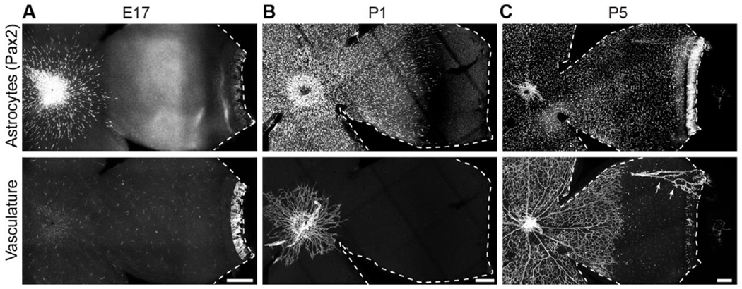
Flat-mounted mouse retinas labeled with markers of astrocyte nuclei (Pax2) and vasculature (A,C: Isolectin B4; B: anti-CD31).
A) Astrocytes begin to enter the retina before birth (E17), prior to endothelial cell entry. No vasculature is present although lectin-labeled microglia are visible.
B) By P1, astrocytes cover over half of the RNFL. Vasculature is just beginning to exit the optic nerve head.
C) At P5, astrocytes have reached the periphery and vasculature covers approximately half of the retina. Arrows, hyaloid vessels that were not completely removed during retinal dissection. Ahead of the angiogenic wavefront astrocyte somata are arranged in a circular pattern, mirroring astrocyte arbors (Fig. 1B).
Dotted lines delineate edge of retinal tissue. Scale bars, 100 μm.