



ABSTRACT
Sapovirus is increasingly recognized as an important cause of acute gastroenteritis (AGE) worldwide; however, studies of sapovirus prevalence, genetic diversity, and strain-specific clinical implications have been scarce. To fill this knowledge gap, we used reverse transcription–real-time PCR and sequencing of the partial major capsid protein VP1 gene to analyze stool specimens and rectal swabs obtained from 3,347 children with AGE and 1,355 asymptomatic controls (all <18 years old) collected between December 2014 and August 2018 in Alberta, Canada. Sapovirus was identified in 9.5% (317/3347) of the children with AGE and 2.9% of controls. GI.1 (36%) was the predominant genotype identified, followed by GI.2 (18%), GII.5 (8%), and GII.3 (6%). Rare genotypes GII.1, GII.2, GV.1, GII.4, GIV.1, GI.3, and GI.7 were also seen. Sapovirus was detected year-round, peaking during the winter months of November to January. The exception was the 2016-2017 season, when GI.2 overtook GI.1 as the predominant strain, with a high detection rate persisting into April. We did not observe significant difference in the severity of gastroenteritis by genogroup or genotype. Repeated infection by sapovirus of different genogroups occurred in three controls who developed AGE later. Our data suggest that sapovirus is a common cause of AGE in children with high genetic diversity.
KEYWORDS: sapovirus, pediatric gastroenteritis, genotype, seasonality, repeated infection
INTRODUCTION
Human sapovirus has been increasingly recognized as an important cause of acute gastroenteritis (AGE) worldwide in both outbreaks and sporadic settings (1). Belonging to the same Caliciviridae family as norovirus, sapovirus typically causes milder yet virtually identical symptoms (2). Molecular assays required to detect sapovirus are not commonly included in commercial gastroenteritis detection panels, nor do clinical laboratory testing algorithms routinely include sapovirus. Consequently, few epidemiological studies of sapovirus have been published.
The sapovirus genome consists of two open reading frames (ORFs). ORF1 encodes 5 nonstructural proteins (NS1 to NS5), the NS6-7 fusion protein, which functions as protease-RNA-dependent RNA polymerase (RdRp), and the major capsid protein, VP1. ORF2 is proposed to encode the minor capsid protein, VP2 (1). The genetic classification of sapovirus is based on VP1 sequences, partial RdRp, or both (1). Based on VP1 nucleotide sequence similarities, sapovirus can be classified into 19 genogroups (GI to GXIX), with genogroups I, II, IV, and V infecting humans; all other genogroups (GIII and GVI to GXIX) have been detected in animals (3). Human sapoviruses are further divided into 18 well-established genotypes: GI.1 to GI.7, GII.1 to GII.8, GIV.1, and GV1 and GV.2, with another tentative GII genotype, GII.NA 1 (not assigned), reported in 2019 (3–6). Similar to norovirus, sapovirus detection peaks during the winter months in temperate regions of the northern hemisphere (7–9).
The severity of symptoms from sapovirus infections varies from mild to severe, with some children requiring emergency department (ED) evaluations and even hospitalization (10, 11). The association of gastroenteritis severity with sapovirus genogroup or genotypes is not well understood. Although all age groups are affected, sapovirus gastroenteritis most commonly occurs in children under 5 years of age (2). Asymptomatic infections have been reported (1, 2). Symptomatic reinfections by sapovirus or different genogroups or genotypes have been identified in Japan and Peru, suggesting that genogroup- and genotype-specific immunity does not offer cross protection (5, 12).
To date, there are limited data on the molecular epidemiology of sapovirus in Canada (13, 14). Understanding sapovirus disease burden, seasonality, genotype distribution, and emergent strains, as well as genogroup- and genotype-specific clinical severity, is crucial to the planning of public health initiatives. Such knowledge can improve our ability to detect circulating strains while providing insight into vaccine development, especially in the post-rotavirus vaccine era, where sapovirus is emerging as an important cause of childhood AGE (15). Moreover, the disease burden of sapovirus pediatric gastroenteritis remains unknown because sapovirus is not included in routine laboratory diagnostic testing protocols, and like norovirus, sapovirus is only generally notifiable during outbreaks. Thus, we sought to fill these knowledge gaps by (i) assessing the prevalence of sapovirus infection in children, (ii) characterizing circulating sapovirus strains and their associated clinical severity, (iii) describing codetections of sapovirus with other enteropathogens, and (iv) quantifying the frequency of recurrent sapovirus infections in children.
MATERIALS AND METHODS
Study participants and specimen acquisition.
Ethics approval was obtained from the University of Alberta and the University of Calgary research ethics boards. The Alberta Provincial Pediatric EnTeric Infection TEam (APPETITE) study protocol has been previously published (16). Briefly, children less than 18 years of age with >3 episodes of vomiting or diarrhea within 24 h and <7 days of symptoms were eligible to participate and were classified as AGE cases. Children were recruited in two urban, pediatric tertiary care emergency departments (ED) (Alberta Children’s Hospital, Calgary, and Stollery Children’s Hospital, Edmonton) and through Health Link (Alberta Health Services), a province-wide nursing advice telephone line serving the province of Alberta, Canada. Controls with no infectious symptoms (i.e., no vomiting, diarrhea, fever, rhinorrhea, or cough) were also enrolled in the ED and at a public health clinic attended by children presenting for their routine infant immunizations in Calgary, Alberta. Consent was obtained from the caregivers of eligible children, along with assent when appropriate. Children enrolled in the previous 14 days, those who could not complete follow-up, and those with preexisting psychiatric illness or neutropenia or in need of emergent medical intervention were ineligible.
Rectal swabs and stool specimens from AGE and control cases were collected between December 2014 and August 2018. FLOQSwabs (Copan Italia, Brescia, Italy) were inserted into the rectum, rotated 360°, and then placed into sterile sheaths. Stool specimens were collected in fecal collectors (V302-F; Starplex Scientific, Ontario, Canada). Specimens collected in the ED were maintained at 4°C and delivered to the laboratory within 24 h of collection and immediately frozen at −70°C. Stool samples not provided prior to discharge from ED, and samples from participants recruited through Health Link and the public health clinic were collected at home and transported by a study-funded courier in coolers with ice packs to the laboratory for processing and freezing within 12 h of collection.
Nucleic acid extraction and molecular pathogen detection.
Total nucleic acid was extracted from a 60% rectal swab or 10% stool suspension in NucliSENS lysis buffer (bioMérieux, Laval, Quebec, Canada). Nucleic acid was extracted and eluted in 70 μl using the NucliSENS easyMag system (bioMérieux, Laval, Quebec, Canada) according to the manufacturer’s instructions. All resultant nucleic acid extracts were immediately stored at −70°C until testing. Viral cDNA was generated with the SuperScript II (Thermo Fisher Scientific, MA, USA) reverse transcription system as previously described (17).
Sapovirus was detected using an in-house multiplex reverse transcription–real-time quantitative PCR (RT-qPCR) assay that also detects norovirus (genogroups I and II), group A rotavirus, adenovirus (all serotypes), and astrovirus (18). The primers and probes for sapovirus detection were originally designed by Oka et al. in 2006 (19). Threshold cycle (CT) values were used as a semiquantitative measure that inversely correlates with viral load in the specimen. The CT value cutoff for a positive result was set at ≤38. Specimens also underwent enteropathogen detection using the Luminex Gastroenteritis Pathogen Panel (GPP), which detects three viruses (norovirus GI and GII, rotavirus, and adenovirus 40/41), nine bacteria (Clostridioides difficile, Escherichia coli O157, enterotoxigenic E. coli [ETEC], Shiga toxin-producing E. coli [STEC], Salmonella, Campylobacter, Vibrio cholerae, Shigella, Yersinia enterocolitica) and three parasites (Cryptosporidium, Giardia, Entamoeba histolytica) (17). Rectal swabs and stool specimens collected from the same patient were tested individually with the Gastroenteritis Virus Panel (GVP) and GPP.
Seasonality was assessed using the following definitions: for winter, 1 December to 28 February; spring, 1 March to 31 May; summer, 1 June to 31 August 31; and autumn, 1 September to 30 November. Average monthly temperature data were obtained by courtesy of Environment and Climate Change of Canada on the Edmonton weatherstat website (https://edmonton.weatherstats.ca/metrics/temperature.html).
Sapovirus genotype determination.
The genotype of sapovirus isolates was determined in only one of the two specimen types (the specimen with the lower CT value) collected from each sapovirus-positive participant. To identify the genotype for each sapovirus-positive sample, the cDNA underwent nested PCR amplification with primers SV-F13, SV-F14, SV-R13, SV-R14, SV-F22, and SV-R2 to generate amplicons of approximately 420 bp (20) (see Table S1 and Fig. S1 in the supplemental material). The PCR products were subjected to gel electrophoresis with 2% agarose followed by excision of the product with the expected size to undergo purification via the Qiaquick gel extraction kit (Qiagen, Germantown, MD, USA). The nucleotide sequences of each sample were retrieved using the BigDye Terminator v3.1 cycle sequencing kit (Life Technologies, Austin, TX, USA). The Sanger chromatograms were analyzed using MEGA X 10.0.5 to obtain the sequences, and the NCBI nucleotide BLAST search engine was used to assign genotypes (21, 22). The sapovirus genotyping results were verified using the Calicivirus Typing Tool (https://norovirus.ng.philab.cdc.gov) (23).
Nucleic acids of sapovirus-positive stool samples that were not successfully genotyped by the above method were reextracted using the ReliaPrep RNA Cell Miniprep system (Promega, Madison, WI, USA). Viral RNA was reverse transcribed using the SuperScriptII reverse transcription system as described above, and the cDNAs were subjected to another heminested PCR amplification using primers SLV5317, SLV5749, SVpol3′-A, and SVpol3′-B to generate amplicons of 371 bp for resequencing through agarose gel electrophoresis and gel purification (24) (Table S1 and Fig. S1).
Disease severity and participant clinical characteristics.
Clinical disease severity was assessed using the modified Vesikari scale (MVS) score (25, 26). The MVS is a composite measure that includes seven components that are each assigned a score based on data collected between symptom onset and resolution. The components include (i) duration of diarrhea, (ii) maximal number of episodes of diarrhea in any given 24-h period, (iii) duration of vomiting, (iv) maximal number of episodes of vomiting in any given 24-h period, (v) maximal participant temperature recorded, (vi) presence and type of unscheduled visits to a health care provider, and (vii) type of treatment, if any, received for gastroenteritis. MVS scores can be further categorized into severity groups as mild (0 to 8), moderate (9 to 10), and severe (≥11) (26).
We compared the MVS scores of participants with different sapovirus genogroups and genotypes. The description of illness collected included the presence of fever, duration of diarrhea and vomiting in hours, modified Vesikari score (MVS) (26), and presence of dehydration measured by clinical dehydration scores reported by parents at the index ED visit. The number of codetected pathogens, age in months, and sex were also included in the analysis.
Phylogenetic analysis of VP1-encoding region.
Partial VP1 sequences that were longer than 210 nucleotides were included in this analysis and compared to 143 near-full-length sapovirus reference sequences that were downloaded from GenBank. All sequences were analyzed and aligned with software package MUSCLE in MEGA X. A phylogenetic dendrogram was constructed using the maximum likelihood method, based on the Kimura two-parameter model with a discrete gamma distribution (1,000 bootstraps) in MEGA version X (21, 22).
Statistical analysis.
Data were summarized with frequencies and percentages for categorical variables and medians and interquartile ranges (IQRs) for continuous variables. Categorical variables were compared using the χ2 test or Fisher’s exact test where appropriate. Assay concordance was analyzed with Cohen’s kappa coefficient. Continuous variables were compared using the Kruskal-Wallis test. We calculated two-tailed P values and set the significance level α at 0.05. Descriptive statistics were computed using the SPSS software 26 (27) and GraphPad Prism v8.4.2 (GraphPad Software, San Diego, California, USA; www.graphpad.com).
Accession number(s).
The partial VP1 sequences described above can be accessed in GenBank under accession no. MZ420980 to MZ421251.
RESULTS
Prevalence and diversity of sapovirus strains in children.
In total, 3,347 children with AGE and 1,355 controls submitted specimens. Sapovirus was detected in 9.5% (317/3347) and 2.9% (39/1355) of participants from each group, respectively (Fig. 1). Partial capsid gene sequencing was successful in 85.2% (270/317) of samples from children with AGE; the remaining 14.8% (47/317) did not yield a successful typing result (Fig. 1). Typing was unsuccessful in 17 (43.6%) controls. All known human-pathogenic genogroups (GI, GII, GIV, and GV) were identified. The most common genogroup detected in children with AGE was GI (56.8%; 180/317); GI and GII were detected with equal frequency (28.8%; 11/39) as the leading sapovirus genogroups among controls. Eleven genotypes were identified in the present study (Table 1). Among children with AGE, sapovirus GI.1 (37.2%; 118/317) was the most common genotype, followed by GI.2 (18.9%; 60/317). All other genotypes detected accounted for <10% of detections (Table 1). The concordance between rectal swabs and stool specimens for sapovirus positivity was high (kappa = 0.907). Norovirus genogroups I and II and rotavirus were detected by both the Luminex GPP and the in-house GVP (kappa = 0.87, 0.94, and 0.98, respectively, indicating excellent agreement between the GPP and GVP). Because the GPP identifies adenovirus 40/41 and the GVP detects all serotypes of adenovirus, it was not possible to directly compare the two in adenovirus detection rate.
FIG 1.

Sapovirus testing results for study participants.
TABLE 1.
Sapovirus genotypes identified in gastroenteritis cases and controls enrolled in APPETITEa
| Genotype | Annual no. of SaV AGE casesb |
Total no. (%) per genotypec |
% analyzed for lineage | ||||
|---|---|---|---|---|---|---|---|
| Early 2015 | 2015–2016 | 2016–2017 | 2017–2018 | AGE | Asymptomatic | ||
| GI.1 | 6 | 46 | 25 | 41 | 118 (37.2) | 7 (17.9) | 97.6 |
| GI.2 | 4 | 3 | 49 | 4 | 60 (18.9) | 4 (10.3) | 96.9 |
| GI.3 | 1 | 0 | 0 | 0 | 1 (0.3) | 0 | 100.0 |
| GI.7 | 0 | 1 | 0 | 0 | 1 (0.3) | 0 | 100.0 |
| GII.1 | 3 | 3 | 2 | 10 | 18 (5.7) | 2 (5.1) | 100.0 |
| GII.2 | 0 | 10 | 2 | 2 | 14 (4.4) | 0 | 71.4 |
| GII.3 | 0 | 0 | 13 | 5 | 18 (5.7) | 6 (15.4) | 62.5 |
| GII.4 | 0 | 1 | 0 | 1 | 2 (0.6) | 0 | 100.0 |
| GII.5 | 3 | 16 | 5 | 0 | 24 (7.6) | 3 (7.7) | 100.0 |
| GIV.1 | 1 | 0 | 0 | 1 | 2 (0.6) | 0 | 100.0 |
| GV.1 | 1 | 2 | 3 | 6 | 12 (3.8) | 0 | 91.7 |
| Untypeable | 5 | 15 | 12 | 15 | 47 (14.8) | 17 (43.6) | NA |
| Total | 24 | 97 | 111 | 85 | 317 | 39 | NA |
Abbreviations: AGE, acute gastroenteritis; SaV, sapovirus; NA, not applicable.
An epidemic year is defined as from July of the previous year to June of the next year. “Early 2015” includes January to June 2015, and the period 2017 to 2018 includes up until July 2018.
Shown is the percentage of each genotype out of the total number of sapovirus-positive cases identified in AGE cases and controls
Among children with AGE, sapovirus was detected all year-round, with positive rates near 20% in the late autumn and winter and close to 2% in the summer (Fig. 2). Sapovirus GI.1 was the predominant strain in 2015 (13%) and 2017 (11%), while genotype GI.2 had a higher prevalence (9%) in 2016, with a high rate of sapovirus AGE persisting into spring 2017 (Fig. 2; Table 1). Common genotypes GI.1, GI.2, GII.1, and GV.1 were detected in all 4 years, while GII.2 and GII.5 circulated in three (Table 1).
FIG 2.

Monthly distribution of sapovirus genotypes in APPETITE children with acute gastroenteritis from January 2015 to July 2018. The percentages of cases infected with different genotypes normalized to the monthly total number of AGE cases are shown as stacked bars. The average monthly temperatures are shown as a smooth line.
Among the 270 samples from children with AGE and 22 from controls that were successfully genotyped, 93.5% (273/292) of the sequences were suitable for phylogenetic analysis, with 92.3% (252/273) and 7.7% (21/273) produced by the methods of Okada et al. (20) and Monica et al. (24), respectively. Success rates in obtaining usable sequences belonging to genogroups I, IV, and V, as well as GII.1 and GII.4, were greater than 90%, whereas the proportions of usable GII.2 and GII.3 sequences were 71.4% and 62.5%, respectively (Table 1). Partial VP1 sequences of GI.1 from this study clustered with reference sequences from east Asia (China, Japan, and South Korea), the Americas (the United States and Peru), and Europe (Germany and the United States) collected between 2013 and 2016, as well as a historical strain from Sapporo, Japan, collected in 1982 (see Fig. S2 in the supplemental material). GI.2 from the present study clustered with reference sequences from Germany, the United States, and Brazil collected in recent years (2009 to 2016) and a Bangladesh sequence from 2005. GI.3 sequences clustered with reference sequences from Japan (2008) and Kenya (2006); GI.7 clustered with strains from Japan (2008). The same multicontinent circulation was observed for the GII.1, GII.2, GII.3, GII.5, and GIV.1 strains. On the other hand, GII.4 was found to cluster with reference sequences from Peru (2016) and Brazil (2011) and GV.1 with reference sequences from the United States (2014) (Fig. S2).
Clinical severity of sapovirus infections in children.
There were no differences in the MVS scores, clinical characteristics, or CT values among the AGE cases when comparing the various sapovirus genogroups and genotypes (Fig. 3A and B; see Fig. S3 and Tables S2 to S4 in the supplemental material). Children with AGE presenting to emergency departments (ED) had more severe gastroenteritis symptoms (P < 0.001) than patients receiving care at home (Fig. 3C; see Table S5 in the supplemental material). Of the 231 cases enrolled at an ED who tested positive for sapovirus, 97.2% were discharged, none was admitted to the intensive care unit (ICU), and there were no deaths. There were no differences in sapovirus gastroenteritis severity between different age groups (Fig. 3D).
FIG 3.
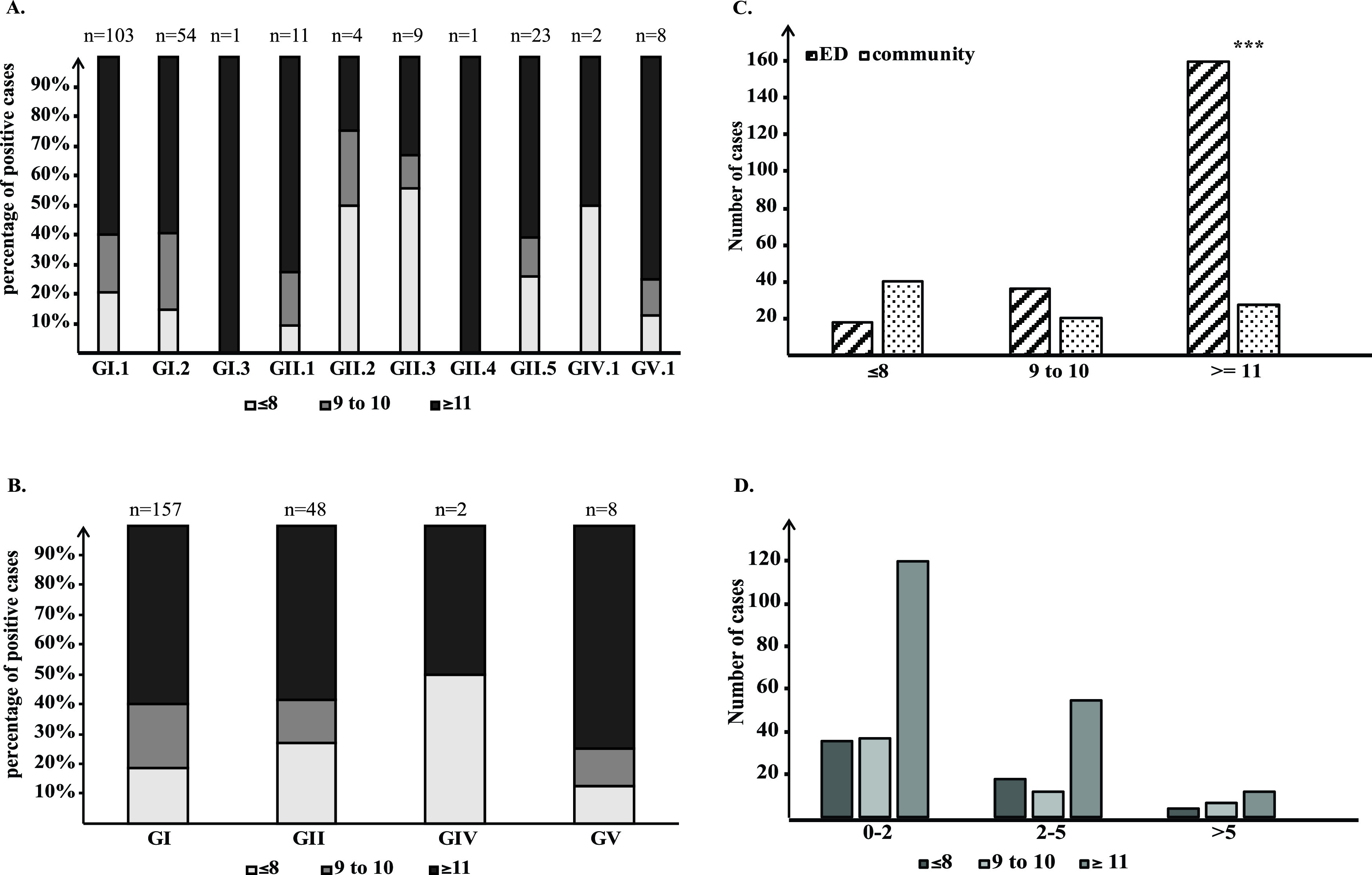
Gastroenteritis severity by sapovirus genogroups, genotypes, or health care settings. MVS ≤ 8, mild; MVS = 9 to 10, moderate; MVS ≥ 11, severe. Cases without an MVS were excluded. (A) AGE severity attributed to different sapovirus genotypes. (B) AGE severity attributed to different sapovirus genogroups. (C) AGE severity in patients who seek ED care versus being managed at home. (D) AGE severity in different age groups.
Codetections of sapovirus with other enteropathogens.
Among children with AGE who tested positive for sapovirus, 35% (111/317) had one or more codetected enteropathogens: there was one other pathogen in 29.3% (93/317), and there were two and four other pathogens in 5.4% (17/317) and 0.3% (1/317), respectively. Adenovirus, norovirus, and rotavirus were the most commonly codetected enteropathogens (Table 2). Codetection of sapovirus and other enteropathogens was found in 35.9% (14/39) of controls, with adenovirus being the most common (Table 2).
TABLE 2.
Sapovirus coinfections in children with acute gastroenteritis and healthy controls
| Infection or coinfection | No. (%) of: |
|
|---|---|---|
| AGE cases (n = 314) | Controls (n = 37) | |
| Sapovirus alone | 203 (64.6) | 28 (75.7) |
| 2 pathogens codetected | 93 (29.6) | 9 (24.3) |
| Sapovirus, norovirus | 20 (6.4) | 1 (2.7) |
| Sapovirus, rotavirus | 19 (6.1) | 1 (2.7) |
| Sapovirus, adenovirus | 47 (15.0) | 6 (16.2) |
| Sapovirus, astrovirus | 1 (0.3) | |
| Sapovirus, ETEC | 1 (0.3) | |
| Sapovirus, Salmonella | 1 (0.3) | |
| Sapovirus, Giardia | 1 (0.3) | 1 (2.7) |
| Sapovirus, Entamoeba | 1 (0.3) | |
| Sapovirus, Aeromonas | 2 (0.6) | |
| 3 pathogens codetected | 17 (5.4) | |
| Sapovirus, norovirus, adenovirus | 10 (3.2) | |
| Sapovirus, norovirus, astrovirus | 1 (0.3) | |
| Sapovirus, norovirus, Salmonella | 1 (0.3) | |
| Sapovirus, rotavirus, adenovirus | 1 (0.3) | |
| Sapovirus, rotavirus, Aeromonas | 1 (0.3) | |
| Sapovirus, adenovirus, E. coli O157 | 1 (0.3) | |
| Sapovirus, adenovirus, Aeromonas | 1 (0.3) | |
| Sapovirus, adenovirus, astrovirus | 1 (0.3) | |
| 5 pathogens codetected | 1 (0.3) | |
| Sapovirus, adenovirus, astrovirus, norovirus, rotavirus | 1 (0.3) | |
Recurrent sapovirus infections in children.
We observed repeated sapovirus infections during the study period in three children who were enrolled initially as controls and subsequently developed AGE (Table 3). Patient 1 was positive for sapovirus with an unknown genotype (CT = 37.7) in October 2015 and had a GI.1 sapovirus (CT = 14.2) detected when symptomatic in November 2016. Patient 2 was positive for GI.1 sapovirus (CT = 32.7) in December 2015 and GII.5 sapovirus (CT = 20.0) in November 2016. Patient 3 was positive for both GII.5 sapovirus (CT = 29.5) and adenovirus species C genotype 2 (28) in December 2015 and GI.2 sapovirus (CT = 15.3) and GII norovirus in November 2016.
TABLE 3.
Repeated sapovirus infection in three participants enrolled as controls who developed acute gastroenteritis subsequently
| Patient | Age group (yr) | Sex | Disease status | Date of collection | Sapovirus genotype | CT value | Enteropathogen codetection | Clinical symptom (s) | MVS |
|---|---|---|---|---|---|---|---|---|---|
| 1 | 0–2 | Male | Control | 23 Oct 2015 | Untypable | 37.7 | |||
| AGE | 11 Mar 2016 | GI.1 | 14.2 | Vomiting, diarrhea, fever | 8 | ||||
| 2 | 0–2 | Female | Control | 24 Dec 2015 | GI.1 | 32.7 | |||
| AGE | 4 Nov 2016 | GII.5 | 20 | Diarrhea | 4 | ||||
| 3 | 0–2 | Male | Control | 24 Dec 2015 | GII.5 | 29.5 | Adenovirus, species C, genotype 2 | ||
| AGE | 4 Nov 2016 | GI.2 | 15.3 | Norovirus GII | Vomiting | 5 | |||
DISCUSSION
The increasing availability of nucleic acid-based detection methods has increased awareness of the presence of sapovirus in people with AGE (2). However, the molecular epidemiology of sapovirus in North America has not been well described. The present study provides comprehensive data on childhood sapovirus AGE over a 4-year period. In keeping with previous reports from other high-income countries (4, 7, 29), the overall sapovirus detection rate in children with AGE was 9.5%, while that of controls was 2.9%. This represents a nearly 4-fold-higher detection rate compared to controls. GI.1 was the most common sapovirus strain identified, followed by GI.2. Sapovirus detection peaked during late autumn and winter, except during the 2016-2017 season, when GI.2 emerged as the predominant strain and a high rate of sapovirus detection continued into April.
Sapovirus prevalence differs across regions and age groups, with reported detection frequencies ranging from 1 to 17% in different settings (15). Previously, sapovirus had been detected in 4.2% of specimens collected from people of all ages hospitalized with sporadic gastroenteritis in the province of Alberta, most frequently in children between the ages of 1 and 2 (18). Two population-based studies of sporadic gastroenteritis were conducted in southwest Alberta. While there were 0.2% confirmed sapovirus/norovirus cases in individuals with gastroenteritis and 0% in healthy individuals found from May to October of 2005, 4.2% sapovirus cases, most prevalent in children aged 1 to 5 years, and 1.2% asymptomatic infections were found from 2008 to 2009 (14, 30). The first study included immunocompromised patients (18), while the second study had a small number of participants (422 gastroenteritis cases and 58 controls), who were recruited in the summer and early autumn, when the sapovirus detection rate was low (30). The third study commented on the underestimation of the true rate due to the limited number of samples being submitted (14). The sapovirus detection rate in children with AGE differed in the present study, likely due to the differences in study population, with the inclusion of patients with isolated vomiting and patients not presenting to health care, in addition to a much higher specimen submission rate due to the study design. Consistent with our findings, Doll et al. studied the etiology of pediatric sporadic gastroenteritis in the province of Quebec, Canada, from 2012 to 2014 and reported sapovirus infections in 6.4% of specimens collected from children aged 8 weeks to less than 3 years (31).
Consistent with previous studies from countries in the northern hemisphere (7, 9, 32), sapovirus circulated year-round in our study, with the highest rate of detection in the winter months. Winter seasonality is a key characteristic of viruses in the Caliciviridae family, including norovirus. Understanding the seasonality of sapovirus transmission patterns can inform diagnostic and infection prevention and control strategies. The high proportion of sapovirus (>20%) seen in February 2015 can be explained by the low number of children with AGE (n = 9) who were enrolled in that month.
A variety of sapovirus strains cocirculated throughout the year. Sapovirus GI.1 was the predominant genotype detected, except in the winter of 2016 to 2017, when GI.2 became predominant. GI.1 has been identified as the most frequently identified genotype in sporadic sapovirus gastroenteritis in several prior studies (7, 9, 33–35). Since 2007, an increase in GI.2 prevalence has been reported, which coincided with an increase of sapovirus outbreaks globally: thus, GI.2 is considered an emerging genotype that has predominated in some regions of the world (32, 36, 37). The rise in GI.2 in our study was associated with a longer sapovirus season that year—possibly due to the absence of preexisting immunity in the population. Interestingly, we observed high detection frequencies of both GI.1 and GI.2 in our study, contrary to trends reported previously that a high GI.1 prevalence usually is observed with a low prevalence of GI.2 and GII.5 and vice versa (15). The cause of this unique trend in our data is unclear. The GVP CT values of GII.2- and GII.3-positive samples were low, indicating high viral load (Fig. S2). This is consistent with the observation of abundant genotyping PCR product on agarose gels (unpublished observation). The GII.2 and GII.3 isolates excluded from the phylogenetic analysis were chosen based on the relatively low quality of sequences. Although GIV was the predominant strain (56.7%) in norovirus-negative outbreaks in Alberta between 2004 and 2007 (13), we did not observe a large number of AGE cases with GIV in our study. Thus, the existence of distinct genotype-specific niches such as settings, age groups, or sampling periods seems to impact sapovirus genotype-specific frequencies.
Codetections of sapovirus and other enteropathogens have been frequently reported (38). Mixed infections were found in 35% of children with AGE with sapovirus and 36% of controls, respectively, in the present study. Adenovirus was identified in many of the codetections; however, a large number of serotypes were not enteric adenovirus (i.e., non-F40/41), suggesting that many of these do not represent pathogens likely to cause acute gastroenteritis (28). Norovirus and rotavirus codetections were also common. These multipathogen mixed infections could potentially lead to more severe gastroenteritis symptoms in children (39).
Three cases of sapovirus symptomatic infection after asymptomatic detection were observed in our cohort. All patients were asymptomatic at the first visit but presented with symptoms later and had a different sapovirus strain identified. Although the observation that sapovirus infections occur most frequently in the first year of life suggested that early infection may induce protective immunity during childhood (1), our data suggest that asymptomatic sapovirus infections offer limited cross-protection against infection by sapovirus of a different genogroup, consistent with a report that demonstrated that protective immunity was short-lived and genogroup specific (12). This may be due to the fact that the sapovirus capsid-encoding VP1 regions are highly diverse at the nucleotide and amino acid levels, resulting in distinct antigenicity among different genogroups and genotypes (40–43).
Our study has some limitations. We employed established primers to identify and sequence the conserved VP1 region. However, because of the high genetic diversity of sapovirus, new strains might not have been detected by our selected primers (19, 44). Diez-Valcarce et al. sequenced full and nearly complete sapovirus genomes and identified several mismatches between the commonly used genotyping primers and 134 VP1 sequences, especially in GII and GIV strains (3). A recent report by Oka et al. revealed that the well-established TaqMan RT-qPCR detection assay failed to detect some GI.2 GI.7, GII.7, GII.8, and GV.2 genotypes (45). In fact, no GI.6, GII.7, GII.8, GII.NA, or GV.2 strains were identified in our study. An updated primer design for sapovirus diagnostic methods is therefore needed. We were also unable to perform a detailed lineage analysis as the partial VP1 sequences (∼400 bp) used for phylogenetic tree construction were insufficiently long. Moreover, we did not employ the partial RNA-dependent RNA polymerase sequences needed to identify recombinant strains (46).
In conclusion, sapovirus was detected in nearly 10% of children with AGE in a Canadian province. Highly diverse sapovirus genogroups and genotypes were identified, and most cases occurred during the winter months. Continuous surveillance and consideration of modification of the diagnostic algorithm are needed to monitor the emergence of new predominant sapovirus strains and improve our understanding of the role sapovirus plays in AGE in children.
ACKNOWLEDGMENTS
We gratefully acknowledge all members of the APPETITE team for their input and support.
This work was supported by the Alberta Provincial Pediatric EnTeric Infection TEam (APPETITE), which was funded by a grant from the Alberta Innovates—Team Collaborative Innovation Opportunity. APPETITE was also supported by the Alberta Children’s Hospital Research Institute (Calgary, Alberta) and the Women and Children’s Health Research Institute (Edmonton, Alberta). S. B. Freedman was supported by the Alberta Children’s Hospital Foundation Professorship in Child Health and Wellness. The Pediatric Emergency Medicine Research Associate Program (PEMRAP) was supported by a grant from the Alberta Children’s Hospital Foundation. In-kind support to enable the conduct of this study was provided by Calgary Laboratory Services, ProvLab Alberta, Luminex Corporation, and Copan Italia.
We declare no conflicts of interest.
Footnotes
Supplemental material is available online only.
Contributor Information
Xiaoli Pang, Email: xiao-li.pang@albertaprecisionlabs.ca.
Daniel J. Diekema, University of Iowa College of Medicine
REFERENCES
- 1.Oka T, Wang Q, Katayama K, Saif LJ. 2015. Comprehensive review of human sapoviruses. Clin Microbiol Rev 28:32–53. 10.1128/CMR.00011-14. [DOI] [PMC free article] [PubMed] [Google Scholar]
- 2.Becker-Dreps S, Bucardo F, Vinje J. 2019. Sapovirus: an important cause of acute gastroenteritis in children. Lancet Child Adolesc Health 3:758–759. 10.1016/S2352-4642(19)30270-6. [DOI] [PMC free article] [PubMed] [Google Scholar]
- 3.Diez-Valcarce M, Montmayeur A, Tatusov R, Vinje J. 2019. Near-complete human sapovirus genome sequences from Kenya. Microbiol Resour Announc 8:e01602-18. 10.1128/MRA.01602-18. [DOI] [PMC free article] [PubMed] [Google Scholar]
- 4.Diez-Valcarce M, Castro CJ, Marine RL, Halasa N, Mayta H, Saito M, Tsaknaridis L, Pan C-Y, Bucardo F, Becker-Dreps S, Lopez MR, Magaña LC, Ng TFF, Vinjé J. 2018. Genetic diversity of human sapovirus across the Americas. J Clin Virol 104:65–72. 10.1016/j.jcv.2018.05.003. [DOI] [PMC free article] [PubMed] [Google Scholar]
- 5.Liu X, Jahuira H, Gilman RH, Alva A, Cabrera L, Okamoto M, Xu H, Windle HJ, Kelleher D, Varela M, Verastegui M, Calderon M, Sanchez G, Sarabia V, Ballard SB, Bern C, Mayta H, Crabtree JE, Cama V, Saito M, Oshitani H. 2016. Etiological role and repeated infections of sapovirus among children aged less than 2 years in a cohort study in a peri-urban community of Peru. J Clin Microbiol 54:1598–1604. 10.1128/JCM.03133-15. [DOI] [PMC free article] [PubMed] [Google Scholar]
- 6.Kagning Tsinda E, Malasao R, Furuse Y, Gilman RH, Liu X, Apaza S, Espetia S, Cama V, Oshitani H, Saito M. 2017. Complete coding genome sequences of uncommon GII.8 sapovirus strains identified in diarrhea samples collected from Peruvian children. Genome Announc 5:e01137-17. 10.1128/genomeA.01137-17. [DOI] [PMC free article] [PubMed] [Google Scholar]
- 7.Mann P, Pietsch C, Liebert UG. 2019. Genetic diversity of sapoviruses among inpatients in Germany, 2008−2018. Viruses 11:726. 10.3390/v11080726. [DOI] [PMC free article] [PubMed] [Google Scholar]
- 8.Pang X, Qiu Y, Gao T, Zurawell R, Neumann NF, Craik S, Lee BE. 2019. Prevalence, levels and seasonal variations of human enteric viruses in six major rivers in Alberta, Canada. Water Res 153:349–356. 10.1016/j.watres.2019.01.034. [DOI] [PubMed] [Google Scholar]
- 9.Pitkänen O, Vesikari T, Hemming‐Harlo M. 2019. The role of the sapovirus infection increased in gastroenteritis after national immunisation was introduced. Acta Paediatr 108:1338–1344. 10.1111/apa.14690. [DOI] [PubMed] [Google Scholar]
- 10.Bucardo F, Reyes Y, Svensson L, Nordgren J. 2014. Predominance of norovirus and sapovirus in Nicaragua after implementation of universal rotavirus vaccination. PLoS One 9:e98201. 10.1371/journal.pone.0098201. [DOI] [PMC free article] [PubMed] [Google Scholar]
- 11.Matussek A, Dienus O, Djeneba O, Simpore J, Nitiema L, Nordgren J. 2015. Molecular characterization and genetic susceptibility of sapovirus in children with diarrhea in Burkina Faso. Infect Genet Evol 32:396–400. 10.1016/j.meegid.2015.03.039. [DOI] [PubMed] [Google Scholar]
- 12.Harada S, Oka T, Tokuoka E, Kiyota N, Nishimura K, Shimada Y, Ueno T, Ikezawa S, Wakita T, Wang Q, Saif LJ, Katayama K. 2012. A confirmation of sapovirus re-infection gastroenteritis cases with different genogroups and genetic shifts in the evolving sapovirus genotypes, 2002–2011. Arch Virol 157:1999–2003. 10.1007/s00705-012-1387-7. [DOI] [PubMed] [Google Scholar]
- 13.Pang XL, Lee BE, Tyrrell GJ, Preiksaitis JK. 2009. Epidemiology and genotype analysis of sapovirus associated with gastroenteritis outbreaks in Alberta, Canada: 2004–2007. J Infect Dis 199:547–551. 10.1086/596210. [DOI] [PubMed] [Google Scholar]
- 14.Leblanc D, Inglis GD, Boras VF, Brassard J, Houde A. 2017. The prevalence of enteric RNA viruses in stools from diarrheic and non-diarrheic people in southwestern Alberta, Canada. Arch Virol 162:117–128. 10.1007/s00705-016-3063-9. [DOI] [PubMed] [Google Scholar]
- 15.Becker-Dreps S, Gonzalez F, Bucardo F. 2020. Sapovirus: an emerging cause of childhood diarrhea. Curr Opin Infect Dis 33:388–397. 10.1097/QCO.0000000000000671. [DOI] [PMC free article] [PubMed] [Google Scholar]
- 16.Freedman SB, Lee BE, Louie M, Pang X-L, Ali S, Chuck A, Chui L, Currie GR, Dickinson J, Drews SJ, Eltorki M, Graham T, Jiang X, Johnson DW, Kellner J, Lavoie M, MacDonald J, MacDonald S, Svenson LW, Talbot J, Tarr P, Tellier R, Vanderkooi OG. 2015. Alberta Provincial Pediatric EnTeric Infection TEam (APPETITE): epidemiology, emerging organisms, and economics. BMC Pediatr 15:89. 10.1186/s12887-015-0407-7. [DOI] [PMC free article] [PubMed] [Google Scholar]
- 17.Zhuo R, Parsons BD, Lee BE, Drews SJ, Chui L, Louie M, Crago B, Freedman SB, Ali S, Pang X. 2018. Identification of enteric viruses in oral swabs from children with acute gastroenteritis. J Mol Diagn 20:56–62. 10.1016/j.jmoldx.2017.09.003. [DOI] [PubMed] [Google Scholar]
- 18.Pang XL, Preiksaitis JK, Lee BE. 2014. Enhanced enteric virus detection in sporadic gastroenteritis using a multi-target real-time PCR panel: a one-year study. J Med Virol 86:1594–1601. 10.1002/jmv.23851. [DOI] [PubMed] [Google Scholar]
- 19.Oka T, Katayama K, Hansman GS, Kageyama T, Ogawa S, Wu F-T, White PA, Takeda N. 2006. Detection of human sapovirus by real-time reverse transcription-polymerase chain reaction. J Med Virol 78:1347–1353. 10.1002/jmv.20699. [DOI] [PubMed] [Google Scholar]
- 20.Okada M, Yamashita Y, Oseto M, Shinozaki K. 2006. The detection of human sapoviruses with universal and genogroup-specific primers. Arch Virol 151:2503–2509. 10.1007/s00705-006-0820-1. [DOI] [PubMed] [Google Scholar]
- 21.Stecher G, Tamura K, Kumar S. 2020. Molecular Evolutionary Genetics Analysis (MEGA) for macOS. Mol Biol Evol 37:1237–1239. 10.1093/molbev/msz312. [DOI] [PMC free article] [PubMed] [Google Scholar]
- 22.Kumar S, Stecher G, Li M, Knyaz C, Tamura K. 2018. MEGA X: Molecular Evolutionary Genetics Analysis across computing platforms. Mol Biol Evol 35:1547–1549. 10.1093/molbev/msy096. [DOI] [PMC free article] [PubMed] [Google Scholar]
- 23.Tatusov RL, Chhabra P, Diez-Valcarce M, Barclay L, Cannon JL, Vinjé J. 2021. Human Calicivirus Typing tool: a web-based tool for genotyping human norovirus and sapovirus sequences. J Clin Virol 134:104718. 10.1016/j.jcv.2020.104718. [DOI] [PMC free article] [PubMed] [Google Scholar]
- 24.Monica B, Ramani S, Banerjee I, Primrose B, Iturriza-Gomara M, Gallimore CI, Brown DW, M F, Moses PD, Gray JJ, Kang G. 2007. Human caliciviruses in symptomatic and asymptomatic infections in children in Vellore, South India. J Med Virol 79:544–551. 10.1002/jmv.20862. [DOI] [PMC free article] [PubMed] [Google Scholar]
- 25.Ruuska T, Vesikari T. 1990. Rotavirus disease in Finnish children: use of numerical scores for clinical severity of diarrhoeal episodes. Scand J Infect Dis 22:259–267. 10.3109/00365549009027046. [DOI] [PubMed] [Google Scholar]
- 26.Schnadower D, Tarr PI, Gorelick MH, O’Connell K, Roskind CG, Powell EC, Rao J, Bhatt S, Freedman SB. 2013. Validation of the modified Vesikari score in children with gastroenteritis in 5 US emergency departments. J Pediatr Gastroenterol Nutr 57:514–519. 10.1097/MPG.0b013e31829ae5a3. [DOI] [PMC free article] [PubMed] [Google Scholar]
- 27.IBM Corp. 2017. IBM SPSS Statistics for Windows, version 25.0. IBM Corp, Armonk, NY. [Google Scholar]
- 28.Pabbaraju K, Tellier R, Pang X-L, Xie J, Lee BE, Chui L, Zhuo R, Vanderkooi OG, Ali S, Tarr PI, Funk A, Freedman SB. 2020. A clinical epidemiology and molecular attribution evaluation of adenoviruses in pediatric acute gastroenteritis: a case-control study. J Clin Microbiol 59:e02287-20. 10.1128/JCM.02287-20. [DOI] [PMC free article] [PubMed] [Google Scholar]
- 29.Pang XL, Zeng SQ, Honma S, Nakata S, Vesikari T. 2001. Effect of rotavirus vaccine on Sapporo virus gastroenteritis in Finnish infants. Pediatr Infect Dis J 20:295–300. 10.1097/00006454-200103000-00015. [DOI] [PubMed] [Google Scholar]
- 30.Inglis GD, Boras VF, Houde A. 2011. Enteric campylobacteria and RNA viruses associated with healthy and diarrheic humans in the Chinook health region of southwestern Alberta, Canada. J Clin Microbiol 49:209–219. 10.1128/JCM.01220-10. [DOI] [PMC free article] [PubMed] [Google Scholar]
- 31.Doll MK, Gagneur A, Tapiéro B, Charest H, Gonzales M, Buckeridge DL, Quach C. 2016. Temporal changes in pediatric gastroenteritis after rotavirus vaccination in Quebec. Pediatr Infect Dis J 35:555–560. 10.1097/INF.0000000000001077. [DOI] [PubMed] [Google Scholar]
- 32.Wang G, Shen Z, Qian F, Li Y, Yuan Z, Zhang J. 2014. Genetic diversity of sapovirus in non-hospitalized adults with sporadic cases of acute gastroenteritis in Shanghai, China. J Clin Virol 59:250–254. 10.1016/j.jcv.2014.01.007. [DOI] [PubMed] [Google Scholar]
- 33.Dey SK, Phathammavong O, Nguyen TD, Thongprachum A, Chan-It W, Okitsu S, Mizuguchi M, Ushijima H. 2012. Seasonal pattern and genotype distribution of sapovirus infection in Japan, 2003–2009. Epidemiol Infect 140:74–77. 10.1017/S0950268811000240. [DOI] [PubMed] [Google Scholar]
- 34.Varela MF, Rivadulla E, Lema A, Romalde JL. 2019. Human sapovirus among outpatients with acute gastroenteritis in Spain: a one-year study. Viruses 11:144. 10.3390/v11020144. [DOI] [PMC free article] [PubMed] [Google Scholar]
- 35.Gelaw A, Pietsch C, Mann P, Liebert UG. 2019. Molecular detection and characterisation of sapoviruses and noroviruses in outpatient children with diarrhoea in Northwest Ethiopia. Epidemiol Infect 147:e218. 10.1017/S0950268819001031. [DOI] [PMC free article] [PubMed] [Google Scholar]
- 36.Iritani N, Yamamoto SP, Abe N, Kubo H, Oka T, Kaida A. 2016. Epidemics of GI.2 sapovirus in gastroenteritis outbreaks during 2012–2013 in Osaka City, Japan. J Med Virol 88:1187–1193. 10.1002/jmv.24451. [DOI] [PubMed] [Google Scholar]
- 37.Svraka S, Vennema H, van der Veer B, Hedlund K-O, Thorhagen M, Siebenga J, Duizer E, Koopmans M. 2010. Epidemiology and genotype analysis of emerging sapovirus-associated infections across Europe. J Clin Microbiol 48:2191–2198. 10.1128/JCM.02427-09. [DOI] [PMC free article] [PubMed] [Google Scholar]
- 38.Makhaola K, Moyo S, Kebaabetswe LP. 2020. Distribution and genetic variability of sapoviruses in Africa. Viruses 12:490. 10.3390/v12050490. [DOI] [PMC free article] [PubMed] [Google Scholar]
- 39.Valentini D, Vittucci AC, Grandin A, Tozzi AE, Russo C, Onori M, Menichella D, Bartuli A, Villani A. 2013. Coinfection in acute gastroenteritis predicts a more severe clinical course in children. Eur J Clin Microbiol Infect Dis 32:909–915. 10.1007/s10096-013-1825-9. [DOI] [PubMed] [Google Scholar]
- 40.Farkas T, Zhong WM, Jing Y, Huang PW, Espinosa SM, Martinez N, Morrow AL, Ruiz-Palacios GM, Pickering LK, Jiang X. 2004. Genetic diversity among sapoviruses. Arch Virol 149:1309–1323. 10.1007/s00705-004-0296-9. [DOI] [PubMed] [Google Scholar]
- 41.Hansman GS, Oka T, Sakon N, Takeda N. 2007. Antigenic diversity of human sapoviruses. Emerg Infect Dis 13:1519–1525. 10.3201/eid1310.070402. [DOI] [PMC free article] [PubMed] [Google Scholar]
- 42.Jiang X, Cubitt WD, Berke T, Zhong W, Dai X, Nakata S, Pickering LK, Matson DO. 1997. Sapporo-like human caliciviruses are genetically and antigenically diverse. Arch Virol 142:1813–1827. 10.1007/s007050050199. [DOI] [PubMed] [Google Scholar]
- 43.Oka T, Mori K, Iritani N, Harada S, Ueki Y, Iizuka S, Mise K, Murakami K, Wakita T, Katayama K. 2012. Human sapovirus classification based on complete capsid nucleotide sequences. Arch Virol 157:349–352. 10.1007/s00705-011-1161-2. [DOI] [PubMed] [Google Scholar]
- 44.Yinda CK, Conceição-Neto N, Zeller M, Heylen E, Maes P, Ghogomu SM, Van Ranst M, Matthijnssens J. 2017. Novel highly divergent sapoviruses detected by metagenomics analysis in straw-colored fruit bats in Cameroon. Emerg Microbes Infect 6:e38. 10.1038/emi.2017.20. [DOI] [PMC free article] [PubMed] [Google Scholar]
- 45.Oka T, Iritani N, Yamamoto SP, Mori K, Ogawa T, Tatsumi C, Shibata S, Harada S, Wu F-T. 2019. Broadly reactive real-time reverse transcription-polymerase chain reaction assay for the detection of human sapovirus genotypes. J Med Virol 91:370–377. 10.1002/jmv.25334. [DOI] [PubMed] [Google Scholar]
- 46.Katayama K, Miyoshi T, Uchino K, Oka T, Tanaka T, Takeda N, Hansman GS. 2004. Novel recombinant sapovirus. Emerg Infect Dis 10:1874–1876. 10.3201/eid1010.040395. [DOI] [PMC free article] [PubMed] [Google Scholar]
Associated Data
This section collects any data citations, data availability statements, or supplementary materials included in this article.
Supplementary Materials
Fig. S1. Download JCM.00986-21-s0001.pdf, PDF file, 0.4 MB (438.4KB, pdf)
Fig. S2. Download JCM.00986-21-s0002.pdf, PDF file, 0.7 MB (722.2KB, pdf)
Fig. S3. Download JCM.00986-21-s0003.pdf, PDF file, 0.4 MB (436.6KB, pdf)
Tables S1 to S5. Download JCM.00986-21-s0004.xlsx, XLSX file, 0.02 MB (16.9KB, xlsx)
References cited in Fig. S1 and Table S1. Download JCM.00986-21-s0005.pdf, PDF file, 0.04 MB (37.6KB, pdf)