


Figure 1:
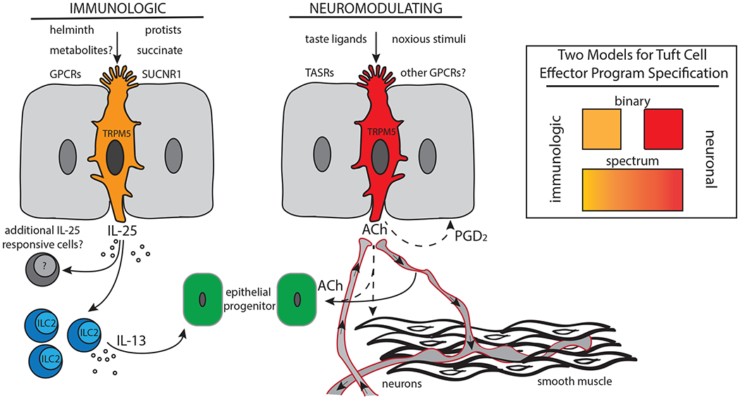
Tuft cells have been found in diverse epithelia where it is thought they play chemosensory and/or immunomodulatory roles enabled by expression of multiple GPCRs to sense luminal stimuli. Tuft cells in the small intestine, for example, are known to respond to luminal helminths and protists like Tritrichomonas. Presence of the latter is sensed by detection of a major metabolic endproduct, succinate, via a tuft cell GPCR, SUCNR1. Subsequently, tuft cells activate tissue resident ILC2s in a TRPM5- and IL-25-dependent fashion to produce effector cytokines like IL-13. Other tuft cell-derived immune mediators, including ALOX5-dependent eicosanoids, may be involved. IL-13 acts on epithelial progenitor cells in the intestinal crypt, promoting dynamic increases in tuft and goblet cell output. In other tissues such as the olfactory epithelium, tuft cells appear to respond to bitter taste ligands, although the physiological ligands are unknown. Here, tuft cells may enact a neuronal effector program, perhaps through secretion of ACh which could act on efferent neurons, smooth muscle, or epithelial progenitor cells. Additional tuft cell effectors, including HPGDS-dependent prostanoids like PGD2, may contribute. Inset: We propose that tuft cells exhibit immunologic and/or neuronal effector states. These effector phenotypes could be mutually exclusive, discreet cell populations, or could represent tuning of fully differentiated cells along a spectrum of function dependent on mutable luminal or niche signals.