


Figure 1. MC38 tumor growth is accelerated by HFD feeding in a CD8+ T cell-dependent manner.
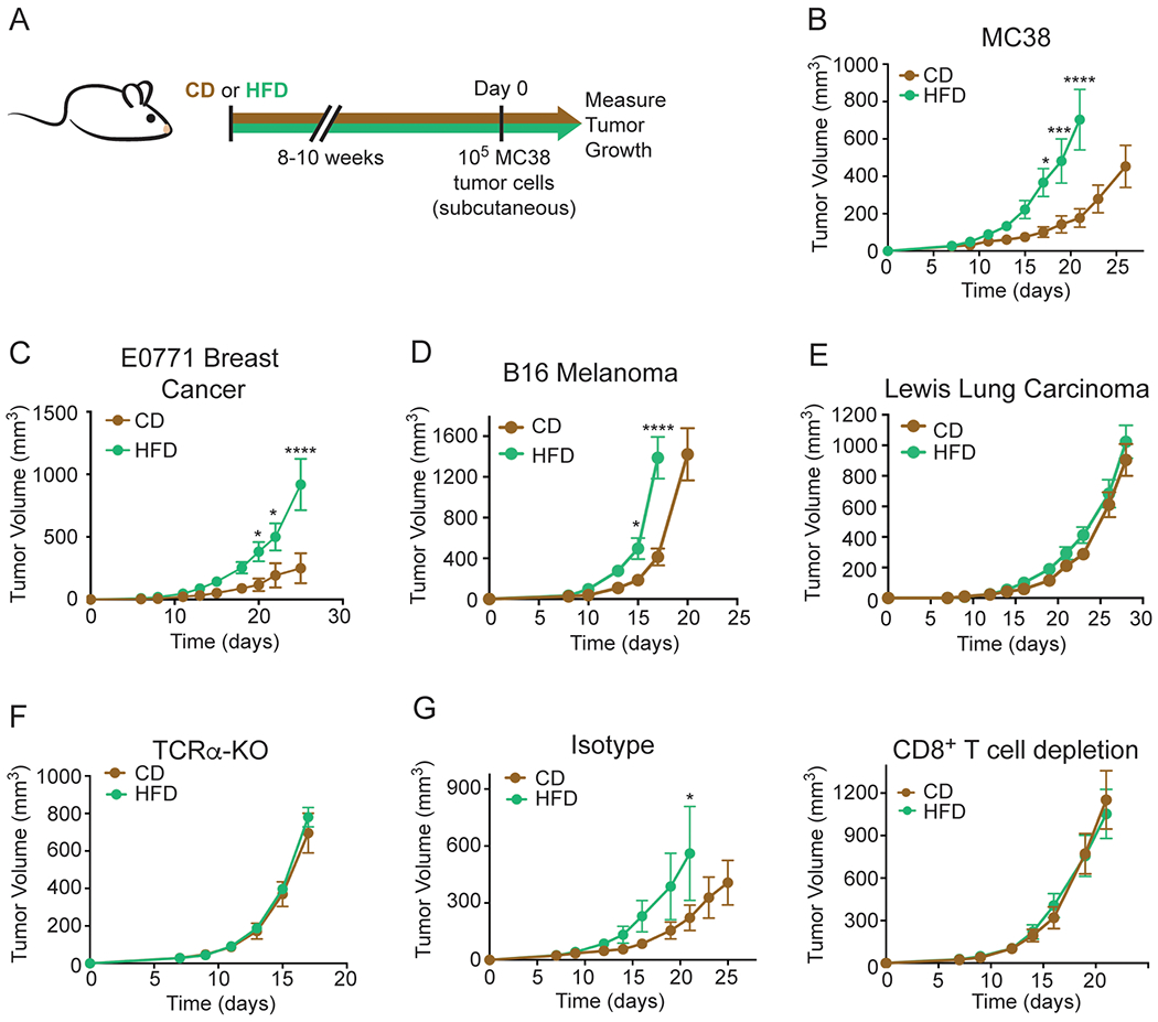
(A) Schematic depicting experimental setup.
(B-E) Tumor growth curves of WT C57BL/6J mice inoculated with 105 MC38 (B), 2x10⁵ E0771 (C), 105 B16 melanoma (D), or 105 Lewis Lung Carcinoma (E) tumor cells.
(F) Tumor growth curves of TCRα-KO mice fed CD or HFD inoculated with 105 MC38 tumor cells.
(G) Tumor growth curves of WT C57BL/6J mice inoculated with 105 MC38 tumor cells and treated with isotype control (left) or depleting anti-CD8 (right) antibodies after CD or HFD feeding for 8-10 weeks.
Data represent ≥ two independent experiments with ≥ 5 mice per group. (*p≤0.05, **p<≤.01, ***p≤0.001, ****p≤0.0001). Graphs display mean +/− SEM (B-G).
See also Figure S1.