



ABSTRACT
Deep sternal wound infection (DSWI) represents a dangerous complication that can follow open‐heart surgery with median sternotomy access. Muscle flaps, such as monolateral pectoralis major muscle flap (MPMF), represent the main choices for sternal wound coverage and infection control. Negative pressure incision management system has proven to be able to reduce the incidence of these wounds' complications. Prevena™ represents one of these incision management systems and we aimed to evaluate its benefits. A total of 78 patients with major risk factors that presented post‐sternotomy DSWI following cardiac surgery was selected. Thrity patients were treated with MPMF and Prevena™ (study group). Control group consisted of 48 patients treated with MPMF and conventional wound dressings. During the follow‐up period, 4 (13%) adverse events occurred in the study group, whereas 18 complications occurred (37·5%) in the control group. Surgical revision necessity and mean postoperative time spent in the intensive care unit were both higher in the control group. Our results evidenced Prevena™ system's ability in improving the outcome of DSWI surgical treatment with MPMF in a high‐risk patient population.
Keywords: Deep sternal wound infection, Pectoralis major muscle flap, Prevena™
Introduction
Open‐heart surgery with median sternotomy access can be followed by severe complications as heart failure, stroke and sternal bone infection 1, 2. Sternal wound infection (SWI) is a complication with a great impact on costs of care and hospitalisation 3, 4.
Obesity [body mass index, (BMI) ≥ 30], diabetes, smoking, age above 66 years and female gender are all considered high‐risk factors for developing postoperative SWI 5, 6, 7.
When the infection is localised in the skin, subcutaneous tissues until the pectoralis fascia, it is called superficial sternal wound infection (SSWI) and its incidence ranges from 0·8% to 8%. When there are clinical evidences of mediastinitis (chest pain, fever, sternal instability and purulent drainage), or an organism is isolated from blood culture or from drainage of mediastinal area 8, 9, 10, there is a deep sternal wound infection (DSWI), which has a lower incidence of up to 5% 11, 12, 13, 14, 15.
‘Conventional’ therapy of DSWI provides debridement and secondary closure or closed catheter irrigation. This leads to mortality rates that range from 5% to 80% 14, 16.
In the last years, however, management of DSWI has developed considerably and mortality rates reduced to a range of 4·8–10·5% with radical debridement and closure of the defects with vascularised flaps 17, 18, 19.
Pectoralis major, rectus abdominis and latissimus dorsi muscle flaps represent the main choices for sternal wound coverage and infection control 12, 17, 18, 19, 20.
Another technique that has proven to be able to reduce the incidence of these wounds' complications is negative pressure incision management system 21. One of these systems is Prevena™ (KCI, San Antonio, TX). Its positive outcomes have been reported in the literature in decreasing wound infections' rate in obese post‐sternotomy patients 22.
In this study, we aimed to evaluate the outcome of Prevena™ Incision Management System after sternotomy and monolateral pectoralis major muscle flap (MPMF) coverage in patients with DSWI major risk factors (obesity, diabetes, smoking, age above 66 years, female gender). The study group results were compared with those achieved in a control group that underwent conventional wound dressings.
Materials and methods
Patients
In this retrospective, single‐centre study, all patients had major risk factors (BMI ≥ 30, diabetics, smokers, age ≥ 66 years, female gender) and presented post‐sternotomy DSWI following cardiac surgery (Figure 1). They were subsequently treated with MPMF in combination with Prevena™ Incision Management System and were evaluated between 2012 and 2016 (study group).
Figure 1.
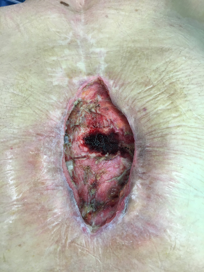
Post‐sternotomy deep sternal wound infection.
A cohort of patients evaluated between 2008 and 2011, also treated with MPMF post‐sternotomy DWSI but without application of Prevena™, served as the control group.
Demographical data and surgical data were assessed routinely from all patients.
Surgical technique
Before surgery, all patients were evaluated to be free from bone and soft tissue infections (biopsies and clinical evaluation). All patients were treated with MPMF. After excision of the wound margins and a deep debridement with resection of all necrotic parts of the sternum and the ribs, the MPMF was raised as described as standard procedure. The muscle monolateral flap was placed upon the sternal defect and fixated without tension (Figure 2). Closure of the wound was performed in a multilayer technique using 2–0 and 3–0 absorbable single stitches sutures (Vicryl®, Ethicon, Norderstedt, Germany) subcutaneously, and 2–0 and 3–0 non‐absorbable (Ethilon®, Ethicon, Norderstedt, Germany) single vertical mattress sutures for skin closure.
Figure 2.
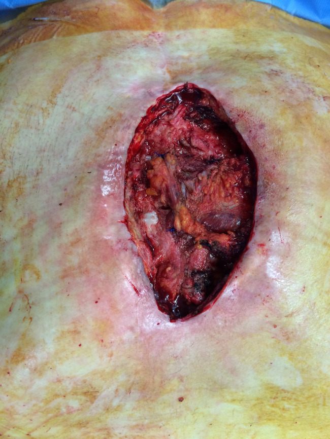
Monolateral pectoralis major muscle flap placed upon the sternal defect and fixated without tension.
In the control group, patients received conventional wound dressing, such as sterile gauzes and elastic bandages.
In the study group, the incisional dressing system was applied immediately after wound closure to reduce wound tension. Standardised 24‐cm‐long Prevena™ foam was placed upon the closed incision, and negative pressure of −125 mmHg was applied (Figure 3).
Figure 3.
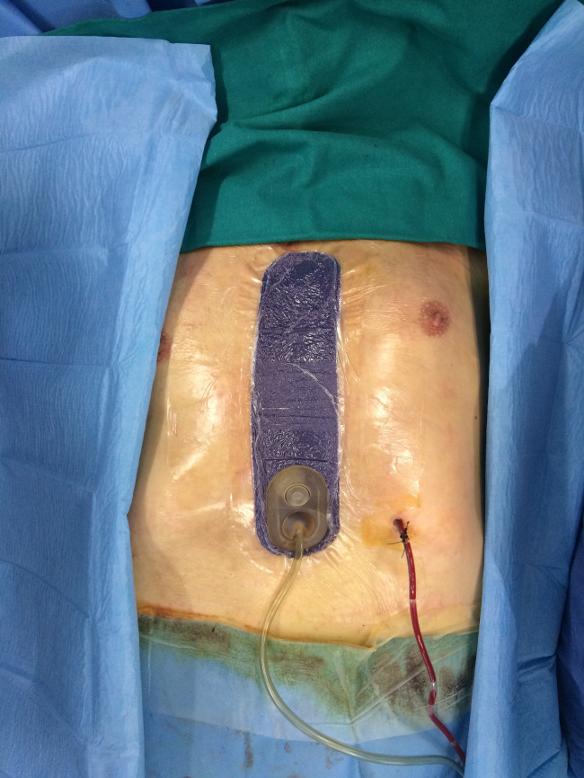
Standardised 24‐cm‐long Prevena™ foam is placed upon the closed incision immediately after surgery.
It was left in place until the built‐in battery had ended its life span (up to 9 days). Wounds were inspected immediately after removal of Prevena™ (Figure 4). Dressings were changed every second day in patients without Prevena™ (control group) and after removal of Prevena™ (Figure 5) (study group), respectively.
Figure 4.
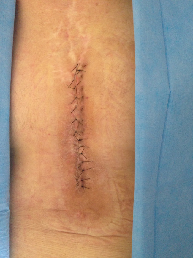
Surgical wound's appearance immediately after removal of Prevena™.
Figure 5.
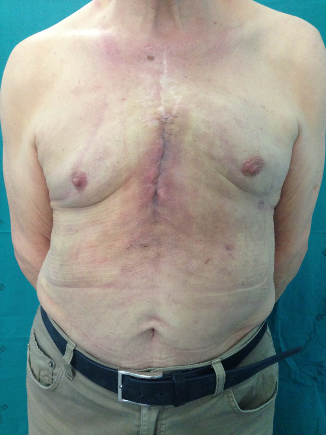
Surgical wound's final appearance after stitches' removal.
Statistical analysis
Statistical analysis was performed using GraphPad Prism5 (GraphPad Software Inc., La Jolla, CA). Fisher's exact two‐tailed test was used to compare postoperative complications (seroma, haematoma, dehiscence, surgical revision). A P‐value of 0·05 was considered statistically significant.
Results
Patient characteristics
Between 2012 and 2016, 30 patients with major risk factors were treated with MPMF and Prevena™ (study group) for DSWI. Control group consisted of 48 patients treated with MPMF and conventional wound dressings between 2008 and 2011.
Before being selected for MPMF surgery, all patients underwent cardiac surgery. In the study group, 21 patients (70%) underwent coronary artery bypass grafting (CABG), 5 (16·6%) underwent a valve replacement procedure and 4(13·3%) underwent both CABG and valve surgery. In the control group, 36 patients (75%) underwent CABG, 6 (16·5%) underwent valve replacement procedures and 6 (16·5%) underwent both surgical procedures. The mean age of the patients in the study group was 68·1 years; patients in the control group had a mean age of 64·2 years. Patient characteristics, heart surgery‐related variables and comorbidities were compared between the two groups. No statistical significant differences could be seen between the two groups.
In the study group, 4 (13%) adverse events occurred, whereas in the control group 18 complications occurred (37·5%). In 7 (15%) of 48 control patients, surgical revision was necessary, while in the study group one patient needed surgical revision (3%; P = 0·1433). Detailed data are listed in Table 1.
Table 1.
Comparison of complications' incidence in study group and control group
| Complications | Study group (n = 30) | Control group (n = 48) | P |
|---|---|---|---|
| Seroma | 1 (3%) | 0 (0%) | 0·3846 |
| Haematoma | 2 (7%) | 4 (8%) | 1 |
| Deishences | 0 (0%) | 7 (15%) | 0·0394 |
| Revision needed | 1 (3%) | 7 (15%) | 0·1433 |
One patient in the study group died during hospitalisation, whereas in the control group no patient died before discharge. The mean postoperative time spent in the intensive care unit (ICU) after pectoralis major flap surgery was 3·7 ± 1·3 days in the study group, compared with 4·2 ± 2 days in the control group. Days until discharge were 15·5 ± 3·4 and 15·8 ± 3, respectively.
Discussion
The Prevena™ Incision Management System consists of a foam placed in wound's bed, connected to suction system that leads to a canister that collects aspirated fluids. This system creates a negative pressure of −125 mmHg in the wound and its dressing contains ionic silver (0·019%) that prevents bacterial growth 22.
Utility of negative wound pressure therapy has been described in complicated wounds' management 21, 22, 23, such as in DSWI 24, 25.
We strongly believe that the use of flaps together with a good debridement of the wound can be helpful in the management of DSWI.
Among the muscle flaps that can be used in these cases, MPMF is described as an effective and rapid technique. It does not depend on intact thoracic arteries that can be damaged during cardiac surgery and it leads to low postoperative morbidity and mortality 26, 27, 28, 29. In cases of DSWIs, after radical debridement, monolateral or bilateral pectoralis major muscle flap surgery represents the standard procedure at our department.
Nevertheless, in the past years, among patients with major risk factors, this technique lead to some complications even in cases where wound's closure was performed in tension‐free manner. In 23% of these cases occurred minor complications, while 15% presented major complications that required surgical revision.
In 2012, we introduced Prevena™ in our department and aimed to exploit its ability to reduce wounds tension in order to decrease complication rates in patients at risk after flap surgery for DSWI. Besides, its election was sustained by contemporary literature's description of how Prevena™ system can reduce from 16% to 4% the incidence of infections (usually sustained by gram‐positive skin flora) in obese patients 30.
The alternative method is to use conventional dressing and elastic bandages positioned in a way to reduce tension of the wound, but it has to be considered that this method can easily fail because of poor patient's compliance.
Our results evidenced Prevena™ advantages: patients who underwent this treatment had a shorter postoperative stay at the ICU (mean 1·7 versus 7·0 days) and a shorter overall stay in hospital (mean 17·3 versus 29·0 days). Moreover, revision surgery was necessary only in 3% of the study group patients, while 15% of the patients in control group needed surgical revision for postoperative wound‐related complications.
The most remarkable result was the difference between incidence of sternum dehiscence: it was observed in 15% of the control group patients and it did not occur in the study group (0%) 31, 32.
In conclusion, we observed the benefits that Prevena™ system carries in managing complicated wounds in a high‐risk patient population. The study presented some limitations: a small cohort of patients that lead to non‐significant statistical results and its retrospective character. Larger numbers of patients should be involved and long‐term results should be collected in further prospective studies.
Despite these limitations, a far lower rate of revision surgery after flap surgery, a shorter stay in the ICU and an overall shorter hospitalisation length were observed and these outcomes lead to lower economic burden to the hospital.
We could demonstrate that Prevena™ can be a useful additional tool in the treatment of high‐risked patients (obese patients BMI ≥ 30, diabetics, smokers, age ≥ 66 years and female gender) with DSWI and subsequent pectoralis major muscle surgery. Further study with a larger cohort of patients will be necessary to confirm our results.
Acknowledgements
None of the authors declares any financial interest regarding this work. Prevena™ was supplied by the ‘Sapienza’ University of Rome, Policlinico Umberto Primo.
References
- 1. Mokhtari A, Sjogren J, Nilsson J, Gustafsson R, Malmsjo M, INgemansson R. The cost of vacuum assisted closure therapy in treatment of deep sternal wound infections. Scand Cardiovasc J 2008;42:85–9. [DOI] [PubMed] [Google Scholar]
- 2. Carlesimo B, Ruggiero M, Lo Torto F, Marcasciano M. Imaging and Surgical Principles of Osteomyelitis and Pressure Ulcers. In: Saba L, Rozen WM, Alonso‐Burgos A, Ribuffo D, editors. Imaging for plastic surgery. Boca Raton, Florida, USA: CRC Press, 2014:793–818. [Google Scholar]
- 3. Lucet JC, Batisse D, Brucker G. Mediastinitis after sternotomy. Mortality and hospital length of stay. Groupe parisien detude des sternotomies. Arch Mal Coeur Vaiss 1997;90:471–5. [PubMed] [Google Scholar]
- 4. Loop FD, Lytle BW, Cosgrove DM, Mahfood S, McHenry MC, Goormastic M, Stewart RW, Golding LA, Taylor PC. J. Maxwell Chamberlain memorial paper. Sternal wound complications after isolated coronary artery bypass grafting: early and late mortality, morbidity, and cost of care. Ann Thorac Surg 1990;49:179–86; discussion 186–7.. [DOI] [PubMed] [Google Scholar]
- 5. Ridderstolpe L, Gill H, Granfeldt H, Ahlfeldt H, Rutberg H. Superficial and deep sternal wound complications: incidence, risk factors and mortality. Eur J Cardiothorac Surg 2001;20:1168–75. [DOI] [PubMed] [Google Scholar]
- 6. Lu JC, Grayson AD, Jha P, Srinivasan AK, Fabri BM. Risk factors for sternal wound infection and mid‐term survival following coronary artery bypass surgery. Eur J Cardiothorac Surg 2003;23:943–9. [DOI] [PubMed] [Google Scholar]
- 7. Buja A, Zampieron A, Cavalet S, Chiffi D, Sandona P, Vinelli A, Baldovin T, Baldo V. An update review on risk factors and scales for prediction of deep sternal wound infections. Int Wound J 2012;9:372–86. [DOI] [PMC free article] [PubMed] [Google Scholar]
- 8. Gabrielli E, Fothergill AW, Brescini L, Sutton DA, Marchionni E, Orsetti E, Staffolani S, Castelli P, Gesuita R, Barchiesi F. Osteomyelitis caused by Aspergillus species: a review of 310 reported cases. Clin Microbiol Infect 2014;6:559–65. [DOI] [PubMed] [Google Scholar]
- 9. Culliford AT, Cunningham JN, Zeff RH, Isom OW, Teiko P, Spencer FC. Sternal and costochondral infections following open heart surgery. A review of 2,594 cases. J Thorac Cardiovasc Surg 1976;72:714–26. [PubMed] [Google Scholar]
- 10. Garner JS, Jarvis WR, Emori TG, Horan TC, Hughes JM. CDC definitions for nosocomial infections, 1988. Am J Infect Control 1988;16:128–40. [DOI] [PubMed] [Google Scholar]
- 11. Beier JP, Arkudas A, Lang W, Weyand M, Horch RE. Sternal osteomyelitis: surgical treatment concepts. Chirurg 2016;87:537–50. [DOI] [PubMed] [Google Scholar]
- 12. Grossi EA, Culliford AT, Krieger KH, Kloth D, Press R, Baumann FG, Spencer FC. A survey of 77 major infectious complications of median sternotomy: a review of 7,949 consecutive operative procedures. Ann Thorac Surg 1985;40:214–23. [DOI] [PubMed] [Google Scholar]
- 13. Losanoff JE, Richman BW, Jones JW. Disruption and infection of median sternotomy: a comprehensive review. Eur J Cardiothorac Surg 2002;21:831–9. [DOI] [PubMed] [Google Scholar]
- 14. Sarr MG, Gott VL, Townsend TR. Mediastinal infection after cardiac surgery. Ann Thorac Surg 1984;38:415–23. [DOI] [PubMed] [Google Scholar]
- 15. Chan M, Yusuf E, Giulieri S, Perrottet N, Von Segesser L, Borens O, Trampuz A. A retrospective study of deep sternal wound infections: clinical and microbiological charcteristics treatment and risk factors for complications. Diagn Microbiol Infect Dis 2016;84:261–5. [DOI] [PubMed] [Google Scholar]
- 16. Bryant LR, Spencer FC, Trinkle JK. Treatment of median sternotomy infection by mediastinal irrigation with an antibiotic solution. Ann Surg 1969;169:914–20. [DOI] [PMC free article] [PubMed] [Google Scholar]
- 17. Carlesimo B, Lo Torto F, Rossi A, Marcasciano M, Ruggiero M. Long‐term result of bilateral pectoralis major muscle advancement flap in median sternotomy wound infections. Eur Rev Med Pharmacol Sci 2014;18:3767–72. [PubMed] [Google Scholar]
- 18. Jurkiewicz MJ, Bostwick J 3rd, Hester TR, Bishop JB, Craver J. Infected median sternotomy wound. Successful treatment by muscle flaps. Ann Surg 1980;191:738–44. [DOI] [PMC free article] [PubMed] [Google Scholar]
- 19. Pairolero PC, Arnold PG, Harris JB. Long‐term results of pectoralis major muscle transposition for infected sternotomy wounds. Ann Surg 1991;213:583–9; discussion 589–90. [DOI] [PMC free article] [PubMed] [Google Scholar]
- 20. Perkins DJ, Hunt JA, Pennington DG, Stern HS. Secondary sternal repair following median sternotomy using interosseous absorbable sutures and pectoralis major myocutaneous advancement flaps. Br J Plast Surg 1996;49:214–9. [DOI] [PubMed] [Google Scholar]
- 21. Lo Torto F, Ruggiero M, Parisi P, Borab Z, Sergi M, Carlesimo B. The effectiveness of negative pressure therapy on infected wounds: preliminary results. Int Wound J 2017; doi: 10.1111/iwj.12725. [DOI] [PMC free article] [PubMed] [Google Scholar]
- 22. Reddy VS. Use of closed incision management with negative pressure therapy for complex cardiac patients. Cureus 2016;8:e506. [DOI] [PMC free article] [PubMed] [Google Scholar]
- 23. Stannard JP, Atkins BZ, O'Malley D, Singh H, Bernstein B, Fahey M, Masden D, Attinger CE. Use of negative pressure therapy on closed surgical incisions: a case series. Ostomy Wound Manage 2009;55:58–66. [PubMed] [Google Scholar]
- 24. Colli A, Camara ML. First experience with a new negative pressure incision management system on surgical incisions after cardiac surgery in high risk patients. J Cardiothorac Surg 2011;6:160. [DOI] [PMC free article] [PubMed] [Google Scholar]
- 25. Grauhan O, Navasardyan A, Tutkun B, Hennig F, Muller P, Hummel M, Hetzer R. Effect of surgical incision management on wound infections in a poststernotomy patient population. Int Wound J 2014;11(1 Suppl):6–9. [DOI] [PMC free article] [PubMed] [Google Scholar]
- 26. Arnold PG, Pairolero PC. Use of pectoralis major muscle flaps to repair defects of anterior chest wall. Plast Reconstr Surg 1979;63:205–13. [DOI] [PubMed] [Google Scholar]
- 27. Ascherman JA, Patel SM, Malhotra SM, Smith CR. Management of sternal wounds with bilateral pectoralis major myocutaneous advancement flaps in 114 consecutively treated patients: refinements in technique and outcomes analysis. Plast Reconstr Surg 2004;114:676–83. [DOI] [PubMed] [Google Scholar]
- 28. Lopez‐Monjardin H, de‐la‐Pena‐Salcedo A, Mendoza‐Munoz M, Lopez‐Yanez‐de‐la‐Pena A, Palacio‐Lopez E, Lopez‐Garcia A. Omentum flap versus pectoralis major flap in the treatment of mediastinitis. Plast Reconstr Surg 1998;101:1481–5. [DOI] [PubMed] [Google Scholar]
- 29. Nahai F, Morales L Jr, Bone DK, Bostwick J 3rd. Pectoralis major muscle turnover flaps for closure of the infected sternotomy wound with preservation of form and function. Plast Reconstr Surg 1982;70:471–4. [DOI] [PubMed] [Google Scholar]
- 30. Grauhan O, Navasardyan A, Hofmann M, Muller P, Stein J, Hetzer R. Prevention of poststernotomy wound infections in obese patients by negative pressure wound therapy. J Thorac Cardiovasc Surg 2013;145:1387–92. [DOI] [PubMed] [Google Scholar]
- 31. Mino J, Remzi FH. Use of the Prevena incision management system as a potential solution for high risk, complicated perineal wounds. Tech Coloproctol 2016;20:601. [DOI] [PubMed] [Google Scholar]
- 32. Horch RE. Incisional negative pressure wound therapy for high‐risk wounds. J Wound Care 2015;24(4 Suppl):21–8. [DOI] [PubMed] [Google Scholar]