


Figure 3. Sequencing of [89Zr]-mCD47 injection relative to FUS BBB/BTB-D markedly impacts antibody accumulation in murine GL261 gliomas.
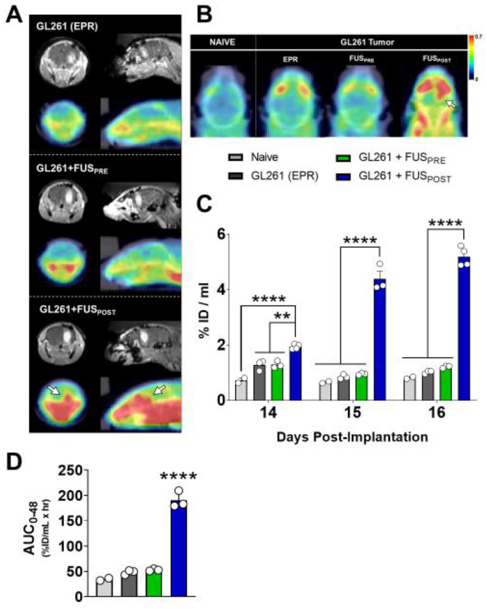
(A) Representative coronal (left) and sagittal (right) contrast-enhanced T1-weighted MR images (top row) and decay-corrected PET/CT images (bottom row) for each experimental group on Day 14. MR images depict a clear, enhancing tumor in the right hemisphere of GL261-bearing mice. (B) Representative axial decay-corrected PET/CT images for each experimental group on Day 14. White arrows denote region of visibly elevated radioactivity at the tumor site targeted with FUS. Scale unit: g/ml (SUVbw). (C) Whole brain standardized [89Zr]-mCD47 uptake values (SUVs) extracted from serial static PET/CT images obtained between days 14 and 16 post-implantation (0 to 2 days post-[89Zr]-mCD47 injection). %ID/mL = % injected dose per mL. **p<0.01, ****p<0.0001 vs. group(s) indicated. Significance assessed by RM mixed effects model implementing restricted maximum likelihood method, followed by Tukey multiple comparison correction. (D) Tumor-drug exposure for [89Zr]-mCD47 in naïve brain or GL261 tumors, based on integration of SUVs from decay-corrected PET/CT images collected between 0 and 48 hours after BBB/BTB-D and/or [89Zr]-mCD47 injection. Significance assessed by one-way ANOVA followed by Tukey multiple comparison correction. ****p<0.0001 vs. all other groups.