


Figure 1. MR-image guided FUS-mediated blood brain/tumor barrier disruption in murine GL261 tumors.
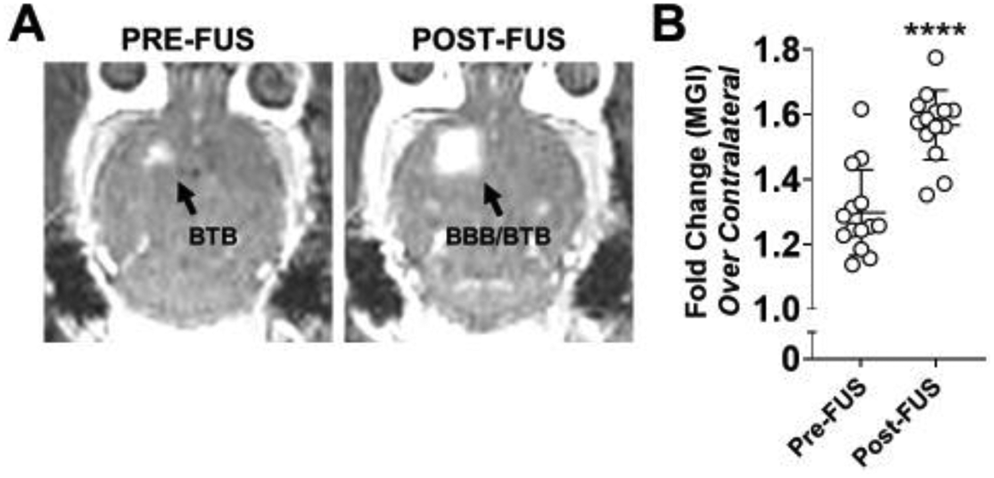
(A) Representative contrast-enhanced T1-weighted MR images of a GL261 tumor-bearing brain pre- and post-FUS. Enhancement in the right cerebral hemisphere on pre-FUS imaging indicates baseline barrier disruption induced by the presence of a brain tumor. Expansion of the enhanced region on post-FUS imaging reflects effective FUS-mediated BBB/BTB disruption. (B) Mean greyscale intensity (MGI) of contrast enhancement pre- and post-FUS BBB/BTB-D. Calculated as fold change over contralateral brain. Mean ± SD. ****p<0.0001. Significance assessed by unpaired t-test.