



Abstract
Much research seeks to articulate the brain structures and pathways implicated in addiction and addiction recovery. Prominent neurobiological models emphasize the interplay between cortical and limbic brain regions as a main driver of addictive processes, but largely do not take into consideration sensory and visceral information streams that link context and state to the brain and behavior. Yet these brain–body information streams would seem to be necessary elements of a comprehensive model of addiction. As a starting point, we describe the overlap between one current model of addiction circuitry and the neural network that not only regulates cardiovascular system activity but also receives feedback from peripheral cardiovascular processes through the baroreflex loop. We highlight the need for neurobiological, molecular, and behavioral studies of neural and peripheral cardiovascular signal integration during the experience of internal states and environmental contexts that drive alcohol and other drug use behaviors. We end with a call for systematic, mechanistic research on the promising, yet largely unexamined benefits to addiction treatment of neuroscience-informed, adjunctive interventions that target the malleability of the cardiovascular system to alter brain processes.
Keywords: autonomic nervous system, central autonomic network, substance use disorder
1 |. INTRODUCTION
Contemporary neurobiological models of addiction coalesce around interactions among specific limbic and cortical brain regions that are thought to control the imbalance of automatic/impulsive relative to deliberate/self-controlled behaviors associated with substance use disorder (SUD). Evidence suggests, however, that information streams from the body link context and bodily states to the brain’s behavioral outputs via both subcortical and cortical paths. The present article argues that the absence of body systems in current neurobiological addiction theories limits their translational and clinical value by missing a fundamental yet malleable source of individual variability. This individual variability can be tied to the seemingly automatic reactions to salient contexts and cues that interfere with more deliberate behavioral responses and can be therapeutically manipulated. We focus on the cardiovascular system as a source of consequential visceral information that is inherently embedded in the experience of addiction.
2 |. THE BODY RELAYS CONTEXT TO THE BRAIN
Alcohol and drug use behaviors are initiated in-the-moment based on internal dynamics, such as cortical and subcortical integration of sensory information with affective states that are often prompted by salient environmental cues.1 Further, body systems can promote addictive behaviors by, for example, their capacity to serve as “silent” drivers of attention allocation and embedded components of associative learning, motivation, and salience.2–4 Theoretical models such as Damasio’s somatic marker hypothesis5 and Robinson and Berridge’s incentive salience hypothesis6 have led to important contributions in this area, but the specific ways in which body systems actively contribute to flexible or inflexible behavior towards alcohol and other drugs continues to receive only limited attention.7–10
The neurophysiology, functional anatomy, and molecular mechanisms of the bidirectional information streams that connect neural and cardiovascular processes have been well characterized.11 From this literature, the central autonomic network (CAN) was described as a coordinated network of forebrain, midbrain, and brain stem structures that centrally meditate autonomic nervous system functioning.12,13 This set of structures bears remarkable overlap with those articulated in various contemporary neurobiological models of addiction, and the structures conceptually implicated in core features of addictive behavior.14–16 Yet, translational addiction models have not gone much beyond a “black box” conceptualization of bodily inputs.
The body continually conveys signals that are components of arousal states and emotional tone to the brain through humoral (e.g., central chemoreceptors in the hypothalamus monitor the hematological milieu), vagal (i.e., cholinergic vagal afferents relay information about physiological states to sensory afferent cell bodies in the nodose ganglia that propagate to the CAN), and spinal pathways (e.g., the lamina 1 spinothalamocortical pathway with its somatotopic organization, conveying visceral information that converges with afferent information from cranial nerves before projecting to the CAN).17
Rather than being static inputs, however, there is marked complexity in how the brain receives and interprets this information. For instance, afferent signals from the heart may be manipulated or optimized at the level of the brain to alter the way affective stimuli are processed during differing psychological and physiological states.18–20 Additionally, meta-analytic evidence points to distinct parasympathetic and sympathetic subnetworks to task negative (i.e., the default mode network) and task positive (i.e., the frontoparietal control network, salience network) networks, respectively,12 which further embeds cardiovascular activation pathways within the functional neural networks that orient an individual towards self-referential thought or externally oriented attention. Moreover, it underscores the importance of the cardiovascular system processes that often operate outside of awareness and contribute to the automaticity of substance use behaviors in addictive disorders.
We therefore posit that CAN-regulated cardiovascular dynamics are not independent parallel processes that regulate physiological processes such as heart rate and blood pressure, but are in fact integrated and necessary components of the brain’s response to any given challenge. To this point, mounting evidence suggests that central control of visceral reactivity is predictive rather than strictly reflexive, such that neural prediction errors contribute to exaggerated and blunted cardiovascular stress reactions that undermine adaptive whole-body responses to challenge.18 This is a conceptual departure from predominant theories such as Thayer’s heuristic neurovisceral integration model21 that have focused on cardiovascular processes as downstream indicators of neural integration, rather than active processes that may enhance neural regulation.
We hypothesize that the neural alterations elicited by addiction also negatively affect central control of cardiovascular processes and arousal. In turn, dysregulation in these processes can then contribute to or exacerbate deficits in brain circuit regulatory mechanisms underlying addiction. A corollary hypothesis discussed in a later section of this article shows that improving central control of cardiovascular processes with interventions such as heart rate variability (HRV) biofeedback provides physiological scaffolding to support behaviorally focused addiction interventions. As a starting point, to illustrate functional overlaps between putative addiction processes and the primary functions of CAN structures (Figure 1), we use Koob and Volkow’s widely discussed neurobiological model of addiction.22 We note, however, that the highlighted functional and structural overlaps are applicable to other neuroscientific models of addiction.15,23,24
FIGURE 1.
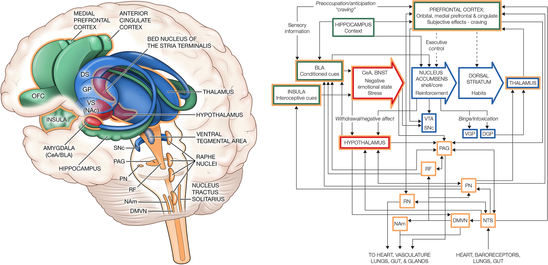
Integration of neural circuitry associated with Koob and Volkow’s addiction model and the central autonomic network (CAN). Koob and Volkow22 mapped specific neural circuits onto a three-stage addiction model that includes a binge/intoxication stage (blue), withdrawal/negative affect stage (red), and preoccupation/anticipation/craving stage (green). These structures are heavily interconnected with the constellation of connected brain structures known as the CAN (gold/gold outline). LEFT: A three-dimensional rendering of the brain showing the integration of the CAN with structures implicated in the binge/intoxication stage (blue), withdrawal/negative affect stage (red), and preoccupation/anticipation/craving stage (green) of addiction. Overlap between these structures and the CAN are demarcated in blue, red, and green with a gold outline. RIGHT: A schematic of the brain to body (descending) and body to brain (ascending) pathways showing connectivity among the structures of the three-stage addiction cycle model, and the structures of the CAN (gold / gold outline). Note: for simplicity, not all projections noted below are shown. During the binge/intoxication stage (blue), the CAN likely participates through ascending pathways that integrate autonomic/visceral information via the thalamus. During the withdrawal/negative affect stage (red), the primary intersection of CAN and addiction circuitry appears to occur via projections to the extended amygdala. The CAN also shares significant overlap with the structures implicated in the preoccupation/anticipation/craving stage (green), including the medial prefrontal, orbitofrontal, insula, and anterior cingulate cortices, as well as the basolateral amygdala. Independent of the proposed neural circuitry of addiction,22 evidence from decades of research on the CAN suggests that brain stem nuclei are key integration centers of autonomic/visceral information, and that these structures are highly interconnected to higher order brain structures. At the bottom right of the schematic is the nucleus tractus solitarius (NTS), the first central relay (ascending) for all medullary reflexes controlling cardiovascular functions (cardiac and baroreflex reflexes) and respiration (carotid chemoreflex and pulmonary mechanoreflexes). It receives and responds to stimuli from the heart, baroreceptors, lungs and gut, and directly communicates to higher brain areas like the hypothalamus and cortex. It further relays information to subcortical and cortical centers via the parabrachial nuclei (PN), periaqueductal gray (PAG) region, and raphe nuclei (RN). The parabrachial nuclei are key regulators of cardiovascular, respiratory, and gastrointestinal functioning with ascending projections to the hypothalamus, amygdala, and insular and infralimbic cortex, as well as descending projections to the nucleus ambiguus (NAm). The parabrachial nuclei also receive converging visceral, nociceptive, and thermoreceptive inputs from the spinal cord and convey this information to the hypothalamus, amygdala, and thalamus. The periaqueductal gray likewise plays a critical role in autonomic regulation, motivated behavior, and behavioral responses to stress, as well as pain modulation via its extensive inputs from higher brain areas including the cortex, amygdala, and hypothalamus, with outputs to brainstem areas like the reticular formation (RF) and raphe nuclei. The raphe nuclei have a diverse range of roles including pain inhibition via the dorsal horn, temperature regulation, and modulation of circadian rhythms and alertness. Finally, the dorsal motor vagal nucleus (DMVN) provides primary descending parasympathetic control of the heart and is the motor operator for cardiac, baroreflex, and pulmonary reflexes; it receives indirect inputs from the cortex as well as direct inputs from the nucleus tractus solitarius. Abbreviations: ACC, anterior cingulate cortex; BLA, basolateral amygdala; BNST, bed nucleus of the stria terminalis; CeA, central amygdala; DS, dorsal striatum; GP, globus pallidus; NAc, nucleus accumbens; OFC, orbitofrontal cortex; RN, raphe nuclei; SNc, substantia nigra pars compacta; VGP, ventral globus pallidus; DGP, dorsal globus pallidus; VS, ventral striatum; VTA, ventral tegmental area
3 |. PERIPHERAL AND CENTRAL CARDIOVASCULAR CONTROL IS ALTERED BY INTOXICATION
In Koob and Volkow’s neurobiological model of addiction,22 the binge/intoxication stage of the addiction cycle is characterized by substance seeking and use, loss of executive control mediated by glutamatergic systems,25 and the manifestation and reinforcement of incentive salience via phasic activation of dopaminergic systems.26 A number of subcortical neural structures, many in the basal ganglia, are implicated as primary drivers of the acutely reinforcing properties of alcohol and other drugs. These include the ventral tegmental area, substantia nigra pars compacta, nucleus accumbens, central nucleus of the amygdala, globus pallidus, and thalamus. As exposure to intoxication–withdrawal cycles builds, the dorsal striatum is recruited to automate stimulus–response habits that drive further substance use related behaviors.
It is noteworthy how these areas, many of which are shared by the CAN, are highly interconnected with brainstem areas that regulate the cardiovascular and respiratory systems. Alterations of cardiovascular processes during the binge/intoxication stage have been extensively documented. HRV (the variability in consecutive R-spike to R-spike intervals in the electrocardiogram waveform), which is commonly used as a biomarker of cardiovascular adaptive capacity, decreases during acute intoxication, as does blood pressure variability.27 Additionally, the sensitivity of the neurally mediated feedback loop that modulates blood pressure known as the baroreflex has also been shown to decrease in response to alcohol intoxication.28 In parallel, high risk drinkers show heightened response of an HRV index that captures baroreflex activation (i.e., the 0.1-Hz HRV index) to alcohol and other drug cues,29 and vascular disruptions have been observed in binge drinkers within the first nine years of drinking.30
This raises the possibility that the allostatic load of repeated intoxication–withdrawal cycles leads to a loss of flexibility in the cardiovascular system in the same way flexibility is thought to be lost in other body systems such as the hypothalamic–pituitary adrenal axis under repeated intoxication–withdrawal.31 Thus, less accurate information about physiological context would be relayed to the brain, further undermining basal ganglionic regulatory process, and hypothetically contributing in discoverable ways to the aberrant cognitive, affective, and behavioral features of addiction.
4 |. CARDIOVASCULAR FEATURES ARE EMBEDDED IN NEGATIVE AFFECTIVE STATES
The aversive experience of substance withdrawal, characterized by substance-specific physiological symptoms and negative affect, is also proposed as essential to the addictive cycle. In the withdrawal/negative affect stage of Koob and Volkow’s model,22 the extended amygdala, through its projections to the hypothalamus and brainstem, and in the case of opioid withdrawal the locus coeruleus,32 drives physiological responses to neurological states such as those arising from withdrawal. Evidence for including peripheral physiological feedback in this stage of addiction is strong because extended amygdala components including the central nucleus of the amygdala, bed nucleus of the stria terminalis, and to a lesser extent, the basolateral amygdala are important components of the CAN,13 and by definition, the neural structures of the CAN serve by merging feedback from the heart and vasculature with higher order cognitive and affective factors derived in midbrain and forebrain structures, as well as with information from other autonomic systems. In doing so, visceral constituents of withdrawal and negative affect become embedded in the subjective affective experience of these states.17 Tachycardia/heart palpitations and diaphoresis, for example, are inextricably linked to the negative affective states that arise during acute alcohol or opioid withdrawal. This aligns with current theory that conceptualizes the structures of the dopaminergic extended amygdala as receivers of information that are responsible for evaluating the emotional content of stimuli or states and allocating attention.9,22
5 |. CARDIOVASCULAR INFORMATION IS INTEGRATED CORTICALLY
Koob and Volkow also propose a substance preoccupation/anticipation stage of addiction characterized by incentive sensitization that is driven by the basolateral amygdala and insular cortex and processing of contextual information represented in the orbitofrontal, prefrontal, and anterior cingulate cortices, as well as the amygdala.22 The insula, prefrontal, and anterior cingulate cortices are reciprocally innervated with midbrain structures, brain stem nuclei, and the periphery in the service of integrating effortful and automatic information processing (Figure 1). To understand how visceral and sensory information become embedded in an addictive process, it is useful to consider the functional roles of these cortical structures, all of which integrate cognitive and emotional information with a variety of bodily sensations. A clear example is the insular cortex, which as the seat of interoception, is considered a primary viscerosensory area that mediates consciousness of feelings through its role in the representation of bodily states and urges.33,34 Large literatures further implicate this structure in emotional awareness, appetitive drive, and urges to use alcohol and other drugs.34,35
Diminished capacity of the executive system structures to regulate attention, salience, and craving is believed to be an essential component of addiction.36,37 The discussion of disruptions in attention and salience, and the experience of craving, raises consideration of the baroreflex feedback loop that delivers cardiovascular information to and from the brain and ensures a continuous bidirectional stream of modulation.38 This recursive feedback loop involves well-documented pathways from the brain to the heart, via the efferent parasympathetic and sympathetic nervous systems, and from the cardiovascular system to the brain (afferent traffic) through baroreceptors—mechanoreceptors in arterial walls that respond to vessel stretching in response to moment-to-moment changes in blood pressure (Figure 2).
FIGURE 2.
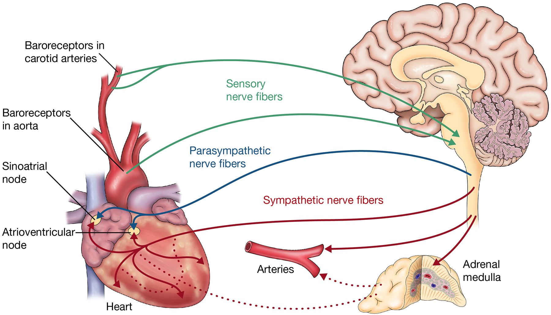
Bidirectional neuro-cardiovascular pathways. Dynamic control of heart rate is maintained via efferent pathways from the brain via cranial (parasympathetic) and sympathetic nerve fibers of the autonomic nervous system. These efferent nerve fibers converge on the heart, mainly at the sinoatrial and atrioventricular nodes, to modulate moment-to-moment heart rate. Dynamic changes in blood pressure are likewise maintained via efferent sympathetic pathways that directly and indirectly control arterial tone (i.e., constricting or dilating blood vessels) through innervation of the arteries, and the secretion of epinephrine and norepinephrine via the adrenal medulla gland. These end organs then convey feedback to the brain via stretch receptors, called baroreceptors, in the aorta and carotid arteries. Activation of baroreceptors is relayed to the nucleus tractus solitarius and higher order central autonomic network (CAN) structures, thereby ensuring real-time information about blood pressure and heart rate is propagated to neural centers that control cardiovascular processes, as well as behavioral responding
Baroreceptors convey information to nucleus tractus solitarius neurons that then propagate information to higher neural centers that control peripheral resistance (i.e., vasculature tone) and heart rate, as well as behavioral responding. The entirety of the baroreflex feedback loop allows us to consider how system variability is generated and used to modulate homeostatic set points and adjust to chronic challenges either adaptively or maladaptively (i.e., allostatic load).39,40 Experimental evidence suggests baroreflex-mediated afferent signaling about cardiovascular variability participates in substance use behavior maintenance through its influence on attention capture by provocative cues, cue salience, and visceral reaction to cues.2 Thus, visceral arousal responses elicited by cues in the environment—such the sight of a regularly attended drinking establishment or substance use location—can influence attention capture and thereby serve to perpetuate or exacerbate preoccupation, salience, anticipation, and craving. Other pathways of heart–brain interaction, such as assessed by heartbeat-evoked potentials41 and direct modulation of neural blood flow and oxygenation,42 further implicate cardiac cycle timing effects and predictive coding in attention and somatosensory perception.
6 |. CLINICAL IMPLICATIONS OF INCLUDING THE CARDIOVASCULAR SYSTEM IN MODELS OF ADDICTION
Cardiovascular disease progression associated with substance use is well studied,43,44 but there is a need for laboratory and experimental study of the mechanistic role of cardiovascular changes in decision making,45 the emotion regulation failures associated with addiction,46 and their potential for rebound during recovery. We previously observed that women receiving a 12-week course of cognitive behavioral therapy for alcohol use disorder experienced improvements in cardiovascular regulation from pretreatment to posttreatment, even without stable abstinence.47 These improvements were observed across measures of metabolic activity, HRV, and baroreflex sensitivity. This study supported other research showing that improved baroreflex sensitivity may signal improved blood pressure regulation and cardio protection,48 as well as increased regulation of emotion, stress, and behavior,49 and aligns with a growing literature showing improvements in cardiovascular processes following mindfulness-based interventions and cognitive behavioral therapy for a range of clinical presentations. It is likely that other treatments like dialectical behavior therapy give rise to similar improvements in cardiovascular functioning. Future clinical trials of psychotherapeutic interventions should include cardiovascular measures to explore such effects. Additionally, measures of cardiovascular regulation could serve as much needed objective measures of treatment response in clinical trials.50
Observed improvements in cardiovascular regulation in individuals receiving cognitive behavioral therapy further support the possibility that improved cardiovascular–brain feedback may serve as a scaffold to reinforce the goals of psychotherapy and behavior change. It is also possible that restoration of autonomic homeostasis may help individuals better buffer stress and mitigate craving. If, in fact, the cardiovascular system is capable of recovery even after years of chronic, heavy substance use,47 treatment adjuncts that directly target cardiovascular regulatory mechanisms such as the baroreflex may accelerate cardiovascular recovery in the service of sustaining substance use behavior change,51 as well as improving health and quality of life.
The idea that improvements in cardiovascular processes may help increase regulation of emotion, stress, and behavior is consistent with the observation that sympatholytic medications affecting the cardiovascular system are known to reduce stress, craving and substance use. Prazosin and doxazosin, two alpha-1 adrenergic inverse agonists commonly referred to as alpha-blockers and widely used to treat hypertension, reduce stress reactivity, craving, and the use of alcohol and other drugs,52–56 potentially by dampening high basal levels of sympathetic arousal commonly observed in individuals with SUD.55,56 Whereas the effects of alpha-blockers on blood pressure are primarily driven by countering norepinephrine-mediated vasoconstriction via alpha-1 adrenergic receptors in the smooth muscle of the vasculature, effects on craving and substance use likely derive from direct actions on alpha-1 adrenergic receptors found throughout the central nervous system.
GABAergic drugs offer another class of medications with the potential to mitigate CAN/autonomic dysregulation in SUD.57 Acamprosate (Campral), for instance, is a centrally acting GABA agonist and glutamate antagonist58 that is hypothesized to attenuate hyper-glutamatergic states associated with craving57 and relatedly effect reductions in psychophysiological arousal, as evidenced by reduced heart rate, galvanic skin conductance,59,60 and blood cortisol.61 Acamprosate may also reduce alcohol and other drug use, although findings to date have been mixed.62
Similar effects can also be achieved with HRV biofeedback, a behavioral intervention that originated from the observation that maximal increases in the amplitude of heart rate oscillation are produced when the cardiovascular system is rhythmically stimulated by paced breathing at a frequency of approximately 0.1 Hz (equivalent to six breaths per minute).63,64 This effect is linked to resonance properties of the cardiovascular system and the physiological phenomenon of respiratory sinus arrhythmia, that is, increases and decreases in heart rate in response to respiratory inhalation and exhalation. HRV biofeedback is empirically supported for treating a variety of disorders and performance enhancement, putatively through effects on baroreceptors and the vagal afferent pathway to frontal brain regions.65 A recent meta-analysis found that HRV biofeedback was associated with large effect size reductions in stress and anxiety that were not moderated by clinical diagnosis.66 A small but growing body of research also suggests that HRV biofeedback may be a useful complement to current first-line treatments for SUD. Preliminary studies suggested that both short67,68 and longer term69,70 courses of HRV biofeedback may help reduce craving levels beyond reductions observed during inpatient addiction treatment as usual, and relapse prevention housing. A recent systematic review suggested that HRV biofeedback and paced breathing proxies were useful for promoting decreases in craving, although larger and better controlled studies are needed.71
Theoretically, HRV biofeedback enhances communication between neural and cardiovascular systems that modulate physiological arousal in response to salient cues and internal states that trigger substance use, and thus may increase behavioral flexibility towards appetitive stimuli, and improve individuals’ capacity to regulate negative affect. Given the strong psychophysiological component of craving72 and HRV biofeedback’s ability to acutely effect major shifts in autonomic activation,65 it is possible that HRV biofeedback may mitigate craving by helping individuals restore autonomic homeostasis or executive control, or reduce autonomic allostatic load during craving states. It is also possible that HRV biofeedback may chronically enhance CAN regulatory capacity to buffer against craving or restore cardiovascular flexibility in the more generic service of supporting new learning. These speculations are empirically testable in human experimental studies designed to capture HRV biofeedback’s effects on craving, emotion, cognition, and behavior at multiple, integrated system levels using neuroimaging, electroencephalogram, electrocardiogram, cognitive tasks, neuropsychological tests, and subjective report. The growing availability and scalability (via mobile devices) of HRV biofeedback provides the means for expanded and ecologically valid study of enhanced baroreflex sensitivity and diminished in-the-moment cardiovascular reactivity to triggers as potential mechanisms that may interrupt substance use behaviors in the natural environment.
A recent proof-of-concept functional magnetic resonance imaging study supports this idea. Bates and colleagues20 exposed alcohol drinkers to two blocks of alcohol picture cues with an experimental group completing 5 min of 0.1-Hz breathing between alcohol cue blocks (equivalent to six breaths per minute; a typical breathing rate in HRV biofeedback at which resonance between respiration and HRV occurs) and controls completing a low-demand cognitive task. During exposure to the second round of alcohol cues, the experimental group exhibited significantly less activation in brain regions involved in visual processing (i.e., left inferior and superior lateral occipital cortices, right inferior lateral occipital cortex, bilateral occipital pole, and temporal occipital fusiform cortices), while activation in brain areas implicated in behavioral control, internally directed cognition, and brain–body integration was increased (i.e., medial prefrontal, anterior and posterior cingulate, and precuneus cortices, paracingulate, and lingual gyri). The control group showed no significant changes in brain activation between alcohol cue exposures. These findings provide preliminary evidence that manipulation of the cardiovascular system with 0.1-Hz breathing alters neural activation in a manner theoretically consistent with a dampening of automatic sensory input and strengthening of higher level cognitive processing.
7 |. CONCLUSIONS
There remains vigorous debate about the neurological processes underlying the acute reinforcement of alcohol and other drugs, the learning and stimulus–response habits that drive further substance use, the aversive experience of substance withdrawal, and perseveration of motivational states (e.g., craving).45 The cardiovascular system, as well as other sensory and visceral systems, importantly contribute to these processes and the experience of addiction by providing contextual information to the brain, often outside of conscious awareness. This information can derive from and support an overlearned behavioral repertoire, even in opposition to conscious efforts to inhibit such responses. Physiological reactivity serves a critical role in survival and adaptation, but can also become maladaptive when reflexive, predictive, or over-learned responses (e.g., changes in heart rate and blood pressure) help to perpetuate SUD.
We suggest that neurobiological models that include sensory and visceral information streams are more complete because their inclusion allows models to more roundly explain the role of context and affective states in addiction and addiction recovery. Decades of psychophysiological research provides tractable experimental manipulations to test the plasticity of the baroreflex loop, and mobile assessment of physiological signals used to study cardiovascular mechanisms in the natural environment are now widely available. As such, as a field, we are well positioned to capture simultaneous visceral and neural response through well-defined and ecologically valid decisional and emotion regulation tasks that will inform more holistic models of addiction and recovery—work that is ongoing in the cognitive and affective neuroscience communities at large.45
Interest is growing in the way the cardiovascular and other physiological systems (e.g., immune and gastrointestinal) are involved in psychopathology, but research in these domains remains “fringe” in both biological and psychological sciences of addiction. In this article, we used Koob and Volkow’s neurobiological model of addiction as an example to point out several natural intersections between the brain, body, and behavior. We emphasized that without sensory and visceral input and reactions, the brain would be disconnected from the bodily context in which it exists. These ideas integrate well with other neuroscientific models and theories of addiction,5,25,73 that provide an additional level for exploring core biological mechanisms underlying addictive disorders through Research Domain Criteria (RDoC).74 These ideas also align with calls for neuro-clinical assessment of individuals with addiction to characterize patients’ individual prognosis and probability of response to behavioral and pharmacological treatments.25
In conclusion, it is difficult to imagine a comprehensive model of addiction that does not consider sensory and visceral information streams and attendant bodily processes as fundamental components of behavioral responding. Expanding existing neurobiological models of addiction to more comprehensively integrate the neurological structures subsumed by the CAN and the sensory and visceral information streams it both controls and is influenced by, as well as connected bodily systems (e.g., cardiovascular, endocrine), will grow the translational and clinical utility of current models. Further, this will support the development of noninvasive, body-based interventions that specifically target the automatic physiological reactivity that so often undermines SUD recovery attempts and serve as a complement to existing cognitive-behavioral and pharmacotherapeutic approaches.
ACKNOWLEDGEMENTS
Preparation of this work was supported by National Institute on Alcohol Abuse and Alcoholism (R01-AA023667, R01-AA027017, K23-AA027577, K02-AA025123, F32-AA025251, and L30-AA026135), and the Pershing Square Venture Fund for Research on the Foundations of Human Behavior at Harvard University. We thank Kristin Zavodjancik for producing the article’s figures.
Funding information
National Institute on Alcohol Abuse and Alcoholism, Grant/Award Numbers: F32-AA025251, K02-AA025123, K23-AA027577, L30-AA026135, R01-AA023667, R01-AA027017; Pershing Square Venture Fund for Research on the Foundations of Human Behavior at Harvard University
Footnotes
CONFLICT OF INTEREST
The authors declare that there is no conflict of interest that could be perceived as prejudicing the impartiality of the research reported.
REFERENCES
- 1.Shiffman S, Stone AA, Hufford MR. Ecological momentary assessment. Annu Rev Clin Psychol. 2008;4(1):1–32. [DOI] [PubMed] [Google Scholar]
- 2.Eddie D, Buckman JF, Mun EY, et al. Different associations of alcohol cue reactivity with negative alcohol expectancies in mandated and inpatient samples of young adults. Addict Behav. 2013;38(4):2040–2043. [DOI] [PMC free article] [PubMed] [Google Scholar]
- 3.Hyman SE. Addiction: a disease of learning and memory. Am J Psychiatry. 2005;162(8):1414–1422. [DOI] [PubMed] [Google Scholar]
- 4.Kalivas PW, Volkow ND. The neural basis of addiction: a pathology of motivation and choice. Am J Psychiatry. 2005;162(8):1403–1413. [DOI] [PubMed] [Google Scholar]
- 5.Damasio AR. The somatic marker hypothesis and the possible functions of the prefrontal cortex. Philos Trans R Soc Lond B Biol Sci. 1996; 351(1346):1413–1420. [DOI] [PubMed] [Google Scholar]
- 6.Robinson TE, Berridge KC. The neural basis of drug craving: an incentive-sensitization theory of addiction. Brain Res Brain Res Rev. 1993;18(3):247–291. [DOI] [PubMed] [Google Scholar]
- 7.Bates ME, Buckman JF. Integrating body and brain systems in addiction neuroscience. In: Miller P, ed. Biological research on addiction: Comprehensive addictive behaviors and disorders. Vol.2 New York, NY: Academic Press; 2013:187–196. [Google Scholar]
- 8.Buckman JF, Vaschillo EG, Fonoberova M, Mezic I, Bates ME. The translational value of psychophysiology methods and mechanisms: multilevel, dynamic, Personalized. J Stud Alcohol Drugs. 2018;79(2): 229–238. [DOI] [PMC free article] [PubMed] [Google Scholar]
- 9.Koob GF, Schulkin J. Addiction and stress: an allostatic view. Neurosci Biobehav Rev. 2018;106:245–262. [DOI] [PubMed] [Google Scholar]
- 10.Milivojevic V, Sinha R. Central and peripheral biomarkers of stress response for addiction risk and relapse vulnerability. Trends Mol Med. 2018;24(2):173–186. [DOI] [PMC free article] [PubMed] [Google Scholar]
- 11.Goldstein DS. The autonomic nervous system in health and disease. New York, NY: Marcel Dekker, Inc.; 2001. [Google Scholar]
- 12.Beissner F, Meissner K, Bar KJ, Napadow V. The autonomic brain: an activation likelihood estimation meta-analysis for central processing of autonomic function. J Neurosci. 2013;33(25):10503–10511. [DOI] [PMC free article] [PubMed] [Google Scholar]
- 13.Benarroch EE. The central autonomic network. In: Low PA, ed. Clinical Autonomic Disorders. 2nd ed. Philadelphia, PA: Lippincott-Raven; 1997:17–23. [Google Scholar]
- 14.Koob GF, Volkow ND. Neurobiology of addiction: a neurocircuitry analysis. Lancet Psychiatry. 2016;3(8):760–773. [DOI] [PMC free article] [PubMed] [Google Scholar]
- 15.Robinson TE, Berridge KC. The psychology and neurobiology of addiction: an incentive-sensitization view. Addiction. 2000;95(Suppl2):S91–S117. [DOI] [PubMed] [Google Scholar]
- 16.Koob GF, Le Moal M. Neurobiological mechanisms for opponent motivational processes in addiction. Philosophical Transactions of the Royal Society B: Biological Sciences. 2008;363(1507):3113–3123. [DOI] [PMC free article] [PubMed] [Google Scholar]
- 17.Critchley HD. Neural mechanisms of autonomic, affective, and cognitive integration. J Comp Neurol. 2005;493(1):154–166. [DOI] [PubMed] [Google Scholar]
- 18.Gianaros PJ, Jennings JR. Host in the machine: a neurobiological perspective on psychological stress and cardiovascular disease. Am Psychol. 2018;73(8):1031–1044. [DOI] [PMC free article] [PubMed] [Google Scholar]
- 19.Critchley HD, Garfinkel SN. Interactions between visceral afferent signaling and stimulus processing. Front Neurosci. 2015;9:286. [DOI] [PMC free article] [PubMed] [Google Scholar]
- 20.Bates ME, Lesnewich LM, Uhouse SG, Gohel S, Buckman JF. Resonance-paced breathing alters neural response to visual cues: proof-of-concept for a neuroscience-informed adjunct to addiction treatments. Front Psych. 2019;10:624. [DOI] [PMC free article] [PubMed] [Google Scholar]
- 21.Thayer JF, Lane RD. A model of neurovisceral integration in emotion regulation and dysregulation. J Affect Disord. 2000;61(3):201–216. [DOI] [PubMed] [Google Scholar]
- 22.Koob GF, Volkow ND. Neurocircuitry of addiction. Neuropsychopharmacology. 2010;35(1):217–238. [DOI] [PMC free article] [PubMed] [Google Scholar]
- 23.Naqvi NH, Bechara A. The hidden island of addiction: the insula. Trends Neurosci. 2009;32(1):56–67. [DOI] [PMC free article] [PubMed] [Google Scholar]
- 24.Lewis M. Addiction and the brain: development, not disease. Neuro-ethics. 2017;10(1):7–18. [DOI] [PMC free article] [PubMed] [Google Scholar]
- 25.Kwako LE, Momenan R, Litten RZ, Koob GF, Goldman D. Addictions neuroclinical assessment: a neuroscience-based framework for addictive disorders. Biol Psychiatry. 2016;80(3):179–189. [DOI] [PMC free article] [PubMed] [Google Scholar]
- 26.Berridge KC. From prediction error to incentive salience: mesolimbic computation of reward motivation. Eur J Neurosci. 2012;35(7):1124–1143. [DOI] [PMC free article] [PubMed] [Google Scholar]
- 27.Buckman JF, Eddie D, Vaschillo EG, Vaschillo B, Garcia A, Bates ME. Immediate and complex cardiovascular adaptation to an acute alcohol dose. Alcohol Clin Exp Res. 2015;39(12):2334–2344. [DOI] [PMC free article] [PubMed] [Google Scholar]
- 28.Abdel-Rahman A, Merrill RH, Wooles WR. Effect of acute ethanol administration on the baroreceptor reflex control of heart rate in normotensive human volunteers. Clin Sci. 1987;72(Pt 1):113–122. [DOI] [PubMed] [Google Scholar]
- 29.Mun EY, von Eye A, Bates ME, Vaschillo EG. Finding groups using model-based cluster analysis: heterogeneous emotional self-regulatory processes and heavy alcohol use risk. Dev Psychol. 2008; 44(2):481–495. [DOI] [PMC free article] [PubMed] [Google Scholar]
- 30.Vaschillo EG, Vaschillo B, Buckman JF, Heiss S, Singh G, Bates ME. Early signs of cardiovascular dysregulation in young adult binge drinkers. Psychophysiology. 2018;55(5):e13036. [DOI] [PMC free article] [PubMed] [Google Scholar]
- 31.Blaine SK, Sinha R. Alcohol, stress, and glucocorticoids: from risk to dependence and relapse in alcohol use disorders. Neuropharmacology. 2017;122:136–147. [DOI] [PMC free article] [PubMed] [Google Scholar]
- 32.Van Bockstaele EJ, Reyes BA, Valentino RJ. The locus coeruleus: a key nucleus where stress and opioids intersect to mediate vulnerability to opiate abuse. Brain Res. 2010;1314:162–174. [DOI] [PMC free article] [PubMed] [Google Scholar]
- 33.Gray MA, Critchley HD. Interoceptive basis to craving. Neuron. 2007; 54(2):183–186. [DOI] [PMC free article] [PubMed] [Google Scholar]
- 34.Paulus MP, Stewart JL. Interoception and drug addiction. Neuropharmacology. 2014;76:342–350. [DOI] [PMC free article] [PubMed] [Google Scholar]
- 35.Naqvi NH, Bechara A. The insula and drug addiction: an interoceptive view of pleasure, urges, and decision-making. Brain Structure and Function. 2010;214(5–6):435–450. [DOI] [PMC free article] [PubMed] [Google Scholar]
- 36.Seo D, Lacadie CM, Tuit K, Hong K-I, Constable RT, Sinha R. Disrupted ventromedial prefrontal function, alcohol craving, and subsequent relapse risk. JAMA Psychiat. 2013;70(7):727–739. [DOI] [PMC free article] [PubMed] [Google Scholar]
- 37.Sinha R. New findings on biological factors predicting addiction relapse vulnerability. Curr Psychiatry Rep. 2011;13(5):398–405. [DOI] [PMC free article] [PubMed] [Google Scholar]
- 38.Vaschillo EG, Vaschillo B, Buckman JF, Pandina RJ, Bates ME. The investigation and clinical significance of resonance in the heart rate and vascular tone baroreflexes. In: Fred A, Filipe J, Gamboa H, eds. Biomedical Engineering Systems and Technologies: Communications in Computer and Information Science 127. Heidelberg, Germany: Springer; 2011:224–237. [Google Scholar]
- 39.Juster RP, McEwen BS, Lupien SJ. Allostatic load biomarkers of chronic stress and impact on health and cognition. Neuroscience & Biobehavioral Reviews. 2010;35(1):2–16. [DOI] [PubMed] [Google Scholar]
- 40.Tracey KJ. Reflex control of immunity. Nat Rev Immunol. 2009;9(6): 418–428. [DOI] [PMC free article] [PubMed] [Google Scholar]
- 41.Al E, Iliopoulos F, Forschack N, et al. Heart-brain interactions shape somatosensory perception and evoked potentials. Proc Natl Acad Sci U S a. 2020;117(19):10575–10584. [DOI] [PMC free article] [PubMed] [Google Scholar]
- 42.Vaschillo EG, Vaschillo B, Buckman JF, Bates ME. New approach for brain stimulation. Brain Stimulation. 2019;12(2):393. [Google Scholar]
- 43.Gardner JD, Mouton AJ. Alcohol effects on cardiac function. Compr Physiol. 2015;5(2):791–802. [DOI] [PubMed] [Google Scholar]
- 44.Eddie D, Greene MC, White WL, Kelly JF. Medical burden of disease among individuals in recovery from alcohol and drug problems in the United States: findings from the National Recovery Survey. J Addict Med. 2019;13(5):385–395. [DOI] [PMC free article] [PubMed] [Google Scholar]
- 45.Ekhtiari H, Victor TA, Paulus MP. Aberrant decision-making and drug addiction—how strong is the evidence? Curr Opin Behav Sci. 2017;13: 25–33. [Google Scholar]
- 46.Quintana DS, Guastella AJ, McGregor IS, Hickie IB, Kemp AH. Heart rate variability predicts alcohol craving in alcohol dependent outpatients: further evidence for HRV as a psychophysiological marker of self-regulation. Drug Alcohol Depend. 2013;132(1–2):395–398. [DOI] [PubMed] [Google Scholar]
- 47.Buckman JF, Vaschillo B, Vaschillo EG, et al. Improvement in women’s cardiovascular functioning during cognitive behavioral therapy for alcohol use disorder. Psychol Addict Behav. 2019;33(8):659–668. [DOI] [PMC free article] [PubMed] [Google Scholar]
- 48.Swenne CA. Baroreflex sensitivity: mechanisms and measurement. Neth Heart J. 2013;21(2):58–60. [DOI] [PMC free article] [PubMed] [Google Scholar]
- 49.Duschek S, Werner NS, Reyes Del Paso GA. The behavioral impact of baroreflex function: a review. Psychophysiology. 2013;50(12):1183–1193. [DOI] [PubMed] [Google Scholar]
- 50.Price CJ, Crowell SE. Respiratory sinus arrhythmia as a potential measure in substance use treatment-outcome studies. Addiction. 2016; 111(4):615–625. [DOI] [PMC free article] [PubMed] [Google Scholar]
- 51.Eddie D, Vaschillo E, Vaschillo B, Lehrer P. Heart rate variability biofeedback: theoretical basis, delivery, and its potential for the treatment of substance use disorders. Addict Res Theory. 2015;23(4): 266–272. [DOI] [PMC free article] [PubMed] [Google Scholar]
- 52.Fox HC, Anderson GM, Tuit K, et al. Prazosin effects on stress-and cue-induced craving and stress response in alcohol-dependent individuals: preliminary findings. Alcohol Clin Exp Res. 2012;36(2): 351–360. [DOI] [PMC free article] [PubMed] [Google Scholar]
- 53.Shorter D, Lindsay JA, Kosten TR. The alpha-1 adrenergic antagonist doxazosin for treatment of cocaine dependence: a pilot study. Drug Alcohol Depend. 2013;131(1–2):66–70. [DOI] [PMC free article] [PubMed] [Google Scholar]
- 54.Kenna GA, Haass-Koffler CL, Zywiak WH, et al. Role of the alpha1 blocker doxazosin in alcoholism: a proof-of-concept randomized controlled trial. Addict Biol. 2016;21(4):904–914. [DOI] [PMC free article] [PubMed] [Google Scholar]
- 55.Verplaetse TL, Weinberger AH, Oberleitner LM, et al. Effect of doxazosin on stress reactivity and the ability to resist smoking. J Psychopharmacol. 2017;31(7):830–840. [DOI] [PMC free article] [PubMed] [Google Scholar]
- 56.Newton TF, De La Garza IIR, Brown G, Kosten TR, Mahoney JJ III, Haile CN. Noradrenergic α1 receptor antagonist treatment attenuates positive subjective effects of cocaine in humans: a randomized trial. PloS One. 2012;7(2):e30854. [DOI] [PMC free article] [PubMed] [Google Scholar]
- 57.Mann K, Kiefer F, Spanagel R, Littleton J. Acamprosate: recent findings and future research directions. Alcohol Clin Exp Res. 2008;32(7): 1105–1110. [DOI] [PubMed] [Google Scholar]
- 58.Rammes G, Mahal B, Putzke J, et al. The anti-craving compound acamprosate acts as a weak NMDA-receptor antagonist, but modulates NMDA-receptor subunit expression similar to memantine and MK-801. Neuropharmacology. 2001;40(6):749–760. [DOI] [PubMed] [Google Scholar]
- 59.Ooteman W, Koeter MWJ, Verheul R, Schippers GM, van den Brink W. The effect of naltrexone and acamprosate on cue-induced craving, autonomic nervous system and neuroendocrine reactions to alcohol-related cues in alcoholics. Eur Neuropsychopharmacol. 2007; 17(8):558–566. [DOI] [PubMed] [Google Scholar]
- 60.Agelink MW, Lemmer W, Malessa R, Zeit T, Majewski T, Klieser E. Improved autonomic neurocardial balance in short-term abstinent alcoholics treated with acamprosate. Alcohol Alcohol. 1998;33(6): 602–605. [DOI] [PubMed] [Google Scholar]
- 61.Hammarberg A, Jayaram-Lindstrom N, Beck O, Franck J, Reid MS. The effects of acamprosate on alcohol-cue reactivity and alcohol priming in dependent patients: a randomized controlled trial. Psycho-pharmacology (Berl). 2009;205(1):53–62. [DOI] [PubMed] [Google Scholar]
- 62.Plosker GL. Acamprosate: a review of its use in alcohol dependence. Drugs. 2015;75(11):1255–1268. [DOI] [PubMed] [Google Scholar]
- 63.Vaschillo EG, Lehrer P, Rishe N, Konstantinov M. Heart rate variability biofeedback as a method for assessing baroreflex function: a preliminary study of resonance in the cardiovascular system. Appl Psychophysiol Biofeedback. 2002;27(1):1–27. [DOI] [PubMed] [Google Scholar]
- 64.Song HS, Lehrer PM. The effects of specific respiratory rates on heart rate and heart rate variability. Appl Psychophysiol Biofeedback. 2003; 28(1):13–23. [DOI] [PubMed] [Google Scholar]
- 65.Lehrer PM, Gevirtz R. Heart rate variability biofeedback: how and why does it work? Front Psychol. 2014;5:756. [DOI] [PMC free article] [PubMed] [Google Scholar]
- 66.Goessl VC, Curtiss JE, Hofmann SG. The effect of heart rate variability biofeedback training on stress and anxiety: a meta-analysis. Psychol Med. 2017;47(15):2578–2586. [DOI] [PubMed] [Google Scholar]
- 67.Eddie D, Kim C, Lehrer P, Deneke E, Bates ME. A pilot study of brief heart rate variability biofeedback to reduce craving in young adult men receiving inpatient treatment for substance use disorders. Appl Psychophysiol Biofeedback. 2014;39(3):181–192. [DOI] [PMC free article] [PubMed] [Google Scholar]
- 68.Penzlin AI, Barlinn K, Illigens BM, Weidner K, Siepmann M, Siepmann T. Effect of short-term heart rate variability biofeedback on long-term abstinence in alcohol dependent patients - a one-year follow-up. BMC Psychiatry. 2017;17(1):325. [DOI] [PMC free article] [PubMed] [Google Scholar]
- 69.Eddie D, Conway FN, Alayan N, Buckman JF, Bates ME. Assessing heart rate variability biofeedback as an adjunct to college recovery housing programs. J Subst Abuse Treat. 2018;92:70–76. [DOI] [PMC free article] [PubMed] [Google Scholar]
- 70.Teeravisutkul P, Chumchua V, Saengcharnchai P, Leelahanaj T. Stress and craving reduction under treatment with heart rate variability biofeedback and the Phramongkutklao model among patients with alcohol use disorder. Psychol Res Behav Manag. 2019;12:619–627. [DOI] [PMC free article] [PubMed] [Google Scholar]
- 71.Alayan N, Eller L, Bates ME, Carmody DP. Current evidence on heart rate variability biofeedback as a complementary anticraving intervention. J Altern Complement Med. 2018;24(11):1039–1050. [DOI] [PMC free article] [PubMed] [Google Scholar]
- 72.Verheul R, van den Brink W, Geerlings P. A three-pathway psychobiological model of craving for alcohol. Alcohol Alcohol. 1999;34(2): 197–222. [DOI] [PubMed] [Google Scholar]
- 73.Hinson JM, Jameson TL, Whitney P. Somatic markers, working memory, and decision making. Cogn Affect Behav Neurosci. 2002;2(4): 341–353. [DOI] [PubMed] [Google Scholar]
- 74.Litten RZ, Ryan ML, Falk DE, Reilly M, Fertig JB, Koob GF. Heterogeneity of alcohol use disorder: understanding mechanisms to advance personalized treatment. Alcohol Clinical and Experimental Research. 2015;39(4):579–584. [DOI] [PubMed] [Google Scholar]