



Abstract
The COVID-19 pandemic caused by the global spread of the SARS-CoV-2 virus has led to a staggering number of deaths worldwide and significantly increased burden on healthcare costs as nations scramble to find mitigation strategies. While significant progress has been made in COVID-19 diagnostics and therapeutics, effective prevention and treatment options remain scarce. Because of the potential for the SARS-CoV-2 infections to cause systemic inflammation and multiple organ failure, it is imperative for the scientific community to evaluate therapeutic options aimed at modulating the causative host immune responses to prevent subsequent systemic complications. Harnessing decades of expertise in the use of natural and synthetic materials for biomedical applications, the biomaterials community has the potential to play an especially instrumental role in developing new strategies or repurposing existing tools to prevent or treat complications resulting from the COVID-19 pathology. Leveraging microparticle and nanoparticle-based technology, especially in pulmonary delivery, biomaterials have demonstrated the ability to effectively modulate inflammation and may be well-suited for resolving SARS-CoV-2-induced effects. Here, we provide an overview of the SARS-CoV-2 virus infection and highlight current understanding of the host’s pulmonary immune response and its contributions to disease severity and systemic inflammation. Comparing to frontline COVID-19 therapeutic options, we highlight the most significant untapped opportunities in immune engineering of the host response using biomaterials and particle technology, which have the potential to improve outcomes for COVID-19 patients, and identify areas needed for future investigations. We hope that this work will prompt preclinical and clinical investigations of promising biomaterials-based treatments to introduce new options for COVID-19 patients.
Keywords: COVID-19, SARS-CoV-2, biomaterials, nanoparticles, inflammation, immune engineering
Graphical Abstract

INTRODUCTION
The emergence of the SARS-CoV-2 pathogen in 2019 has prompted the scientific community to work tirelessly in search for therapeutic solutions to slow or stop the COVID-19 pandemic. While significant progress has been made to characterize the effects of SARS-CoV-2 with new information on the pathogen and associated host response continuing to emerge daily, there are still outstanding mechanistic questions that have yet to be addressed regarding the COVID-19 pathology and its long-term implications. SARS-CoV-2 is primarily a respiratory infection; however, severe COVID-19 can present with multiple organ involvement, including cardiovascular,1 neurological,2 and renal3 damage, that is largely driven by dysregulated host immune response.4 Therefore, successful modulation of the immune environment is a critical therapeutic target for improving patient outcomes, especially for critically ill COVID-19 patients. Several therapeutic options modulating host immune-specific targets have been developed or repurposed for mitigating COVID-19 symptoms, and many clinical trials are underway; however, current attempts to resolve hyperinflammatory states mostly rely on system-wide immunomodulatory strategies that may benefit from a more targeted approach. Therefore, particle engineering solutions that target the host immune response, particularly in affected organs, are desperately needed to avoid potential off-target effects of potent immunomodulatory treatments.
The goal of this article is to summarize the current clinical understanding of immune response to SARS-CoV-2 along with key host immunological factors implicated in COVID-19 which will inform immune engineering approaches that hold therapeutic potential for mitigating COVID-19 responses occurring in the lung, among other organs. We point to lessons learned from therapeutic approaches in the fields of biomaterials and nanomedicine that have demonstrated robustness in inducing anti-inflammatory and immunomodulatory effects, particularly in the context of airway inflammation. Biomaterial-inspired drug delivery designs offer many opportunities for tunable immune modulation as tolerogenic therapeutics or prophylactic/therapeutic vaccines5 because of their advantageous properties. Engineered particle formulations can be delivered via inhalation or injection with customizable biodegradability that can take advantage of either extracellular or intracellular cues alongside properties including passive or active targeting to lung immune cells, detailed surface functionalization for precise stimulation of innate immune cells, and high loading capacities of cargo with varied properties.6 Collectively, these considerations lead to highly modular systems capable of meeting the drug delivery and immune-modifying needs of COVID-19-related complications. We provide a potential road map for modulating the pulmonary immune response experienced during COVID-19 and seek to answer one key question: what are untapped immune engineering approaches to control unwanted immune responses in the lung as a result of COVID-19?
INFECTION, INFLAMMATION, AND HOST FACTORS IN COVID-19 PATHOPHYSIOLOGY
Understanding the structure of the SARS-CoV-2 virus and the mechanism of infection as well as lessons from other closely related coronavirus infections is critical for development of prophylactic vaccine candidates and treatment options for mitigating severe symptoms. Innate and adaptive immune responses play an instrumental role in recovery and disease severity. Furthermore, emerging patterns in biological factors including age, sex, and pre-existing health conditions are reported with regard to their impact on SARS-CoV-2 infection and disease severity. In this section, we give an overview of the SARS-CoV-2 pathogenic mechanism of host cell invasion as well as a summary of immune responses and biological factors that result from infection and which, based on current understanding, impact the severity of the COVID-19 pathology.
SARS-CoV-2 etiology and host cell invasion
Coronaviruses are viral pathogens that belong to the Coronaviridae family which are enveloped, single-stranded, positive-sense RNA viruses with a genome length of ~30 kb.7, 8 Structurally, coronaviruses have four main proteins including spike (S) glycoproteins, small envelope (E) glycoproteins, membrane (M) glycoproteins, and nucleocapsid (N) proteins, along with several other proteins.9 The spike glycoprotein is a transmembrane, trimeric glycoprotein that facilitates binding and invasion into the host cell.10 Techniques like electron microscopy have identified SARS-CoV-2 to be a round or oval shaped virus with an approximate diameter of 60–140 nm and a crown-shaped appearance.11 SARS-CoV-2 is closely related to two other coronaviruses, severe acute respiratory syndrome coronavirus (SARS-CoV-1) and Middle East respiratory syndrome coronavirus (MERS-CoV), which have the potential to replicate in the lower respiratory tract and result in fatal pneumonia.12 SARS-CoV-2 shares 79% genome sequence identity with SARS-CoV-1, another human coronavirus, and has approximately 98% genomic similarity to the bat coronavirus, RaTG13.13, 14
SARS-CoV-2 is primarily transmitted via exhaled aerosol particulates containing virus that cause infection following inhalation.15 Upon infection with SARS-CoV-2, the median incubation period is around 4–5 days16, 17 followed by symptom onset within 11.5 days.17 Invasion of SARS-CoV-2 into the host cell initiates with viral binding to angiotensin-converting enzyme 2 (ACE2) receptor (Figure 1A).10, 18 Previous studies with SARS-CoV-1 have identified increased ACE2 receptor expression in pulmonary type II epithelial cells, making them likely targets for invasion.19–21 ACE2 expression decreases from higher amounts in the upper airways to lower expression in the lower respiratory tract, with high SARS-CoV-2 infectivity in the nose compared to the alveolar region.22 The spike (S) protein on the viral surface is comprised of two functional subunits: S1 and S2. S1 subunits facilitate binding with the host cell receptor and comprises an amino-terminal domain and a receptor-binding domain (RBD).23 The RBD binds to the ACE2 receptor followed by endocytosis of viral components into the host cell.24, 25 Following the binding of SARS-CoV-2 to ACE2 and endocytosis, the spike protein undergoes protease cleavage, exposing the S2 site, which results in membrane fusion and release of viral contents into the cell cytoplasm.26 Studies have identified increased affinity of ACE2 receptor with RBD of SARS-CoV-2 relative to SAR-CoV-1, which might explain the greater relative infectivity of SARS-CoV-2.24, 27 The role of the cellular serine protease TMPRSS2 was also identified in promoting host cell entry of the virus in SARS-CoV-2 infections.28 In addition to TMPRSS2, recent literature has implicated other proteases such as TMPRSS4, Mpro, PLpro and RdRp, which might play a role in host cell invasion.29, 30
Figure 1. Immune landscape of the alveolar region in healthy, mild, and severe states during COVID-19.
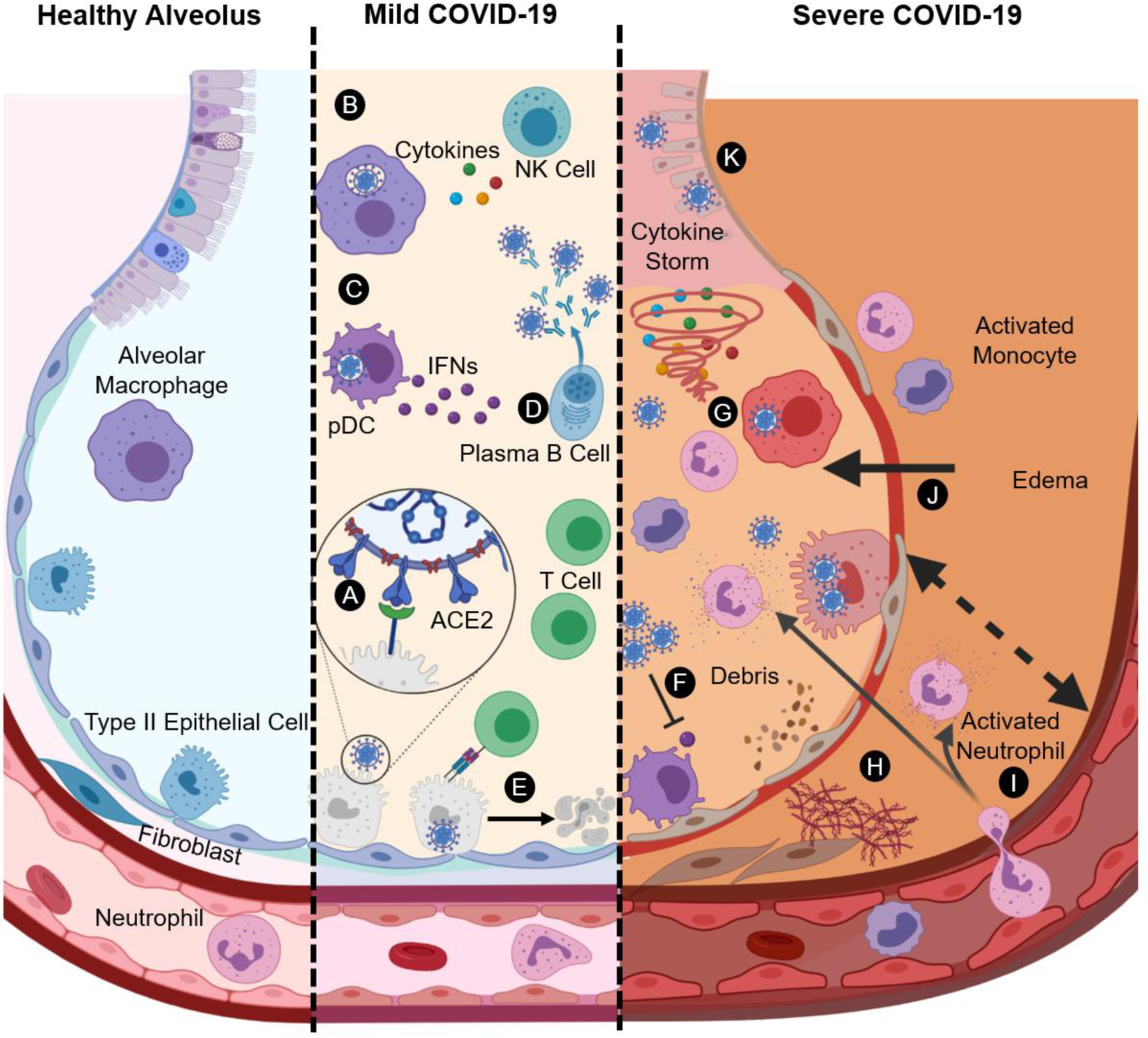
A) Infection of type II alveolar epithelial cells with SARS-CoV-2 through the ACE2 receptor B) Activation of alveolar macrophages following infection/recognition of virus and release of pro-inflammatory cytokines C) Type I IFN response initiated by plasmatocytoid dendritic cells (pDCs) and recruitment of lymphocytes D) Plasma B cells producing SARS-CoV-2-specific antibodies following maturation and priming by innate immune cells E) SARS-CoV-2-specific CD8+ T cells causing apoptosis of infected cells F) Impaired type I IFN response G) Cytokine storm by activated macrophages and recruited inflammatory leukocytes H) Deposition of fibrous strands and extracellular matrix leading to fibrosis and scarring I) Degranulation of activated neutrophils recruited from the bloodstream J) Fluid buildup in the alveolus from edemous tissue and widening of interstisium K) Shedding of ciliated epithelial lining and formation of debris.
Host response to SARS-CoV-2
Both innate and adaptive immune responses are engaged to neutralize the SARS-CoV-2 infection; however, an impaired immune response can result in severe lung pathology and infection manifestations in multiple organs,4 including coagulation abnormalities and thromboembolic events that are common in COVID-19 deaths.31, 32 The severity of initial infection and viral load, combined with the timing and extent of the host immune response, can result in symptoms ranging from asymptomatic or mild to severe pneumonia and acute respiratory distress syndrome (ARDS) with multiple organ failure, as discussed in the following sections. It is critical to increase our understanding of the kinetics, progression, and timing of both healthy and improper host immune involvement prior to developing immune-modifying therapeutics.
Innate Immune Response:
The first stage of host immune involvement predominantly comes from the innate immune system. During healthy lung function, normal alveolar immune landscape mainly consists of alveolar macrophages residing at the air-liquid interface in addition to small numbers of granulocytes, including basophils and eosinophils.33 Alveolar macrophages and dendritic cells (DCs) are key antigen presenting cells (APCs) of the local pulmonary innate immune system, with the mucosal lining, epithelial cells, and the complement cascade providing an essential passive barrier to viral infection.34 As SARS-CoV-2 targets and invades epithelial cells using ACE2 receptors, viral infection spreads, resulting in injury and death of virus-infected cells. Apoptotic cells release pathogen-associated molecular patterns (PAMPs), such as viral RNA, and damage-associated molecular patterns (DAMPs) which are recognized by innate cells using a variety of pattern-recognition receptors (PRRs). Apoptotic epithelial cell components are mainly internalized and cleared by resident pulmonary macrophages and recruited leukocytes to minimize the infection until adaptive immunity is developed.35 PRR signaling within innate immune cells leads to robust pro-inflammatory signaling and cellular recruitment to the lung (Figure 1B). Pro-inflammatory soluble factors such as interleukin (IL)-1β, IL-8, and IL-6 are elevated in both plasma and bronchoalveolar lavage fluid (BALF36) of COVID-19 patients37, 38 and increased levels of tumor necrosis factor (TNF)-α, granulocyte macrophage-colony stimulating factor (GM-CSF), as well as other cytokines and chemokines are elevated in plasma.39 Studies have identified that elevated levels of IL-6 correlate with the severity of the COVID-19 disease,40 with critically ill patients having IL-6 levels ten times higher than those of severe patients in some cases.41 Another study complemented these results by demonstrating that risk of respiratory failure for patients with IL-6 levels of greater than 80 pg/mL was 22 times higher compared to patients with lower IL-6 levels.42 Such increased cytokine presence in the lung microenvironment could potentially result in cytokine-mediated lung damage, respiratory distress, and further organ failure.39 PRR signaling also promotes recruitment of immune cells such as monocytes, neutrophils, and T cells to the site of the infection. Upregulation of chemokines is reported in BALF of COVID-19 patients including CCL2, CCL7, and CXCL8, which are critical for the recruitment of monocytes and neutrophils.37, 43
Importantly, PRR signaling activates production of antiviral type I interferons (IFNs).44 Recent clinical data supports the role of IFN response as a possible indicator of disease severity.43 Type I IFNs are produced by plasmacytoid DCs and are critical to antiviral responses (Figure 1C).44 IFNs activate phagocytosis in macrophages as well as the Janus kinase-signal transducer and activator of transcription (JAK-STAT) pathway, which is essential in polarizing antiviral helper T cells.45 With a robust and early IFN response, SARS-CoV-2 infections remain mild or asymptomatic, while a delayed and/or impaired IFN response leads to increasingly severe disease.46–48 Given the essential antiviral function of IFNs and the JAK-STAT pathway, many viruses have evolved mechanisms to suppress or inhibit functions throughout the IFN and JAK-STAT pathway. Evidence from both SARS-CoV-1 and MERS-CoV viruses imply that SARS-CoV-2 may also have functionality that directly diminishes host IFN response.43
APCs specifically provide the bridge between innate and adaptive immune response. Resident pulmonary DCs are likely involved with priming the adaptive response through exposure to cytokines and SARS-CoV-2 antigens. There are generally two classes of resident pulmonary DCs responsible for antigen presentation: CD103+ and CD11b+, where CD103+ DCs are the main migratory subset. Upon infection and activation, DCs migrate to lymph nodes (LNs) and interface with T and B cells to present processed SARS-CoV-2 antigens through immunological synapses.
Adaptive Immune Response:
Defense by the innate immune system allows for the initiation of a robust adaptive immune response. In mild COVID-19 cases, patients not only have an early and robust IFN response and highly functional innate response, but also a strong, multifaceted adaptive immune response to manage and clear viral infection (Figure 1D). Virus-specific antibodies generated by plasma B cells and CD8+ (cytotoxic) T lymphocytes are educated by activated DCs draining from the lung to nearby LNs. SARS-CoV-2 neutralizing antibodies are found in plasma of patients and have strong affinity to RBD of S protein, inhibiting ACE2 binding.49 Antibodies with specificity to N protein are also detected and IgA, IgG, and IgM classes of antibodies were found,50 similar to anti-RBD antibodies.51 Clinically, a large proportion of the infiltrating adaptive immune cells are T cells, where both CD4+ and CD8+ T cells play a critical role. CD4+ T cells promote B cell activation and immune cell recruitment through cytokine production and are essential for production of neutralizing antibodies. CD8+ T cells are recruited to the airways to trigger apoptosis of infected cells (Figure 1E). Collectively, through robust neutralizing antibody and diverse T cell responses, a proper host immune system is able to clear the SARS-CoV-2 infection and develop robust serological and mucosal immune memory. Recent studies reveal the robust formation of B and T cell memory following SARS-CoV-2 infection, indicating the potential for long-term protection from future infections despite decay in neutralizing antibody titers.52, 53 Detailed clinical studies are needed to investigate whether memory B and T cells are able to protect against SARS-CoV-2 reinfection and prevent severe pathology in the absence of an acute humoral response. Interestingly, computerized tomography (CT) scans of COVID-19 infected patients showed temporal changes and ground glass opacification in the lungs regardless of disease severity,54 indicating broad pathological impacts even in asymptomatic patients and implying untold long-term implications that may extend past recovery from infection.55
Immune Manifestations in Severe Disease:
A delayed and/or impaired IFN response allows for an initial immunodeficient state (Figure 1F) followed by a hyperresponsive, runaway inflammatory positive feedback loop. This is characterized by enhanced recruitment of inflammatory infiltrates and can result in cytokine storm (Figure 1G), with elevated levels of IL-6 and TNF-α being hallmarks and strong predictors of severe COVID-19.56, 57 A hyperinflammatory state may also be responsible for deposition of extracellular matrix and fibrous strands, ultimately leading to fibrosis and scarring (Figure 1H).58, 59 The upregulation of neutrophil chemoattractant IL-8, which may account for the increased neutrophil counts observed in the lungs of COVID-19 patients (Figure 1I), can cause further inflammation through release of neutrophil extracellular traps (NETs) that can induce lung injury.60 In severe COVID-19, patients show high levels of NET components including cell-free DNA and DNA complexes, as well as citrullinated histone H3.60, 61 The impaired immune response, which includes cytokine storm, otherwise known as hypercytokinemia, can result in plasma leakage, vascular permeability, and edema, all of which are commonly observed in severe COVID-19 patients (Figure 1J, K). Cytokine storm and the extent of recruitment of inflammatory cells is indicative of the severity of the COVID-19 infection. Elevated levels of inflammatory cytokines, D-dimers, C-reactive protein (CRP), and other inflammatory factors, as well as lymphopenia or depletion of antiviral lymphocytes are associated with severe COVID-19 infections.39, 62 The cytokine storm in COVID-19 is unique but similar to that in cytokine release syndrome (CRS),63–65 which is mainly induced by viral infections and can result in systemic inflammation, hypercytokinemia, and multiple organ failure.
Importantly, T cell numbers are directly correlated with disease severity; lymphopenia, or low lymphocyte counts, have been commonly reported in severe COVID-19 cases.66 This phenomenon has been attributed to apoptosis caused by cytokine storm66 and infection of T cells with SARS-CoV-2, which ultimately leads to T cell death.67 Furthermore, T cell exhaustion markers were detected in severe COVID-19.66 However, recent reports show evidence for reversal of lymphopenia after recovery.68 In addition, reduced natural killer (NK) counts and the emergence of adaptive NK subsets are associated with severe disease.69 Some clinical data suggests that there may be relatively high antibody titers associated with severe COVID-19;70 while antibodies have a critical role in neutralizing virus to control infections, activation of Fcγ receptors, degranulation of phagocytes, and priming release of cytokines may also contribute to the cytokine storm. Projections from SARS-CoV-1 studies and some reports with COVID-19 patients show elevated levels of complement components in serum and post mortem autopsy lung sections, indicating the potential for complement activation to increase the severity of inflammation and to serve as a host mediator of the resulting ARDS.71–73
Lung-resident alveolar macrophages are not thought to be main drivers of inflammation; infiltrating pro-inflammatory, monocyte-derived macrophages as well as some subsets of wound healing macrophages are found in analysis of BALF cells of COVID-19 patients with varying severity and are expected to contribute to cytokine storm.38 Single cell analysis on BALF revealed the presence of inflammatory macrophages in severe COVID-19.38 Furthermore, studies have observed increased presence of CD14+/CD16+ inflammatory monocytes and the presence of a highly inflammatory, monocyte-derived FCN1+ macrophage population in the COVID-19 patients, which explains the increased pro-inflammatory response observed in the advanced stages of the disease.74, 75 Possible mechanisms of continued cell activation include direct infection and PAMP toll-like receptor (TLR) signaling,44 inflammasome activation,76 interaction with immune complex-opsonized virus,77 and engagement of chemokine and cytokine receptors for soluble factors, as well as delay of Type I IFN response.46 Collectively, the overabundance of activated innate immune cells (neutrophils, monocytes, macrophages) and the scarcity of antigen-specific T cells (lymphopenia) and NK cells creates an ideal environment for a cytokine storm, which may result in sudden clinical deterioration.
Role of biological factors, pre-existing conditions, and lifestyle in COVID-19 host response
The severity of COVID-19 is primarily dependent on the host immune system.78 Many factors play a role into the background immune state of the host including genetics, pre-existing conditions, environmental factors, and even circadian rhythms.79–81 We summarize the current state of understanding for each of these factors as they relate to COVID-19 to identify design criteria for therapeutic intervention.
Age quickly emerged as a major biological risk factor linked to increased susceptibility to COVID-19 infections. Large-sample studies in China, along with other countries, have observed a higher rate of infection in the elderly population (65 or older) along with higher mortality rates.82–85 One potential reason for the increased mortality might be age-related impairment of the innate and adaptive immune systems, termed immunosenescence.86, 87 Type I IFN production in response to viral infection is reduced with age, making elderly patients more susceptible to severe COVID-19 infection.48, 88 Furthermore, increased inflammation with age or “inflammaging” may be attributed to a heightened innate inflammatory baseline in older patients, resulting in worse clinical outcomes and increased severity relative to the younger population.89–92 However, reports of COVID-19 manifestations in children and adolescents have included multisystem inflammatory syndrome and Kawasaki-like symptoms lasting a considerable period after infection, but mechanistic understandings of these phenomena remain unclear.93, 94 Sex disparities have also been found in COVID-19, with increased incidence and mortality in males compared to females.84, 85, 95–97 Broadly, females and males have been observed to exhibit differences in immunological responses to foreign and self antigens, where differences in both genes and hormones are thought to play a role.98 Increased susceptibility of COVID-19 in the male population could be because of the presence of the ACE2 gene on the X-chromosome and higher circulating ACE2 levels in men relative to women.99 Males have also been found to demonstrate increased production of pro-inflammatory cytokines such as IL-8 and IL-18 in addition to worse T cell responses in males,100 indicating the potential need for different treatment options based on patients’ sex.
Along with biological factors, certain pre-existing health conditions increase infection risk and fatality rates. As the lungs are the primary site of infection for COVID-19, respiratory patients with pre-existing health conditions such as asthma, chronic obstructive pulmonary disease (COPD), cystic fibrosis, and pulmonary fibrosis are also at an increased risk.101 COPD, a chronic inflammation disease associated with limitation in airflow, was identified to increase COVID-19 infection risk by a factor of over 4 times and correlated with increased disease severity.32, 102–104 Pulmonary fibrosis, a chronic fibrotic disorder resulting in scarring of the lungs, shares similar risk factors associated with COVID-19.105 ARDS, which is observed in close to 40% of COVID-19 cases, is often linked as a precursor to pulmonary fibrosis.106 Conversely, pre-existing dysregulated wound healing in idiopathic pulmonary fibrosis can increase the risk of contracting COVID-19 infection.105 Currently, the functional relationship between chronic pulmonary conditions and COVID-19 remains speculative, and further studies would be required to formally investigate pulmonary fibrosis as a COVID-19 risk factor. Other health conditions including hypertension, chronic respiratory disease, diabetes, and cardiovascular diseases have also been identified as potential risk factors.32, 106, 107
Furthermore, recent evidence suggests that approximately 10% of people diagnosed with COVID-19 experience symptoms months after testing negative for the virus.108, 109 These people, termed “COVID-19 long haulers,” experience a wide range of symptoms which, while inconsistent between individuals, include coughing, fatigue, body and joint pain, headache, and brain fog among many others. The existence of these long haulers potentially implicates permanent changes in the immune response and lung microenvironment, indicating incomplete recovery from the virus.109, 110 One study from Italy found that approximately 87% of recovered COVID-19 patients aged 19–84 reported at least one persistent symptom, particularly fatigue or dyspnea.111 Another study from a Paris hospital reported a high daily average number of COVID-19 long hauler patients with an average patient age around 40 years and a 4:1 ratio of women to men. Their study suggested that the long term impacts of COVID-19 may be experienced by people as a function of age, sex, and/or disease severity.112, 113 The reasons for the long-term health impacts of COVID-19 are still not clear; however, permanent organ damage from the cytokine storm,114 persistent inflammation,23 and inadequate/variable memory antibody response115 might contribute to long-term impairment of innate and adaptive immune response in COVID-19 long haulers. A recent study showed higher impairment of natural killer T cells (NKT cells) in individuals recovering from COVID-19 resulting in reduced immune response116. Further, similar phenomena were observed with recovered SARS-CoV-1 patients, who experienced fatigue months or even years after recovery, which was hypothesized to result from persistent low-level inflammation caused by the infection.117–119 Further studies would be critical in understanding the relationship between COVID-19 and other chronic diseases along with various biological factors, which will help in guiding therapeutic advances toward the COVID-19 disease both in the short and long terms.
CURRENT THERAPEUTIC PARADIGM IN PULMONARY HOST MODULATION
There are numerous methods being explored for the prevention of SARS-CoV-2 infection that are related to the manners in which the disease accesses cells, triggers host inflammation, and avoids detection. While several specific SARS-CoV-2 antiviral therapeutics have been studied throughout the course of the pandemic, to date, limited efficacy has been demonstrated in mitigating SARS-CoV-2 spread or improving outcomes of COVID-19 patients. Therapeutic targets specific to viral function have included inhibitors of viral fusion (Chloroquine, Hydroxychloroquine), viral proteases (Lopinavir, Ritonavir), and viral polymerases (Remdesivir, Favipiravir). Of these, some promising results of Remdesivir usage have been reported,120 with placebo-controlled trials ongoing; however, conflicting reports of efficacy, especially in severe COVID-19 patients, have also emerged,121 and questions of achieving critical concentrations in the airways have been raised.122 Given the challenges associated with controlling viral entry, the high infectivity of the SARS-CoV-2 virus, and the emergence of COVID-19 pathology despite declining viral load, therapeutic approaches capable of modulating the host response are imperative to halting disease progression and reversing the impacts of SARS-CoV-2 pathology.23 Here, we provide examples of the current therapeutic platforms for managing the COVID-19 manifestations and the resultant inflammation and we point the reader to excellent reviews with more exhaustive discussions of treatment options and clinical trials.123–125
Corticosteroids
Corticosteroids have been widely used for inflammatory and autoimmune diseases in addition to treating pneumonia symptoms and other respiratory conditions, such as asthma and COPD, and can be administered through oral, intravenous, or inhalable routes.126–128 Corticosteroids exert anti-inflammatory effects through glucocorticoid receptor binding and interference with inflammatory gene transcription factors (e.g. NF-κB).129 Because of the hyperinflammatory state caused by the SARS-CoV-2 infection and other viruses causing pneumonia and ARDS, corticosteroids have been proposed to mitigate pulmonary and systemic inflammation caused by cytokine storm. However, the delivery of corticosteroids has had mixed results, with studies indicating that the dosage, timing, and type of corticosteroid used may be important to achieving positive outcomes.130–132 Corticosteroids have previously been evaluated in SARS-CoV-1 and MERS-CoV; treatment showed limited success with some possible harmful effects in SARS-CoV-1 patients,133 while some improvement was observed with methylprednisolone treatment in patients with ARDS.134 There is a strong case against the use of certain corticosteroids for COVID-19 complications based on clinical data from MERS-CoV and SARS-CoV-1, with patients receiving corticosteroids becoming more likely to be placed on mechanical ventilation and showing delayed recovery relative to patients that had not received corticosteroid treatment.135 Several studies investigating methylprednisolone, including a randomized, double blind, placebo-controlled trial, found no significant prevention of deaths or improvement of clinical outcomes following a short-course treatment,131, 136, 137 while others showed modest improvement.138 Budesonide has recently been shown to reduce the number of deaths in patients with severe COVID-19 as compared to untreated patients.139, 140 A recent study with a relatively greater number of participants has shown efficacy with dexamethasone with a six mg daily oral or intravenous dose reducing the number of deaths and requirement of invasive mechanical ventilation in hospitalized COVID-19 patients.141 While these results indicate the potential for some corticosteroids improving outcomes for COVID-19 patients, more studies are required to elucidate the mechanism of dexamethasone compared to other corticosteroids in preventing clinical deterioration in COVID-19. Because of the widespread availability of corticosteroids and the potential utility by physicians for COVID-19 patients, caution must be exercised to avoid undesirable and potentially harmful side effects. The main concern with systemically administered corticosteroids is the immunosuppressive effects that could hinder the host’s innate antiviral responses. In some studies, 50% of non-surviving COVID-19 patients had secondary infections,32 indicating that broad immunosuppressive strategies may be detrimental to the host’s ability to fight infections. While evaluation of corticosteroids in SARS-CoV-1, MERS-CoV, and, in many cases, SARS-CoV-2 has shown disappointing results partly because of the lack of randomized and properly controlled trials, the recent, promising results with dexamethasone treatment emerging from a thorough design and a large number of participants have sparked cautious optimism and pushed dexamethasone into becoming a new standard of care for treating COVID-19. More studies are required to confirm these preliminary results and investigate critical factors such as dosage, frequency, route of administration, and timing of treatment.
Pro-inflammatory cytokine blockade and pathway inhibition
Given the overabundance of pro-inflammatory signaling molecules, approaches to dampen or regulate the immune response and prevent cytokine storm have been attempted in various clinical trials. Unlike corticosteroids, blocking specific cytokine targets and pathways does not risk broad immunosuppression and will likely mitigate individual pro-inflammatory responses without impairing the host’s ability to clear the SARS-CoV-2 virus. It is important to point out that most of these treatments are still under investigation for potential COVID-19 complications, and further trials are required before approval.
IL-6 has been identified as a primary pro-inflammatory marker in COVID-19 and represents a key therapeutic target. Tocilizumab is a recombinant monoclonal antibody (mAb) formulation designed to block both membrane-bound and soluble forms of the IL-6 receptor (IL-6R). It has previously been used for rheumatoid arthritis142 and is approved in the United States for treatment of cytokine storms that occur as a result of CAR-T cell therapy.143 In studies of patients with severe COVID-19, intravenous or subcutaneous administration of Tocilizumab resulted in some improvements in the context of reduction in the instances of invasive mechanical ventilation or death,144 in addition to normalization of body temperature, lymphocyte counts, and C-reactive protein (CRP) levels, indicating the potential for IL-6R blockade to improve clinical symptoms in severe COVID-19.145 However, because there are no clinical guidelines or thresholds for cytokine storm or IL-6 levels, more studies are needed to determine proper dosage and examine the effectiveness of treatment for different levels of disease severity. Preliminary studies with sarilumab, an anti-IL-6Rα mAb, had mixed results, with some studies showing clinical improvements,146, 147 while others did not observe significantly improved outcomes compared to standard of care.146 These studies highlight the importance of dosage and timing of administration, two factors that must be investigated to thoroughly evaluate the efficacy of IL-6R blockade for COVID-19. In addition to IL-6R blockade, siltuximab, a mAb against IL-6 itself has been preliminarily investigated in COVID-19 patients with respiratory failure. A reduction in serum CRP levels and likelihood of death were observed following one or two treatments of siltuximab.148 Results showing lower than expected efficacy could potentially signal the need for localized treatment to the lung, as opposed to systemic IL-6 or IL-6R blockade, in an effort to relieve local hyperinflammatory regions at the primary SARS-CoV-2 infection site. More randomized, placebo-controlled studies are required to validate these findings and further elucidate the role of IL-6 neutralization in mitigating the effects of cytokine storm and ARDS. Furthermore, with IL-6 serving a role in fighting infections, total hindrance or blockage may harm the host’s ability to clear the SARS-CoV-2 infection, indicating the need for more studies to optimize dosage, route of administration, and timing of treating with IL-6 and IL-6R blocking strategies.149
GM-CSF is a cytokine responsible for maintaining homeostasis in the lungs through regulation of immune cell proliferation and differentiation of immune cells, especially macrophages and neutrophils.150 GM-CSF targeting has emerged as a possible therapeutic strategy because of the high numbers of infiltrating inflammatory cells in the lungs and elevated blood levels of GM-CSF in severe COVID-19.39 Aberrant GM-CSF levels may enhance the activation of inflammatory phagocytes and exacerbate cytokine storm by stimulating the secretion of other pro-inflammatory cytokines.151 A study investigating mavrilimumab, a mAb against the GM-CSF receptor (GM-CSFRα), has shown promising results in patients with severe COVID-19. Some of these improvements include faster recovery of respiratory function and clinical improvement as well as reduced CRP levels after a single intravenous dose. Furthermore, administration of three doses of lenzilumab, a mAb targeting GM-CSF itself, has shown notable improvements including lower CRP and pro-inflammatory cytokine levels and higher absolute lymphocyte counts, indicating potential recovery.152
Inflammasome activation as a result of the SARS-CoV-2 infection results in increased secretion of IL-1β, which has been observed in severe COVID-19.76 Anakinra is a mAb with specificity to the IL-1 receptor (IL-1R) and blocks the binding of IL-1α and IL-1β, thereby preventing the propagation of pro-inflammatory cascades, including the expression of IL-6 as a result of IL-1β secretion.153 High-dose intravenous administration of anakinra, in combination with antivirals, promoted higher survival and lower risk for invasive mechanical ventilation in addition to improvement in CRP levels and respiratory function.154 However, in the same study, a subcutaneously-administered, lower dose was not associated with any indicators of clinical improvement and, as a result, the subcutaneous administration study was ended prior to completion. Another study investigating daily intravenous anakinra treatment without the use of antivirals found a rapid response characterized by sharp reduction in fever and CRP within 48 hours of administration.155
In addition to IL-1β, TNF-α is present in blood and lungs of patients in severe cases of COVID-19 and is a common inflammatory agent in cytokine storm. Anti-TNF-α mAbs have been widely used to alleviate inflammation in many autoimmune and inflammatory diseases.156 Infliximab, an anti-TNF-α mAb was used to treat a Crohn’s disease pediatric patient suffering from Kawasaki-like symptoms as a result of severe COVID-19.157
The JAK-STAT pathway is responsible for the transcription of multiple pro-inflammatory cascades158 and is hypothesized to be activated as a result of the SARS-CoV-2 infection.159 JAK inhibitors such as ruxolitinib have been used to treat hemophagocytic lymphohistiocytosis,160 an inflammatory disease caused by the cytokine-mediated hyperactivation of immune cells, and is similar to the inflammatory profile in severe COVID-19.161 Therefore, the use of JAK inhibitors has been proposed to control the runaway inflammation in severe COVID-19. Baricitinib, a potent and specific inhibitor of JAK 1 and 2, has shown promising signs of clinical improvement in a preliminary, non-controlled, retrospective study with a resolution of fever and a reduction of CRP levels,162 indicating potential for expansive investigation of baricitinib to treat COVID-19 complications. In addition to disrupting transcription of pro-inflammatory cytokines, baricitinib is hypothesized to block the cellular entry of SARS-CoV-2, although robust clinical evidence is required to support this hypothesis.163 Another small, non-controlled study supported similar conclusions, with baricitinib treatment shown to promote improvements in respiratory function and reduction of CRP levels within one to two weeks of treatment.164 Ruxolitinib, another potent JAK1 and JAK2 inhibitor, has also been investigated for mitigating the effects of cytokine storm in severe COVID-19 patients. In a small study without a randomized control group, researchers investigated compassionate use of ruxolitinib in older and high risk COVID-19 patients. Treatment showed notable improvement in respiratory function and led to eventual discharge of a large majority of patients.165 Furthermore, higher lymphocyte counts and lower expression of serum inflammatory cytokines was observed 14 days after treatment, and a majority of patients tested negative for SARS-CoV-2 at the end of the study. These results were supported by another small study that reported a sharp decrease in CRP and IL-6 levels as well as a general recovery in lymphocyte counts after treatment with ruxolitinib.166 A single-blind, randomized controlled study with a 5 mg daily, oral dose of ruxolitinib showed significant recovery from lymphopenia and reduction in levels of pro-inflammatory cytokines including IL-6,167 which supports the conclusion from other studies discussed here. Albeit small in sample size, the study is the first randomized controlled trial evaluating ruxolitinib for severe COVID-19.
Overall, preliminary results of various cytokine blockades are promising, and more studies, including placebo-controlled studies, are warranted to confirm the observed trends and investigate potential systemic and pulmonary immunomodulatory effects of blocking these molecules. A major concern regarding the use of inhibitors is the broad suppression of the expression of other pro-inflammatory cytokines including interferons;168 specific cytokine targets must be thoroughly investigated to avoid these undesirable side effects. Administration at incorrect timing could also disrupt the host antiviral response and impair the ability to clear the SARS-CoV-2 infection, while increasing susceptibility to secondary infections.169 Current evaluations are often limited with small sample sizes. Additionally, general impact on antiviral IFNs and host safety must be evaluated in larger cohorts to determine whether there should be wider use of these inhibitors for severe COVID-19 treatment.
Exogenous Interferons
Because of the impaired type I IFN response in severe COVID-19, IFN administration has been proposed as a possible therapeutic strategy to overcome viral immune evasion and promote immune-mediated antiviral pathways. In a prospective non-controlled trial with a small cohort, severe COVID-19 patients that received subcutaneous injections of a type I IFN (IFN-β−1a) showed some improvements in fever, viral clearance, and reduction in airway pathology in chest X-rays and CT scans.170 However, another study found no significant improvement in survival in severe COVID-19 patients receiving subcutaneous IFN-β−1b with or without hydroxychloroquine or other protease inhibitors relative to the control group.171 In a third randomized, open-label trial without placebo groups, subcutaneous administration of IFN-β−1b in combination with protease inhibitors yielded significantly faster viral clearance and suppression of IL-6 levels compared to the control group receiving protease inhibitors only. Relative to the aforementioned studies, this trial showed encouraging results at earlier stages of the disease, indicating the importance of early intervention with antiviral therapies and IFNs. These conflicting results warrant the investigation of factors that affect treatment, especially disease severity and timing of treatment, because the strength of the early IFN response may determine the severity of the disease and clinical outcome.172 Inhalable IFN-α−2b has preliminarily demonstrated potential in enhancing viral clearance and reduction of IL-6 and CRP levels in moderate COVID-19 patients,173 indicating that the route of administration may be important in the efficacy of immunomodulatory treatments.
Immunoglobulins and Convalescent Plasma
Intravenous immunoglobulins (IVIGs) are used to intercept cytokines, antibodies, and inflammatory factors especially in autoimmune diseases and other inflammatory dysfunctions through non-specific interactions.174 IVIGs are also used to block Fcγ receptors on macrophages and other phagocytic cells to prevent involvement of inflammatory cells.175 Some hypotheses are suggesting that a possible route for immune cell infection with SARS-CoV-2 is through Fcγ receptor-mediated internalization of active SARS-CoV-2 bound to antibodies or immune complexes, which could ultimately result in infection and activation of macrophages and inflammatory cells.176 Therefore, there is potential for using IVIGs to block Fcγ receptors on infiltrating inflammatory cells in the lung, while simultaneously depleting some aberrant cytokines that contribute to cytokine storm. High dose IVIG in three patients with severe COVID-19 resulted in a rapid drop in fever and recovery of respiratory function.177 However, a retrospective trial did not find significant improvement in survival or inflammatory response as a result of IVIG treatment,178 although it is unclear whether the lack of effectiveness was a result of the treatment or more severe disease in the IVIG cohort. A study investigating timing of IVIG administration showed evidence suggesting that early treatment (within 48 hours of admission) resulted in better survival than delayed treatment in severe or critical COVID-19 patients.179 Altogether, the conflicting studies from IVIG treatment warrant more robust and larger controlled studies to elucidate conclusions for treatment effectiveness and safety in COVID-19.
Unlike IVIG, which provides non-specific protection, convalescent plasma is derived from plasma of patients with high titers of antibodies specific to SARS-CoV-2 and has recently emerged as a possible therapeutic avenue for COVID-19.180 In an open-label randomized trial of severe and critical COVID-19 patients, convalescent plasma treatment did not result in significant clinical improvement or survival within 28 days, possibly because treatment was initiated at least 14 days after onset of symptoms, indicating the potential importance of early clinical intervention for more effective results.181 A larger, multicenter open-label study demonstrated strong evidence for improved outcomes for patients receiving convalescent plasma treatment within three days or a delayed administration after four days of COVID-19 diagnosis, with a significant improvement in survival in earlier administration.182 Furthermore, the study evaluated the effect of antibody titers and showed higher survival in patients receiving plasma with high antibody titers compared to those receiving plasma with lower titers, indicating a concentration-dependent effect on recovery and showing no evidence of worsening because of antibody-dependent enhancement (ADE). These promising results must be validated in randomized, placebo-controlled trials to further assess its effectiveness as well as quantify secondary clinical outcomes including systemic inflammation and respiratory function.
Prophylactic vaccine strategies
No discussion of immunotherapies for COVID-19 could be complete without acknowledging the heroic effort behind the highly accelerated SARS-CoV-2 vaccine development. An impressive number of potential SARS-CoV-2 vaccine candidates are currently being investigated in accelerated clinical trials.183–185 Several SARS-CoV-2 vaccine formats are currently in development including whole virus, subunit, and nucleic acid vaccines.186 Formats involving whole viruses under investigation include live-attenuated virus vaccine,187 inactivated virus vaccine,188, 189 and adenovirus-based vaccines.190, 191 Subunit vaccines have also had promising results with some progressing to ongoing clinical studies.192 Nucleic acid-based vaccines have recently come into the spotlight with Moderna’s mRNA vaccine showing promising results193 and progressing rapidly in the development pipeline. Other nucleic acid-based (DNA) candidates are also currently under investigation.194 Although there is much optimism surrounding the rapid vaccine development and early positive results, caution is warranted to avoid taking shortcuts. Robust clinical trials must be performed to ensure that vaccines promote protective and proper immune responses in healthy individuals from all ages, genders, races, and genetic backgrounds and avoid potential ADE from non-neutralizing antibodies that exacerbates cell activation and inflammatory response.195, 196
BIOMATERIALS & PARTICLE-BASED IMMUNE ENGINEERING OPPORTUNITIES FOR COVID-19
Though the previously mentioned therapeutics are making inroads in the treatment of COVID-19 and the alleviation of its symptoms, there are many areas of improvement that can enhance these treatments, including reducing off-target effects, more effectively directing immune modulation, and promoting localized action to affected regions. Employing lessons from the fields of drug delivery, biomaterials, and particle engineering offers opportunities to enhance or augment the current treatment paradigm. Utilizing biomaterial therapeutics takes advantage of tailorable building blocks that can favorably interact with immune cells and simultaneously deliver cargo directly to the lungs to avoid off-target effects. One of the primary advantages of particle engineering with biomaterials is that they can be synthesized to fall into the ideal size range for cellular targeting and internalization to critical cellular targets for COVID-19 therapeutic treatment, including innate immune cells, in addition to having controlled release properties.62, 197 Particle engineering can leverage a wide range of formulation compositions, with polymers and lipids allowing for design flexibility (Figure 2A) and size selection during synthesis and processing.198, 199 This tailorability also extends to surface functionalization, which can be used to add active targeting ligands or stimulus-responsive moieties in addition to antibodies or drugs for interfacing with target cells or proteins (Figure 2B).200–202 Engineering particle properties including size, shape, and porosity can also be used to control delivery to the lungs via either pulmonary inhalation or vascular delivery (Figure 2D).203, 204 Furthermore, many biomaterials including poly(lactic-co-glycolic acid) (PLGA),205 poly(ethylene glycol) (PEG),205 and lipids,206 among many others,207 have inherent low cytotoxicity and high biocompatibility, essential attributes for translation for COVID-19 treatment.23
Figure 2. Potential particle engineering features for host immune modulation in COVID-19.
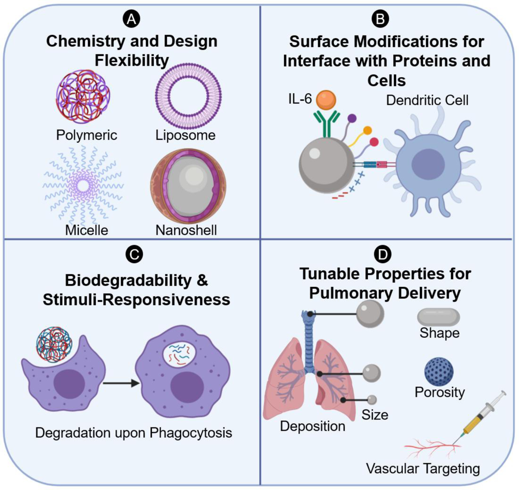
A) Tailorability of design and building blocks to accommodate a wide range of applications and cargo loading and ensure biocompatibility. B) Modular surface design with functional ligands and receptors that are capable of interacting with high specificity with lung immune cells or proteins. C) Controlled degradation and cargo release in low pH environments upon internalization by lung phagocytes. D) Tunability of nanomaterial properties for optimal deposition in the airways and targeting through vasculature.
Particle engineering approaches can be used for treatment strategies including synthetic vaccines, virus decoys, cytokine sequestration, macrophage repolarization, and immune cell distraction, as discussed in the following section. They can also promote the delivery of therapeutics to treat COVID-19, including those listed in the prior section. In this section, we highlight recent advances that improve drug delivery to the lung, identify particle engineering approaches to regulate immune pulmonary responses, and point to untapped opportunities for the use of biomaterials and particle engineering for the development of novel COVID-19 therapies. While these immunomodulatory particle-based strategies represent a promising avenue for regulation of COVID-19 inflammation, significant preclinical and clinical studies considering specific aspects of SARS-CoV-2 and the COVID-19 inflammatory landscapes will be needed for clinical translation.
Localizing Therapeutics to the Lung
Orchestrating the host response in the lung requires consideration of the unique pulmonary microenvironment as well as physiological barriers that arise from both the air and vascular sides of the lung. Delivery of agents which need to act in the lung can be accomplished by systemic delivery (primarily oral or intravenous)208, 209 and diffusion into the tissue. This delivery is non-specific and delivery to the lungs likely should be enhanced through the use of active targeting ligands to aid in crossing the endothelium into lung tissue.210 Efforts to target nanoparticle drug carriers to the inflamed pulmonary vasculature intravenously can be achieved with ligand functionalization that targets upregulated receptors in the leukocyte adhesion cascade occurring during pulmonary infiltration and by tuning carrier modulus.211, 212 Alternatively, high drug concentrations can be achieved through oral inhalation via aerosols, reducing off-target effects associated with systemic delivery routes while increasing the effective dose to affected lung tissue.6, 213 Here, particle size dominates in terms of effect on deposition location and therapeutic efficacy.203 Particles with aerodynamic diameters significantly smaller than 1 μm or larger than 5 μm will have deposition fractions below 0.5 in the alveolar region of the lung, our primary therapeutic target region for treatment and/or prevention of COVID-19.203, 214 Accordingly, it is critical to achieve aerodynamic particle sizes within the 1 to 5 μm range, along with low dispersity, for effective, consistent dosage.203 In the case of aerosol delivery, the delivery device also can be impactful on delivery because of the formulated particle size and patient compliance. For the case of COVID-19 treatment, nebulizers have the advantages of delivering large dosages without particular breathing techniques or coordination by patients that may have difficulty breathing and also allow for ease of formulation chemistry, as the nebulizer will generate aerosols in the desired size range.6, 215 Other delivery devices such as dry powder inhalers and metered dose inhalers have the advantages of higher delivery efficiency and lower cost, but have drawbacks of potential aggregation, which could cause reduced deposition in the alveoli, and requirement of coordinated inspiration by the patient.215 Regardless of the delivery method, pulmonary delivery of biomaterials can offer great advantages in increasing dosage to the lungs and avoiding off-target, systemic effects, while simultaneously targeting immune cells in the lung through size and charge considerations or avoid clearance through the same parameters;216, 217 smaller (50 to a few hundred nm) nanoparticles will be preferentially internalized by alveolar macrophages, while larger (greater than 10 μm) particles will be more likely to avoid internalization. However, to induce immunomodulatory effects and achieve high delivery efficiency to diseased areas and immune cells, pulmonary delivery of therapeutic particles must overcome mucosal and epithelial barrierss.218 Hydrophilic and neutral/positively charged moieties may be incorporated in the particle design to avoid entrapment by mucus and promote localization to APCs.219 Because of the tailorability of biomaterial-based particles, these parameters can be factored into their design and increase the effectiveness of potential therapeutic treatments of SARS-CoV-2.
Biomaterials in vaccine development
Nanoparticles and microparticles have distinct advantages in terms of delivery of vaccines, as they can be passively targeted to immune cells because of their size and can either co-deliver adjuvants or act as their own adjuvants.220, 221 Furthermore, the modular nature of many nanoparticles, whether liposomes, polymer nanoparticles, or others, allows for the design of smart materials to actively target immune cells222 and be biodegradable.205 Some nanovaccines, termed as virus-like particles, utilize components or structures of viruses to achieve effective vaccination owing to their size and ability to enter cells effectively to deliver vaccine components.223 Several virus-like particle vaccines for COVID-19 are rapidly progressing through the pipeline.224 Nanovaccine formulations can have controlled degradation based on nanoparticle properties such as swellability and modulus,225 and adjusted immunogenicity and localization as a function of particle properties such as charge and density of antigen for efficient dendritic cell association and lymph node trafficking (Figure 3A).226–228 Nanoparticles, such as those based on gold,229 lipids (liposomes),230 PLGA,222 and HPMA/NIPAM231 among many others, have been used previously for vaccine approaches. These approaches have been described in depth in previous reviews regarding the use of biomaterials for vaccine applications.218, 222, 232–237 Additionally, we point readers to excellent works reviewing other considerations in design of particle-based vaccines.238–240
Figure 3: Summary of potential particle-based pulmonary immune engineering approaches for COVID-19.
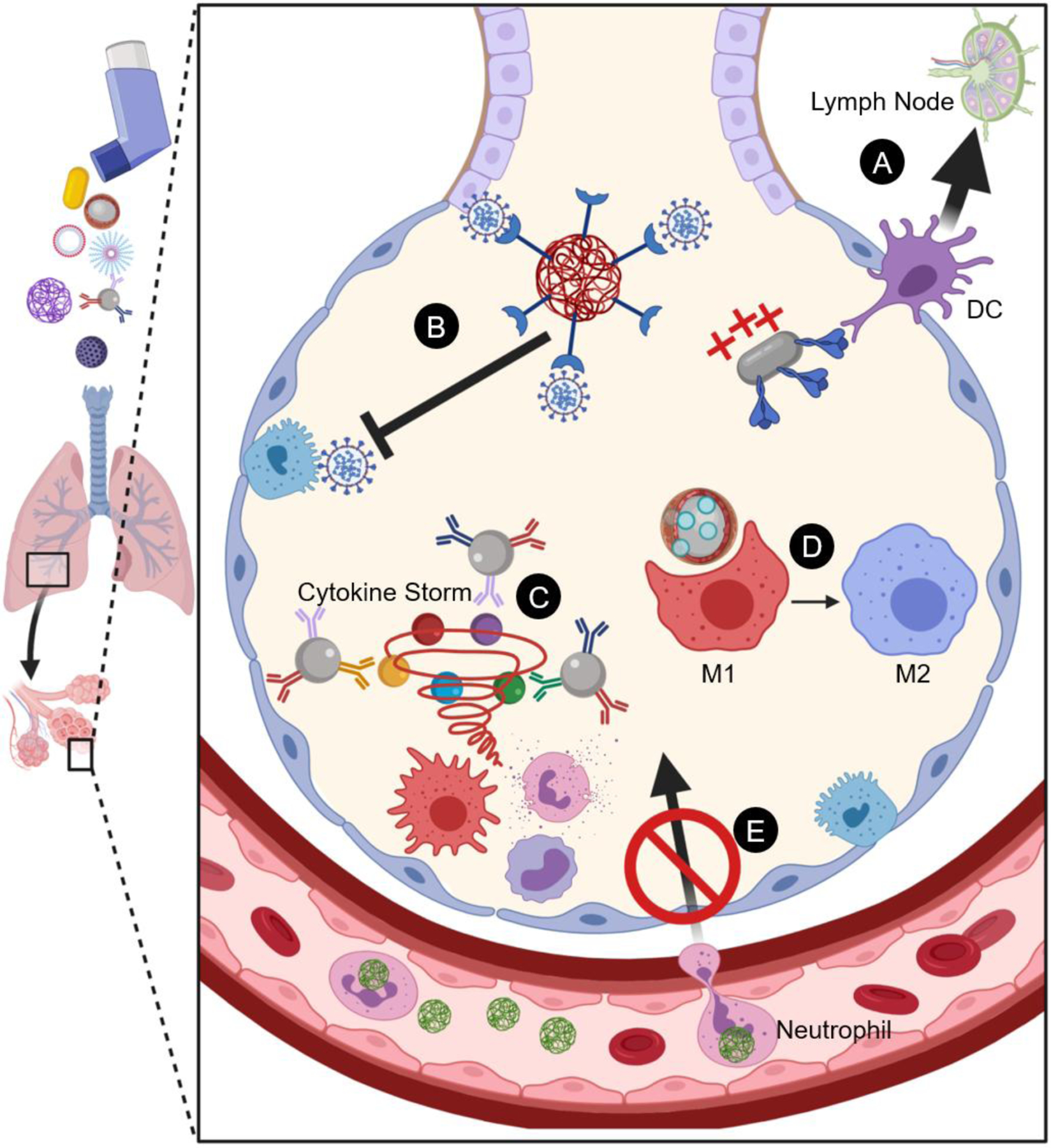
A) Engineering nanoparticle vaccines for enhanced association with migratory dendritic cells and increased lymph node trafficking to prime adaptive immunity. B) Nanoparticles as viral decoys displaying ACE2 to bind SARS-CoV-2 and inhibit infection of alveolar cells. C) Nanoparticles with functional ligands to sequester pro-inflammatory cytokines and prevent cytokine storm. D) Use of nanoparticles and loaded cargo to drive polarization of inflammatory M1 macrophages towards anti-inflammatory M2 macrophages. E) Phagocyte distraction with injected nanoparticles.
Particle-based formulations are rising to the forefront of vaccine approaches to combat COVID-19, with Moderna’s lipid nanoparticle-formulated mRNA vaccine currently in Phase III clinical trials, having shown significant promise in non-human primates.241 The vaccine delivers mRNA in a four-lipid nanoparticle which encodes for the SARS-CoV-2 spike glycoprotein in its prefusion conformation.193, 242 In addition, other leading vaccine candidates including the Pfizer and BioNTech formulations also utilize mRNA technology with lipid nanoparticle designs.243 Respiratory infections like SARS-CoV-2 could benefit from inhalable vaccine strategies to achieve high mucosal immunity in the lungs, the primary target for SARS-CoV-2. Therefore, particle-based, inhalable formulations could be a significant, untapped opportunity that could improve current vaccines in development.
Engineered viral decoys
Engineered particles offer opportunities as novel antivirals by blocking SARS-CoV-2 binding to host ACE2 receptors through host-cell mimicry.140 Delivery of free ACE2 is currently being pursued in clinical trials,244 which would occupy RBD to limit viral binding to host cells. This decoy approach would benefit from nanoparticle-mediated delivery, particularly to the lung, to prevent or abate infection. Nanoparticles can be decorated with ACE2-mimicing moieties that will bind to SARS-CoV-2 RBD and limit viral binding to host cells (Figure 3B). Engineered biomaterials are an ideal class of materials for this purpose, as they can be delivered to the pulmonary space, and can be designed to display high densities of ACE2.245, 246 For example, PLGA polymer nanoparticles could be functionalized and decorated with the aforementioned ACE2 receptor proteins. Recently, Zhang, et al. developed a method to distract SARS-CoV-2 and reduce infectivity using PLGA nanoparticles coated with membranes from alveolar epithelial cells and macrophages, respectively.247 Both of the membrane-coated nanoparticles were able to neutralize infectivity of SARS-CoV-2 in a dose dependent manner up to ~80% at treatments of 5 mg/mL of the nanoparticles in an infection model using Vero E6 cells using authentic SARS-CoV-2 viruses.44 Other promising attempts include using extracellular vesicles containing ACE2 as decoys to block infection by a lentivirus containing SARS-CoV-2 spike protein248 or gene delivery approaches that package ACE2 mRNA inside lipid nanoparticles.249 Collectively, these nanoparticle platforms perform critical host-mimicry to distract the SARS-CoV-2 virus from host binding.
Drug delivery approaches to cytokine sequestration and cytokine storm abatement
As mentioned previously, a number of cytokines and factors have been identified as contributors to the inflammation and severe symptoms of COVID-19 including IL-1β, IL-2, IL-6, IL-18, and granulocyte colony-stimulating factor (G-CSF), among others.23, 140 Treatments have been proposed for the sequestration of IL-6, IL-18, and IL-1β through the use of systemically-delivered antibodies.140, 250, 251 Possible downsides of this approach may be the lack of specificity to the lung space, leading to body or system-wide immune suppression, reducing the recruitment of antiviral immune cells in other instances in which it is desired or necessary. Accordingly, localized delivery to the lung and efficient sequestration of inflammatory cytokines is a potentially untapped opportunity for pulmonary delivery of biomaterials and therapeutic particles. Similar to some of the prophylactic treatments in which ACE2 can be conjugated to the surface of a biomaterial-based nanoparticle, antibodies against one or multiple inflammatory molecules involved in the cytokine storm can be conjugated to the surface of the biomaterial-based nanoparticles to allow for efficient and effective removal of the markers to alleviate the burden caused by the cytokine storm (Figure 3C). Some previously employed strategies have included chitosan and hyaluronic acid-based, PLGA-based, and mesoporous silica nanoparticles to sequester IL-6,252–254 all of which can be adapted for delivery to the lung. The mesoporous silica nanoparticles in particular were designed for aerosol delivery for an asthma model and may have capacity to be translated to cytokine storm abatement during SARS-CoV-2 infection.254 The incorporation strategies utilized, including carbodiimide conjugation of antibodies and formulation of nanoparticles with loaded antibodies, can be utilized in addition to other synthetic strategies, such as thiol-Michael click chemistry to incorporate antibodies into nanoparticles for delivery.252–254 Simultaneous delivery of multiple cues is a secondary advantage through conjugation of multiple antibodies to sequester cytokines, inclusion of active-targeting moieties, and co-delivery of anti-inflammatory agents to limit activated macrophage polarization.222, 255–258 All of these strategies could improve COVID-19 treatment through more efficient, localized delivery, reduction in off-target effects, and simultaneous delivery of anti-inflammatory agents alongside particles sequestering inflammatory molecules.
Delivery of anti-inflammatory agents for repolarization of immune cells
Another approach of promise for the treatment of the COVID-19 pathology is the repolarization of inflammatory macrophages to prevent or abate their cytokine contributions. Macrophages fall along a spectrum of activation, traditionally described as skewing toward either classically activated pro-inflammatory (M1) or alternatively activated anti-inflammatory (M2) phenotypes,259 although the reality of macrophage phenotype is much more fluid. M1 macrophages are abundant in the SARS-CoV-2-infection following monocyte infiltration and cell differentiation in the lung. When present, they cause the further release of inflammatory cytokines including IL-6 and exacerbate existing inflammation.260 Accordingly, therapeutic strategies that aid in the repolarization of macrophages from the M1-like phenotype toward the M2 phenotype (Figure 3D) could be useful in the treatment of COVID-19 symptoms by reducing the cycle of inflammation caused by release of the aforementioned cytokines and further recruitment of inflammatory leukocytes.261 M2 macrophages not only reduce secretion of inflammatory cytokines such as IL-6, TNF-α, and IL-1β,262 but promote secretion of anti-inflammatory factors such as IL-10 that may restore the microenvironment homeostasis.259 When introduced at the right time during COVID-19 host response, macrophage repolarization at the air-liquid interface using inhaled, engineered particles may allow for local restoration of host immune response and mitigation of subsequent inflammatory airway pathologies.
Biomaterials are particularly suited for this purpose because they often do not persist in the body, can deliver cytokines and other materials to aid in the polarization, can be targeted to immune cells like macrophages, and can, themselves, repolarize macrophages.262 A plethora of strategies have been developed to influence inflammatory macrophage response. Some examples include an alginate-based nanoparticle system was developed to deliver IL-10 plasmid DNA to repolarize M1 macrophages to M2 macrophages,263 polyethyleneimine nanoparticles were used to deliver the gene for CD163, an M2 macrophage marker,264 and PLGA nanoparticles have been used to mimic apoptotic bodies via a phospholipid membrane coating supplemented with phosphatidylserine to trigger the memory of uptake of apoptotic bodies, inducing an M2 polarization.265 All of these strategies led to decreases in the release of inflammatory cytokines including IL-6, IL-1β, IFN-γ, TNF-α, and MCP-1 and led to the repolarization of macrophages toward the M2 phenotype. These materials and methods were used in vitro, but their resultant reduction in the release of inflammatory cytokines and macrophage repolarization indicates their potential to be used to reduce or eliminate the inflammation induced by macrophages during SARS-CoV-2 infection.
Another way in which biomaterials could be used for the therapeutic treatment of COVID-19 is the delivery of anti-inflammatory cytokines such as IL-4, IL-10, and IL-13. IL-4 has previously been administered both in vitro and in vivo via conjugation to gold nanoparticles266 and within mesoporous silica nanoparticles.267 Both treatments led to an increase in the population of M2 macrophages alongside a corresponding reduction in the population of M1 macrophages. The silica nanoparticles would also have the benefit of larger aerodynamic diameters than their 180 nm geometric diameter, making them good candidates for pulmonary delivery of IL-4 for the treatment of COVID-19, which, as noted previously, would aid in minimizing off-target effects and increasing efficacy of treatment.213, 267, 268 Other studies have also indicated that mesoporous silica nanoparticles can themselves stimulate the immune system, leading to the production of IFN-γ, IL-2, IL-4, and IL-10, and are preferentially internalized by M1 macrophages.269, 270 This may lead to greater efficiency of treatment relative to other materials, as the M1 macrophages, primary drivers of inflammation and thus key therapeutic targets, will internalize the silica nanoparticles more than other cells, which are less desirable targets. Similar treatments with IL-10 have been developed using nanoparticles including the previously mentioned alginate nanoparticles that deliver genes encoding for IL-10263 and nanoparticulate polyester (PLGA and PEG) polymers nanoprecipitated with IL-10.271 Many of these studies have demonstrated efficacy in vivo, giving more credence to their potential for combatting COVID-19. Many of the nanoparticles have potential for pulmonary delivery as well, which would make them even better candidates for combatting the inflammation associated with COVID-19.
Delivery of corticosteroids can also impact macrophage polarization and thus are another category of therapeutics that may be of use for COVID-19 treatment. A primary concern of corticosteroid use is systemic immunosuppression, which could lead to impaired antiviral response.140 In this regard, pulmonary drug delivery could be advantageous, as particle platforms have already been developed for corticosteroid delivery and can likely be adapted to the use of particular steroids proven to be effective, such as budesonide and dexamethasone.272 For example, budesonide has been loaded into PLGA nanoparticles with 90% encapsulation efficiency and high drug loading of relevant sizes for cellular internalization.273 Similarly, metal-organic framework (MOF) nanoparticles were loaded with dexamethasone and evaluated for pulmonary delivery.274 Some other methods that have been developed to deliver corticosteroids include delivery of solid lipid nanoparticles loaded with budesonide for pulmonary drug delivery275 and dexamethasone for potential lung targeting,276 itraconazole nanoparticle-based dry powders for aerosol delivery of budesonide,277 crosslinked copolymers of methyl methacrylate, N,N-dimethylaminoethyl methacrylate, and butyl methacrylate monomers designed to load hydrophobic corticosteroids.278 Notably, dexamethasone-loaded PLGA microparticles were utilized to polarize macrophages towards an anti-inflammatory phenotype and resulted in a significant suppression of pro-inflammatory genes and TNF-α secretion.279, 280 All of these methods may be viable options to deliver these anti-inflammatory agents, especially if they can be formulated and delivered directly to the lungs to reduce or preclude systemic immunosuppression that may result in worse outcomes for patients treated with corticosteroids.
In addition to the delivery of anti-inflammatory agents, biomaterials, themselves, can be used to either induce or reduce inflammation. Their properties of immune skewing, particularly of macrophages, depend on the size, surface chemistry, and hydrophobicity of the material, in addition to other properties.262 In particular, larger materials (greater than 10 μm in at least one dimension), particles in particular, tend to induce an M1 phenotype in macrophages because of frustrated phagocytosis that occurs when macrophages are unable to effectively engulf the large particles.281 Accordingly, smaller particles would be desired for treatment of COVID-19 to reduce inflammation, as was the case for folate-conjugated silicate and calcium-based nanoparticles282 and RGD-conjugated gold nanorods,283 which both utilized sub-250 nm sized nanoparticles and showed anti-inflammatory properties of the respective nanoparticles. In both of these cases, surface modifications were necessary to aid in M2 polarization; however, there are instances demonstrating non-functionalized materials aiding in the polarization toward M2 phenotypes, such as non-functionalized PLGA nanoparticles and microparticles that reduce the expression of M1 markers and the secretion of pro-inflammatory cytokines, including IL-6 in vitro.284, 285 It has also been shown that titanium implants with calcium and strontium surface modifications and a particular nanotopography can induce an M2 immune response, as were some titania nanoparticles,286 which may be another material type that could be explored to reduce inflammation in the form of titanium or titania nanoparticles containing these ions.287 Any of the aforementioned systems could be used to aid in inducing anti-inflammatory phenotypes in macrophages as part of COVID-19 treatment, though they lack distinct advantages over polymeric biomaterials capable of delivering anti-inflammatory agents and ensuring intracellular degradation. In both cases, active targeting ligands for macrophages may be beneficial to ensure uptake or to increase their anti-inflammatory effects. For example, clustering mannose receptors on macrophages through carbohydrate inclusion can aid in their M2 polarization,288 while conjugation of dextran ligands can target M1 macrophages for delivery of cargo and re-education toward an M2 phenotype.289 Functionalization with molecular “markers of self”, such as CD47 or phosphatidylserine can also promote macrophage polarization for localized immune modulation.290, 291
Phagocyte Distraction
Neutrophils, in addition to macrophages, serve as part of the innate immune response to SARS-CoV-2 infection, contributing significantly to the aforementioned cycle of inflammation and cytokine storm.23 A potential approach to abating the cycle of inflammation in COVID-19 is to direct inflammatory phagocytic cells away from the lung and reduce the number of recruited leukocytes from vasculature or surrounding tissue (Figure 3E). Vascular drug delivery systems have previously been used to distract neutrophils from sites of inflammation, including previous work which demonstrated that intravenous injections of 2 and 0.5 μm carboxylated polystyrene nanoparticles can distract neutrophils from infiltrating into the lung as part of an acute lung injury model.228 Instead, the neutrophils are diverted to the liver, reducing the pulmonary inflammation associated with their response. In addition, negatively charged 500 nm polystyrene and PLGA particles were used to limit trafficking of inflammatory monocytes to inflammation loci in several murine inflammation models including West Nile virus encephalitis.292 Similar methods have been employed for the distraction of monocytes and neutrophils using intravenous injections of PLGA nanorods and nanoparticles.293, 294 PLGA-based nanorods have been shown to be preferentially internalized by neutrophils, which can be used to distract said neutrophils during inflammation to improve pathogenesis, while PLGA nanoparticles have been used to reduce the recruitment of both monocytes and neutrophils to the site of injury using a spinal cord injury model.293, 294 In the latter case, the recruitment of all innate immune cells at the site of injury was reduced by 75%, allowing for much more rapid recovery from injury. Recent work has shown that asymmetric lysozyme-dextran nanogels and crosslinked albumin nanoparticles with agglutinated proteins in amorphous nanostructures particularly target neutrophils during lung injury as demonstrated both in vivo in mice and also using human lungs, which could be critically valuable for selective treatment and/or distraction of neutrophils during COVID-19.295 These examples demonstrate that inflammation can be reduced and innate immune cells can be redirected from the sites of injury, which could be very valuable in the therapeutic treatment of COVID-19, especially in light of increased neutrophil-lymphocyte ratios found in 80% of patients infected with SARS-CoV-2.32 These distraction methods can also potentially be used in tandem with previously discussed drug delivery approaches, leveraging vascular targeting.
Opportunities for biomaterials-based model system development for addressing long-term effects of COVID-19
As noted above, COVID-19 infection can have long-term health implications in addition to short-term difficulties. These effects are attributed to permanent changes in the lung microenvironment and altered innate and adaptive immunity. These long-term issues can be further exacerbated by age, pre-existing health conditions, and COVID-19 infection severity.113 While there is a push to find vaccine candidates and clinical treatments for COVID-19 in the short term, there is also an emerging need to better understand the long-term ailments associated with COVID-19 and develop therapeutics for persistent health conditions for millions of recovering patients worldwide. Screening and developing such new therapeutics will require advances in mechanistic understanding of the resultant multicellular crosstalk involved in COVID-19 resolution as well as novel testing platforms able to recapitulate these effects. Traditional in vitro culture models (i.e., tissue culture plastic systems)296 and in vivo models (e.g., murine animal models)297 may be useful for initial hypothesis testing and translation of winnowed drug candidates; however, such systems alone may limit the development of effective therapeutics for COVID-19 long haulers because of anatomical inaccuracies, lack of complexity, and/or species-related differences. In that vein, biomaterials-based model systems that integrate human cells, including those from each sex, allow for an interesting opportunity to recapitulate the permanent changes in the lung microenvironment such as increased tissue stiffness, altered extracellular matrix composition, and a modified cellular microenvironment. These model systems would enable hypothesis testing in more physiologically relevant conditions that could help in understanding aspects of how the immune response is altered in the long term and consequently allow for the development and testing of therapeutics. Work from several groups, including ours, is utilizing such biomimetic model systems to reproduce key aspects of pulmonary fibrosis, which shares similar pathophysiology to COVID-19, which both cause organ damage and dysregulated wound healing.298–303 Additionally, there exist underlying opportunities in utilizing organ-on-a-chip model systems to study the effects on other organs systems in recovered COVID-19 patients.304–306 Incorporating these model systems along with primary cells derived from recovered patients would facilitate a hypothesis-based approach to understanding post-COVID-19 symptoms and recovery in a physiological manner and could allow for pre-screening of therapeutics in a microenvironment resembling that of COVID-19 long haulers.
OUTLOOK
This work is intended to be a guide to the biomaterials community to consider applying biomaterials-based principles toward COVID-19 and other pulmonary inflammations. We hope that the immune engineering strategies highlighted in this review will lead to more preclinical and clinical investigations of emerging biomaterials and particle-based therapies for COVID-19 and other infections that contribute to pulmonary and systemic inflammations. While the main focus of this review was to highlight opportunities for COVID-19-specific pathologies and complications, the proposed therapeutic strategies could certainly be applicable to other infections and diseases that cause inflammation in the lung. As indicated in various instances in this review, thorough clinical investigations are needed to reveal and characterize the involvement of key immune players in SARS-CoV-2-related infection, inflammation, and immunity. Additional biomaterials and particle-based therapeutic options may be proposed and investigated as a more detailed, mechanistic understanding of COVID-19 pathophysiology is discovered. As prophylactic vaccines are developed, particle-based designs, as well as the pulmonary route of administration, should be considered to enhance the safety and efficacy of the required response. However, it is imperative to characterize potential disadvantages associated with the proposed novel strategies including side effects, toxicity, long-term fate, and costs prior to clinical translation. Finally, approaches suggested for COVID-19 may find significant translation to other respiratory pathogens and are worthy of pursuit as platform technologies to mitigate the global burden of respiratory viral infections.
Acknowledgements
Figures were created using BioRender.com
Funding Information
Authors of this publication were supported by the National Institutes of Health and the State of Delaware under Award Numbers P20GM104316 and P20GM103446, as well as a Research Starter Grant in Pharmaceutics from the PhRMA foundation; Z.S.S. was supported by a T32GM008550 training grant. The content is solely the responsibility of the authors and does not necessarily represent the official views of the National Institutes of Health.
References
- 1.Bansal M, Cardiovascular disease and COVID-19. Diabetes & Metabolic Syndrome: Clinical Research & Reviews 2020, 14 (3), 247–250. [DOI] [PMC free article] [PubMed] [Google Scholar]
- 2.Mao L; Jin H; Wang M; Hu Y; Chen S; He Q; Chang J; Hong C; Zhou Y; Wang D; Miao X; Li Y; Hu B, Neurologic Manifestations of Hospitalized Patients With Coronavirus Disease 2019 in Wuhan, China. JAMA Neurology 2020, 77 (6), 683–690. [DOI] [PMC free article] [PubMed] [Google Scholar]
- 3.Velez JCQ; Caza T; Larsen CP, COVAN is the new HIVAN: the re-emergence of collapsing glomerulopathy with COVID-19. Nature Reviews Nephrology 2020. [DOI] [PMC free article] [PubMed] [Google Scholar]
- 4.Gupta A; Madhavan MV; Sehgal K; Nair N; Mahajan S; Sehrawat TS; Bikdeli B; Ahluwalia N; Ausiello JC; Wan EY; Freedberg DE; Kirtane AJ; Parikh SA; Maurer MS; Nordvig AS; Accili D; Bathon JM; Mohan S; Bauer KA; Leon MB; Krumholz HM; Uriel N; Mehra MR; Elkind MSV; Stone GW; Schwartz A; Ho DD; Bilezikian JP; Landry DW, Extrapulmonary manifestations of COVID-19. Nature Medicine 2020, 26 (7), 1017–1032. [DOI] [PMC free article] [PubMed] [Google Scholar]
- 5.Jarai BM; Kolewe EL; Stillman ZS; Raman N; Fromen CA, Chapter 18 - Polymeric Nanoparticles. In Nanoparticles for Biomedical Applications, Chung EJ; Leon L; Rinaldi C, Eds. Elsevier: 2020; pp 303–324. [Google Scholar]
- 6.Patil JS; Sarasija S, Pulmonary drug delivery strategies: A concise, systematic review. Lung India 2012, 29 (1), 44–49. [DOI] [PMC free article] [PubMed] [Google Scholar]
- 7.Yang P; Wang X, COVID-19: a new challenge for human beings. Cellular & Molecular Immunology 2020, 17 (5), 555–557. [DOI] [PMC free article] [PubMed] [Google Scholar]
- 8.Weiss SR; Navas-Martin S, Coronavirus Pathogenesis and the Emerging Pathogen Severe Acute Respiratory Syndrome Coronavirus. Microbiology and Molecular Biology Reviews 2005, 69 (4), 635. [DOI] [PMC free article] [PubMed] [Google Scholar]
- 9.Zumla A; Chan JFW; Azhar EI; Hui DSC; Yuen K-Y, Coronaviruses — drug discovery and therapeutic options. Nature Reviews Drug Discovery 2016, 15 (5), 327–347. [DOI] [PMC free article] [PubMed] [Google Scholar]
- 10.Walls AC; Park Y-J; Tortorici MA; Wall A; McGuire AT; Veesler D, Structure, Function, and Antigenicity of the SARS-CoV-2 Spike Glycoprotein. Cell 2020, 181 (2), 281–292.e6. [DOI] [PMC free article] [PubMed] [Google Scholar]
- 11.Zhu N; Zhang D; Wang W; Li X; Yang B; Song J; Zhao X; Huang B; Shi W; Lu R; Niu P; Zhan F; Ma X; Wang D; Xu W; Wu G; Gao GF; Tan W, A Novel Coronavirus from Patients with Pneumonia in China, 2019. New England Journal of Medicine 2020, 382 (8), 727–733. [DOI] [PMC free article] [PubMed] [Google Scholar]
- 12.Su S; Wong G; Shi W; Liu J; Lai ACK; Zhou J; Liu W; Bi Y; Gao GF, Epidemiology, Genetic Recombination, and Pathogenesis of Coronaviruses. Trends in Microbiology 2016, 24 (6), 490–502. [DOI] [PMC free article] [PubMed] [Google Scholar]
- 13.Gorbalenya AE; Baker SC; Baric RS; de Groot RJ; Drosten C; Gulyaeva AA; Haagmans BL; Lauber C; Leontovich AM; Neuman BW; Penzar D; Perlman S; Poon LLM; Samborskiy DV; Sidorov IA; Sola I; Ziebuhr J; Coronaviridae Study Group of the International Committee on Taxonomy of, V., The species Severe acute respiratory syndrome-related coronavirus: classifying 2019-nCoV and naming it SARS-CoV-2. Nature Microbiology 2020, 5 (4), 536–544. [DOI] [PMC free article] [PubMed] [Google Scholar]
- 14.Zhou P; Yang X-L; Wang X-G; Hu B; Zhang L; Zhang W; Si H-R; Zhu Y; Li B; Huang C-L; Chen H-D; Chen J; Luo Y; Guo H; Jiang R-D; Liu M-Q; Chen Y; Shen X-R; Wang X; Zheng X-S; Zhao K; Chen Q-J; Deng F; Liu L-L; Yan B; Zhan F-X; Wang Y-Y; Xiao G-F; Shi Z-L, A pneumonia outbreak associated with a new coronavirus of probable bat origin. Nature 2020, 579 (7798), 270–273. [DOI] [PMC free article] [PubMed] [Google Scholar]
- 15.Mason RJ, Pathogenesis of COVID-19 from a cell biology perspective. European Respiratory Journal 2020, 55 (4), 2000607. [DOI] [PMC free article] [PubMed] [Google Scholar]
- 16.Guan W.-j.; Ni Z.-y.; Hu Y; Liang W.-h.; Ou C.-q.; He J.-x.; Liu L; Shan H; Lei C.-l.; Hui DSC; Du B; Li L.-j.; Zeng G; Yuen K-Y; Chen R.-c.; Tang C.-l.; Wang T; Chen P.-y.; Xiang J; Li S.-y.; Wang J.-l.; Liang Z.-j.; Peng Y.-x.; Wei L; Liu Y; Hu Y.-h.; Peng P; Wang J.-m.; Liu J.-y.; Chen Z; Li G; Zheng Z.-j.; Qiu S.-q.; Luo J; Ye C.-j.; Zhu S.-y.; Zhong N.-s., Clinical Characteristics of Coronavirus Disease 2019 in China. New England Journal of Medicine 2020, 382 (18), 1708–1720. [DOI] [PMC free article] [PubMed] [Google Scholar]
- 17.Lauer SA; Grantz KH; Bi Q; Jones FK; Zheng Q; Meredith HR; Azman AS; Reich NG; Lessler J, The Incubation Period of Coronavirus Disease 2019 (COVID-19) From Publicly Reported Confirmed Cases: Estimation and Application. Annals of Internal Medicine 2020, 172 (9), 577–582. [DOI] [PMC free article] [PubMed] [Google Scholar]
- 18.Letko M; Marzi A; Munster V, Functional assessment of cell entry and receptor usage for SARS-CoV-2 and other lineage B betacoronaviruses. Nature Microbiology 2020, 5 (4), 562–569. [DOI] [PMC free article] [PubMed] [Google Scholar]
- 19.Xu H; Zhong L; Deng J; Peng J; Dan H; Zeng X; Li T; Chen Q, High expression of ACE2 receptor of 2019-nCoV on the epithelial cells of oral mucosa. International Journal of Oral Science 2020, 12 (1), 8. [DOI] [PMC free article] [PubMed] [Google Scholar]
- 20.Jia HP; Look DC; Shi L; Hickey M; Pewe L; Netland J; Farzan M; Wohlford-Lenane C; Perlman S; McCray PB, ACE2 Receptor Expression and Severe Acute Respiratory Syndrome Coronavirus Infection Depend on Differentiation of Human Airway Epithelia. Journal of Virology 2005, 79 (23), 14614. [DOI] [PMC free article] [PubMed] [Google Scholar]
- 21.Hamming I; Timens W; Bulthuis MLC; Lely AT; Navis GJ; van Goor H, Tissue distribution of ACE2 protein, the functional receptor for SARS coronavirus. A first step in understanding SARS pathogenesis. The Journal of Pathology 2004, 203 (2), 631–637. [DOI] [PMC free article] [PubMed] [Google Scholar]
- 22.Hou YJ; Okuda K; Edwards CE; Martinez DR; Asakura T; Dinnon KH; Kato T; Lee RE; Yount BL; Mascenik TM; Chen G; Olivier KN; Ghio A; Tse LV; Leist SR; Gralinski LE; Schäfer A; Dang H; Gilmore R; Nakano S; Sun L; Fulcher ML; Livraghi-Butrico A; Nicely NI; Cameron M; Cameron C; Kelvin DJ; de Silva A; Margolis DM; Markmann A; Bartelt L; Zumwalt R; Martinez FJ; Salvatore SP; Borczuk A; Tata PR; Sontake V; Kimple A; Jaspers I; O’Neal WK; Randell SH; Boucher RC; Baric RS, SARS-CoV-2 Reverse Genetics Reveals a Variable Infection Gradient in the Respiratory Tract. Cell 2020. [DOI] [PMC free article] [PubMed] [Google Scholar]
- 23.Tay MZ; Poh CM; Rénia L; MacAry PA; Ng LFP, The trinity of COVID-19: immunity, inflammation and intervention. Nature Reviews Immunology 2020, 20 (6), 363–374. [DOI] [PMC free article] [PubMed] [Google Scholar]
- 24.Chen Y; Guo Y; Pan Y; Zhao ZJ, Structure analysis of the receptor binding of 2019-nCoV. Biochemical and Biophysical Research Communications 2020, 525 (1), 135–140. [DOI] [PMC free article] [PubMed] [Google Scholar]
- 25.Ou X; Liu Y; Lei X; Li P; Mi D; Ren L; Guo L; Guo R; Chen T; Hu J; Xiang Z; Mu Z; Chen X; Chen J; Hu K; Jin Q; Wang J; Qian Z, Characterization of spike glycoprotein of SARS-CoV-2 on virus entry and its immune cross-reactivity with SARS-CoV. Nature Communications 2020, 11 (1), 1620. [DOI] [PMC free article] [PubMed] [Google Scholar]
- 26.Yuki K; Fujiogi M; Koutsogiannaki S, COVID-19 pathophysiology: A review. Clinical Immunology 2020, 215, 108427. [DOI] [PMC free article] [PubMed] [Google Scholar]
- 27.Wrapp D; Wang N; Corbett KS; Goldsmith JA; Hsieh C-L; Abiona O; Graham BS; McLellan JS, Cryo-EM structure of the 2019-nCoV spike in the prefusion conformation. Science 2020, 367 (6483), 1260. [DOI] [PMC free article] [PubMed] [Google Scholar]
- 28.Hoffmann M; Kleine-Weber H; Schroeder S; Krüger N; Herrler T; Erichsen S; Schiergens TS; Herrler G; Wu NH; Nitsche A; Müller MA; Drosten C; Pöhlmann S, SARS-CoV-2 Cell Entry Depends on ACE2 and TMPRSS2 and Is Blocked by a Clinically Proven Protease Inhibitor. Cell 2020, 181 (2), 271–280.e8. [DOI] [PMC free article] [PubMed] [Google Scholar]
- 29.McKee DL; Sternberg A; Stange U; Laufer S; Naujokat C, Candidate drugs against SARS-CoV-2 and COVID-19. Pharmacol Res 2020, 157, 104859–104859. [DOI] [PMC free article] [PubMed] [Google Scholar]
- 30.Zang R; Castro MFG; McCune BT; Zeng Q; Rothlauf PW; Sonnek NM; Liu Z; Brulois KF; Wang X; Greenberg HB; Diamond MS; Ciorba MA; Whelan SPJ; Ding S, TMPRSS2 and TMPRSS4 promote SARS-CoV-2 infection of human small intestinal enterocytes. Science Immunology 2020, 5 (47), eabc3582. [DOI] [PMC free article] [PubMed] [Google Scholar]
- 31.Merrill JT; Erkan D; Winakur J; James JA, Emerging evidence of a COVID-19 thrombotic syndrome has treatment implications. Nature Reviews Rheumatology 2020, 16 (10), 581–589. [DOI] [PMC free article] [PubMed] [Google Scholar]
- 32.Zhou F; Yu T; Du R; Fan G; Liu Y; Liu Z; Xiang J; Wang Y; Song B; Gu X; Guan L; Wei Y; Li H; Wu X; Xu J; Tu S; Zhang Y; Chen H; Cao B, Clinical course and risk factors for mortality of adult inpatients with COVID-19 in Wuhan, China: a retrospective cohort study. The Lancet 2020, 395 (10229), 1054–1062. [DOI] [PMC free article] [PubMed] [Google Scholar]
- 33.Martin TR; Frevert CW, Innate immunity in the lungs. Proc Am Thorac Soc 2005, 2 (5), 403–411. [DOI] [PMC free article] [PubMed] [Google Scholar]
- 34.See H; Wark P, Innate immune response to viral infection of the lungs. Paediatr Respir Rev 2008, 9 (4), 243–250. [DOI] [PMC free article] [PubMed] [Google Scholar]
- 35.Whitsett JA; Alenghat T, Respiratory epithelial cells orchestrate pulmonary innate immunity. Nature Immunology 2015, 16 (1), 27–35. [DOI] [PMC free article] [PubMed] [Google Scholar]
- 36.Zhou Z; Ren L; Zhang L; Zhong J; Xiao Y; Jia Z; Guo L; Yang J; Wang C; Jiang S; Yang D; Zhang G; Li H; Chen F; Xu Y; Chen M; Gao Z; Yang J; Dong J; Liu B; Zhang X; Wang W; He K; Jin Q; Li M; Wang J, Overly Exuberant Innate Immune Response to SARS-CoV-2 Infection. SSRN Electronic Journal 2020. [Google Scholar]
- 37.Zhou Z; Ren L; Zhang L; Zhong J; Xiao Y; Jia Z; Guo L; Yang J; Wang C; Jiang S; Yang D; Zhang G; Li H; Chen F; Xu Y; Chen M; Gao Z; Yang J; Dong J; Liu B; Zhang X; Wang W; He K; Jin Q; Li M; Wang J, Heightened Innate Immune Responses in the Respiratory Tract of COVID-19 Patients. Cell Host Microbe 2020, 27 (6), 883–890.e2. [DOI] [PMC free article] [PubMed] [Google Scholar]
- 38.Liao M; Liu Y; Yuan J; Wen Y; Xu G; Zhao J; Cheng L; Li J; Wang X; Wang F; Liu L; Amit I; Zhang S; Zhang Z, Single-cell landscape of bronchoalveolar immune cells in patients with COVID-19. Nature Medicine 2020, 26 (6), 842–844. [DOI] [PubMed] [Google Scholar]
- 39.Huang C; Wang Y; Li X; Ren L; Zhao J; Hu Y; Zhang L; Fan G; Xu J; Gu X; Cheng Z; Yu T; Xia J; Wei Y; Wu W; Xie X; Yin W; Li H; Liu M; Xiao Y; Gao H; Guo L; Xie J; Wang G; Jiang R; Gao Z; Jin Q; Wang J; Cao B, Clinical features of patients infected with 2019 novel coronavirus in Wuhan, China. The Lancet 2020, 395 (10223), 497–506. [DOI] [PMC free article] [PubMed] [Google Scholar]
- 40.Ulhaq ZS; Soraya GV, Interleukin-6 as a potential biomarker of COVID-19 progression. Med Mal Infect 2020, 50 (4), 382–383. [DOI] [PMC free article] [PubMed] [Google Scholar]
- 41.Chen X; Zhao B; Qu Y; Chen Y; Xiong J; Feng Y; Men D; Huang Q; Liu Y; Yang B; Ding J; Li F, Detectable Serum Severe Acute Respiratory Syndrome Coronavirus 2 Viral Load (RNAemia) Is Closely Correlated With Drastically Elevated Interleukin 6 Level in Critically Ill Patients With Coronavirus Disease 2019. Clinical Infectious Diseases 2020. [DOI] [PMC free article] [PubMed] [Google Scholar]
- 42.Herold T; Jurinovic V; Arnreich C; Hellmuth JC; von Bergwelt-Baildon M; Klein M; Weinberger T, Level of IL-6 predicts respiratory failure in hospitalized symptomatic COVID-19 patients. medRxiv 2020, 2020.04.01.20047381. [Google Scholar]
- 43.Jamilloux Y; Henry T; Belot A; Viel S; Fauter M; El Jammal T; Walzer T; François B; Sève P, Should we stimulate or suppress immune responses in COVID-19? Cytokine and anti-cytokine interventions. Autoimmunity Reviews 2020, 19 (7), 102567. [DOI] [PMC free article] [PubMed] [Google Scholar]
- 44.Li G; Fan Y; Lai Y; Han T; Li Z; Zhou P; Pan P; Wang W; Hu D; Liu X; Zhang Q; Wu J, Coronavirus infections and immune responses. Journal of Medical Virology 2020, 92 (4), 424–432. [DOI] [PMC free article] [PubMed] [Google Scholar]
- 45.Seif F; Khoshmirsafa M; Aazami H; Mohsenzadegan M; Sedighi G; Bahar M, The role of JAK-STAT signaling pathway and its regulators in the fate of T helper cells. Cell Communication and Signaling 2017, 15 (1), 23. [DOI] [PMC free article] [PubMed] [Google Scholar]
- 46.Channappanavar R; Fehr Anthony R.; Vijay R; Mack M; Zhao J; Meyerholz David K.; Perlman S, Dysregulated Type I Interferon and Inflammatory Monocyte-Macrophage Responses Cause Lethal Pneumonia in SARS-CoV-Infected Mice. Cell Host Microbe 2016, 19 (2), 181–193. [DOI] [PMC free article] [PubMed] [Google Scholar]
- 47.Lokugamage KG; Hage A; Schindewolf C; Rajsbaum R; Menachery VD, SARS-CoV-2 is sensitive to type I interferon pretreatment. bioRxiv 2020, 2020.03.07.982264. [DOI] [PMC free article] [PubMed] [Google Scholar]
- 48.Hadjadj J; Yatim N; Barnabei L; Corneau A; Boussier J; Pere H; Charbit B; Bondet V; Chenevier-Gobeaux C; Breillat P; Carlier N; Gauzit R; Morbieu C; Pene F; Marin N; Roche N; Szwebel T-A; Smith N; Merkling S; Treluyer J-M; Veyer D; Mouthon L; Blanc C; Tharaux P-L; Rozenberg F; Fischer A; Duffy D; Rieux-Laucat F; Kerneis S; Terrier B, Impaired type I interferon activity and exacerbated inflammatory responses in severe Covid-19 patients. medRxiv 2020, 2020.04.19.20068015. [DOI] [PMC free article] [PubMed] [Google Scholar]
- 49.Ju B; Zhang Q; Ge J; Wang R; Sun J; Ge X; Yu J; Shan S; Zhou B; Song S; Tang X; Yu J; Lan J; Yuan J; Wang H; Zhao J; Zhang S; Wang Y; Shi X; Liu L; Zhao J; Wang X; Zhang Z; Zhang L, Human neutralizing antibodies elicited by SARS-CoV-2 infection. Nature 2020, 584 (7819), 115–119. [DOI] [PubMed] [Google Scholar]
- 50.Isho B; Abe KT; Zuo M; Jamal AJ; Rathod B; Wang JH; Li Z; Chao G; Rojas OL; Bang YM; Pu A; Christie-Holmes N; Gervais C; Ceccarelli D; Samavarchi-Tehrani P; Guvenc F; Budylowski P; Li A; Paterson A; Feng Yun Y; Marin LM; Caldwell L; Wrana JL; Colwill K; Sicheri F; Mubareka S; Gray-Owen SD; Drews SJ; Siqueira WL; Barrios-Rodiles M; Ostrowski M; Rini JM; Durocher Y; McGeer AJ; Gommerman JL; Gingras A-C, Mucosal versus systemic antibody responses to SARS-CoV-2 antigens in COVID-19 patients. medRxiv 2020, 2020.08.01.20166553. [DOI] [PMC free article] [PubMed] [Google Scholar]
- 51.To KK-W; Tsang OT-Y; Leung W-S; Tam AR; Wu T-C; Lung DC; Yip CC-Y; Cai J-P; Chan JM-C; Chik TS-H; Lau DP-L; Choi CY-C; Chen L-L; Chan W-M; Chan K-H; Ip JD; Ng AC-K; Poon RW-S; Luo C-T; Cheng VC-C; Chan JF-W; Hung IF-N; Chen Z; Chen H; Yuen K-Y, Temporal profiles of viral load in posterior oropharyngeal saliva samples and serum antibody responses during infection by SARS-CoV-2: an observational cohort study. The Lancet Infectious Diseases 2020, 20 (5), 565–574. [DOI] [PMC free article] [PubMed] [Google Scholar]
- 52.Juno JA; Tan H-X; Lee WS; Reynaldi A; Kelly HG; Wragg K; Esterbauer R; Kent HE; Batten CJ; Mordant FL; Gherardin NA; Pymm P; Dietrich MH; Scott NE; Tham W-H; Godfrey DI; Subbarao K; Davenport MP; Kent SJ; Wheatley AK, Humoral and circulating follicular helper T cell responses in recovered patients with COVID-19. Nature Medicine 2020. [DOI] [PubMed] [Google Scholar]
- 53.Sekine T; Perez-Potti A; Rivera-Ballesteros O; Strålin K; Gorin J-B; Olsson A; Llewellyn-Lacey S; Kamal H; Bogdanovic G; Muschiol S; Wullimann DJ; Kammann T; Emgård J; Parrot T; Folkesson E; Rooyackers O; Eriksson LI; Sönnerborg A; Allander T; Albert J; Nielsen M; Klingström J; Gredmark-Russ S; Björkström NK; Sandberg JK; Price DA; Ljunggren H-G; Aleman S; Buggert M, Robust T cell immunity in convalescent individuals with asymptomatic or mild COVID-19. bioRxiv 2020, 2020.06.29.174888. [DOI] [PMC free article] [PubMed] [Google Scholar]
- 54.Long Q-X; Tang X-J; Shi Q-L; Li Q; Deng H-J; Yuan J; Hu J-L; Xu W; Zhang Y; Lv F-J; Su K; Zhang F; Gong J; Wu B; Liu X-M; Li J-J; Qiu J-F; Chen J; Huang A-L, Clinical and immunological assessment of asymptomatic SARS-CoV-2 infections. Nature Medicine 2020. [DOI] [PubMed] [Google Scholar]
- 55.Greenhalgh T; Knight M; A’Court C; Buxton M; Husain L, Management of post-acute covid-19 in primary care. BMJ 2020, 370, m3026. [DOI] [PubMed] [Google Scholar]
- 56.Herold T; Jurinovic V; Arnreich C; Lipworth BJ; Hellmuth JC; von Bergwelt-Baildon M; Klein M; Weinberger T, Elevated levels of IL-6 and CRP predict the need for mechanical ventilation in COVID-19. Journal of Allergy and Clinical Immunology 2020, 146 (1), 128–136.e4. [DOI] [PMC free article] [PubMed] [Google Scholar]
- 57.Del Valle DM; Kim-Schulze S; Huang H-H; Beckmann ND; Nirenberg S; Wang B; Lavin Y; Swartz TH; Madduri D; Stock A; Marron TU; Xie H; Patel M; Tuballes K; Van Oekelen O; Rahman A; Kovatch P; Aberg JA; Schadt E; Jagannath S; Mazumdar M; Charney AW; Firpo-Betancourt A; Mendu DR; Jhang J; Reich D; Sigel K; Cordon-Cardo C; Feldmann M; Parekh S; Merad M; Gnjatic S, An inflammatory cytokine signature predicts COVID-19 severity and survival. Nature Medicine 2020. [DOI] [PMC free article] [PubMed] [Google Scholar]
- 58.Fang Y; Zhou J; Ding X; Ling G; Yu S, Pulmonary fibrosis in critical ill patients recovered from COVID-19 pneumonia: Preliminary experience. The American Journal of Emergency Medicine. [DOI] [PMC free article] [PubMed] [Google Scholar]
- 59.Schaefer I-M; Padera RF; Solomon IH; Kanjilal S; Hammer MM; Hornick JL; Sholl LM, In situ detection of SARS-CoV-2 in lungs and airways of patients with COVID-19. Modern Pathology 2020. [DOI] [PMC free article] [PubMed] [Google Scholar]
- 60.Barnes BJ; Adrover JM; Baxter-Stoltzfus A; Borczuk A; Cools-Lartigue J; Crawford JM; Daßler-Plenker J; Guerci P; Huynh C; Knight JS; Loda M; Looney MR; McAllister F; Rayes R; Renaud S; Rousseau S; Salvatore S; Schwartz RE; Spicer JD; Yost CC; Weber A; Zuo Y; Egeblad M, Targeting potential drivers of COVID-19: Neutrophil extracellular traps. J Exp Med 2020, 217 (6), e20200652. [DOI] [PMC free article] [PubMed] [Google Scholar]
- 61.Zuo Y; Yalavarthi S; Shi H; Gockman K; Zuo M; Madison JA; Blair C; Weber A; Barnes BJ; Egeblad M; Woods RJ; Kanthi Y; Knight JS, Neutrophil extracellular traps in COVID-19. JCI Insight 2020, 5 (11). [DOI] [PMC free article] [PubMed] [Google Scholar]
- 62.Cao X, COVID-19: immunopathology and its implications for therapy. Nature Reviews Immunology 2020, 20 (5), 269–270. [DOI] [PMC free article] [PubMed] [Google Scholar]
- 63.Zhang C; Wu Z; Li J-W; Zhao H; Wang G-Q, Cytokine release syndrome in severe COVID-19: interleukin-6 receptor antagonist tocilizumab may be the key to reduce mortality. International Journal of Antimicrobial Agents 2020, 55 (5), 105954. [DOI] [PMC free article] [PubMed] [Google Scholar]
- 64.Wan S; Yi Q; Fan S; Lv J; Zhang X; Guo L; Lang C; Xiao Q; Xiao K; Yi Z; Qiang M; Xiang J; Zhang B; Chen Y, Characteristics of lymphocyte subsets and cytokines in peripheral blood of 123 hospitalized patients with 2019 novel coronavirus pneumonia (NCP). medRxiv 2020, 2020.02.10.20021832. [Google Scholar]
- 65.Moore JB; June CH, Cytokine release syndrome in severe COVID-19. Science 2020, 368 (6490), 473. [DOI] [PubMed] [Google Scholar]
- 66.Diao B; Wang C; Tan Y; Chen X; Liu Y; Ning L; Chen L; Li M; Liu Y; Wang G; Yuan Z; Feng Z; Zhang Y; Wu Y; Chen Y, Reduction and Functional Exhaustion of T Cells in Patients With Coronavirus Disease 2019 (COVID-19). Frontiers in Immunology 2020, 11 (827). [DOI] [PMC free article] [PubMed] [Google Scholar]
- 67.Xiong Y; Liu Y; Cao L; Wang D; Guo M; Jiang A; Guo D; Hu W; Yang J; Tang Z; Wu H; Lin Y; Zhang M; Zhang Q; Shi M; Liu Y; Zhou Y; Lan K; Chen Y, Transcriptomic characteristics of bronchoalveolar lavage fluid and peripheral blood mononuclear cells in COVID-19 patients. Emerging Microbes & Infections 2020, 9 (1), 761–770. [DOI] [PMC free article] [PubMed] [Google Scholar]
- 68.Kuri-Cervantes L; Pampena MB; Meng W; Rosenfeld AM; Ittner CAG; Weisman AR; Agyekum RS; Mathew D; Baxter AE; Vella LA; Kuthuru O; Apostolidis SA; Bershaw L; Dougherty J; Greenplate AR; Pattekar A; Kim J; Han N; Gouma S; Weirick ME; Arevalo CP; Bolton MJ; Goodwin EC; Anderson EM; Hensley SE; Jones TK; Mangalmurti NS; Luning Prak ET; Wherry EJ; Meyer NJ; Betts MR, Comprehensive mapping of immune perturbations associated with severe COVID-19. Science Immunology 2020, 5 (49), eabd7114. [DOI] [PMC free article] [PubMed] [Google Scholar]
- 69.Maucourant C; Filipovic I; Ponzetta A; Aleman S; Cornillet M; Hertwig L; Strunz B; Lentini A; Reinius B; Brownlie D; Cuapio A; Ask EH; Hull RM; Haroun-Izquierdo A; Schaffer M; Klingström J; Folkesson E; Buggert M; Sandberg JK; Eriksson LI; Rooyackers O; Ljunggren H-G; Malmberg K-J; Michaëlsson J; Marquardt N; Hammer Q; Strålin K; Björkström NK, Natural killer cell immunotypes related to COVID-19 disease severity. Science Immunology 2020, 5 (50), eabd6832. [DOI] [PMC free article] [PubMed] [Google Scholar]
- 70.Long Q-X; Liu B-Z; Deng H-J; Wu G-C; Deng K; Chen Y-K; Liao P; Qiu J-F; Lin Y; Cai X-F; Wang D-Q; Hu Y; Ren J-H; Tang N; Xu Y-Y; Yu L-H; Mo Z; Gong F; Zhang X-L; Tian W-G; Hu L; Zhang X-X; Xiang J-L; Du H-X; Liu H-W; Lang C-H; Luo X-H; Wu S-B; Cui X-P; Zhou Z; Zhu M-M; Wang J; Xue C-J; Li X-F; Wang L; Li Z-J; Wang K; Niu C-C; Yang Q-J; Tang X-J; Zhang Y; Liu X-M; Li J-J; Zhang D-C; Zhang F; Liu P; Yuan J; Li Q; Hu J-L; Chen J; Huang A-L, Antibody responses to SARS-CoV-2 in patients with COVID-19. Nature Medicine 2020, 26 (6), 845–848. [DOI] [PubMed] [Google Scholar]
- 71.Gralinski LE; Sheahan TP; Morrison TE; Menachery VD; Jensen K; Leist SR; Whitmore A; Heise MT; Baric RS, Complement Activation Contributes to Severe Acute Respiratory Syndrome Coronavirus Pathogenesis. mBio 2018, 9 (5), e01753–18. [DOI] [PMC free article] [PubMed] [Google Scholar]
- 72.Gao T; Hu M; Zhang X; Li H; Zhu L; Liu H; Dong Q; Zhang Z; Wang Z; Hu Y; Fu Y; Jin Y; Li K; Zhao S; Xiao Y; Luo S; Li L; Zhao L; Liu J; Zhao H; Liu Y; Yang W; Peng J; Chen X; Li P; Liu Y; Xie Y; Song J; Zhang L; Ma Q; Bian X; Chen W; Liu X; Mao Q; Cao C, Highly pathogenic coronavirus N protein aggravates lung injury by MASP-2-mediated complement over-activation. medRxiv 2020, 2020.03.29.20041962. [Google Scholar]
- 73.Cugno M; Meroni PL; Gualtierotti R; Griffini S; Grovetti E; Torri A; Panigada M; Aliberti S; Blasi F; Tedesco F; Peyvandi F, Complement activation in patients with COVID-19: A novel therapeutic target. J Allergy Clin Immunol 2020, 146 (1), 215–217. [DOI] [PMC free article] [PubMed] [Google Scholar]
- 74.Zhou Y; Fu B; Zheng X; Wang D; Zhao C; Qi Y; Sun R; Tian Z; Xu X; Wei H, Pathogenic T-cells and inflammatory monocytes incite inflammatory storms in severe COVID-19 patients. National Science Review 2020, 7 (6), 998–1002. [DOI] [PMC free article] [PubMed] [Google Scholar]
- 75.Zhang F; Mears JR; Shakib L; Beynor JI; Shanaj S; Korsunsky I; Nathan A; Donlin LT; Raychaudhuri S, IFN-γ and TNF-α drive a CXCL10 + CCL2 + macrophage phenotype expanded in severe COVID-19 and other diseases with tissue inflammation. bioRxiv 2020, 2020.08.05.238360. [DOI] [PMC free article] [PubMed] [Google Scholar]
- 76.Yap JKY; Moriyama M; Iwasaki A, Inflammasomes and Pyroptosis as Therapeutic Targets for COVID-19. The Journal of Immunology 2020, ji2000513. [DOI] [PMC free article] [PubMed] [Google Scholar]
- 77.Liu L; Wei Q; Lin Q; Fang J; Wang H; Kwok H; Tang H; Nishiura K; Peng J; Tan Z; Wu T; Cheung KW; Chan KH; Alvarez X; Qin C; Lackner A; Perlman S; Yuen KY; Chen Z, Anti-spike IgG causes severe acute lung injury by skewing macrophage responses during acute SARS-CoV infection. JCI Insight 2019, 4 (4). [DOI] [PMC free article] [PubMed] [Google Scholar]
- 78.Cully M, Immune status could determine efficacy of COVID-19 therapies. Nat Rev Drug Discov 2020, 19 (7), 431–434. [DOI] [PubMed] [Google Scholar]
- 79.Rod JE; Oviedo-Trespalacios O; Cortes-Ramirez J, A brief-review of the risk factors for covid-19 severity. Rev Saude Publica 2020, 54, 60–60. [DOI] [PMC free article] [PubMed] [Google Scholar]
- 80.Wolff D; Nee S; Hickey NS; Marschollek M, Risk factors for Covid-19 severity and fatality: a structured literature review. Infection 2020. [DOI] [PMC free article] [PubMed] [Google Scholar]
- 81.Patin E; Hasan M; Bergstedt J; Rouilly V; Libri V; Urrutia A; Alanio C. й.; Scepanovic P; Hammer C; J├╢nsson F; Beitz B. о.; Quach H. й. и.; Lim YW; Hunkapiller J; Zepeda M; Green C; Piasecka B; Leloup C; Rogge L; Huetz F. з.; Peguillet I; Lantz O; Fontes M; Di Santo JP; Thomas S. й.; Fellay J; Duffy D; Quintana-Murci L. н.; Albert ML; Abel L; Alcover A; Astrom K; Bousso P; Bruhns P; Cumano A; Demangel C; Deriano L; Di Santo JP; Dromer F. з.; Duffy D; Eberl G. й.; Enninga J; Fellay J; Freitas A; Gelpi O; Boneca IG; Hercberg S; Lantz O; Leclerc C; Mouquet H; Patin E; Pellegrini S; Pol S; Rogge L; Sakuntabhai A; Schwartz O; Schwikowski B; Shorte S; Soumelis V; Tangy F. й. й.; Tartour E; Toubert A; Ungeheuer M.-N. л.; Quintana-Murci L. н.; Albert ML; The Milieu Int├йrieur, C., Natural variation in the parameters of innate immune cells is preferentially driven by genetic factors. Nature Immunology 2018, 19 (3), 302–314. [DOI] [PubMed] [Google Scholar]
- 82.Onder G; Rezza G; Brusaferro S, Case-Fatality Rate and Characteristics of Patients Dying in Relation to COVID-19 in Italy. JAMA 2020, 323 (18), 1775–1776. [DOI] [PubMed] [Google Scholar]
- 83.The Novel Coronavirus Pneumonia Emergency Response Epidemiology, T., The Epidemiological Characteristics of an Outbreak of 2019 Novel Coronavirus Diseases (COVID-19) — China, 2020. 2020, 2 (8), 113–122. [PMC free article] [PubMed] [Google Scholar]
- 84.Livingston E; Bucher K, Coronavirus Disease 2019 (COVID-19) in Italy. JAMA 2020, 323 (14), 1335–1335. [DOI] [PubMed] [Google Scholar]
- 85.Severe Outcomes Among Patients with Coronavirus Disease 2019 (COVID-19) — United States, February 12–March 16, 2020. MMWR Morb Mortal Wkly Rep 2020, 69, 343–346. [DOI] [PMC free article] [PubMed] [Google Scholar]
- 86.Nikolich-Zugich J; Knox KS; Rios CT; Natt B; Bhattacharya D; Fain MJ, SARS-CoV-2 and COVID-19 in older adults: what we may expect regarding pathogenesis, immune responses, and outcomes. Geroscience 2020, 42 (2), 505–514. [DOI] [PMC free article] [PubMed] [Google Scholar]
- 87.Murray MA; Chotirmall SH, The Impact of Immunosenescence on Pulmonary Disease. Mediators of Inflammation 2015, 2015, 692546. [DOI] [PMC free article] [PubMed] [Google Scholar]
- 88.Agrawal A, Mechanisms and Implications of Age-Associated Impaired Innate Interferon Secretion by Dendritic Cells: A Mini-Review. Gerontology 2013, 59 (5), 421–426. [DOI] [PubMed] [Google Scholar]
- 89.Bonanad C; García-Blas S; Tarazona-Santabalbina F; Sanchis J; Bertomeu-González V; Fácila L; Ariza A; Núñez J; Cordero A, The Effect of Age on Mortality in Patients With COVID-19: A Meta-Analysis With 611,583 Subjects. Journal of the American Medical Directors Association 2020, 21 (7), 915–918. [DOI] [PMC free article] [PubMed] [Google Scholar]
- 90.Du Y; Tu L; Zhu P; Mu M; Wang R; Yang P; Wang X; Hu C; Ping R; Hu P; Li T; Cao F; Chang C; Hu Q; Jin Y; Xu G, Clinical Features of 85 Fatal Cases of COVID-19 from Wuhan. A Retrospective Observational Study. American Journal of Respiratory and Critical Care Medicine 2020, 201 (11), 1372–1379. [DOI] [PMC free article] [PubMed] [Google Scholar]
- 91.Riccardo F; Ajelli M; Andrianou X; Bella A; Del Manso M; Fabiani M; Bellino S; Boros S; Mateo Urdiales A; Marziano V; Rota MC; Filia A; Ancona F; Siddu A; Punzo O; Trentini F; Guzzetta G; Poletti P; Stefanelli P; Castrucci MR; Ciervo A; Di Benedetto C; Tallon M; Piccioli A; Brusaferro S; Rezza G; Merler S; Pezzotti P, Epidemiological characteristics of COVID-19 cases in Italy and estimates of the reproductive numbers one month into the epidemic. medRxiv 2020, 2020.04.08.20056861. [Google Scholar]
- 92.Davies NG; Klepac P; Liu Y; Prem K; Jit M; Pearson CAB; Quilty BJ; Kucharski AJ; Gibbs H; Clifford S; Gimma A; van Zandvoort K; Munday JD; Diamond C; Edmunds WJ; Houben RMGJ; Hellewell J; Russell TW; Abbott S; Funk S; Bosse NI; Sun YF; Flasche S; Rosello A; Jarvis CI; Eggo RM; group C. C. w., Age-dependent effects in the transmission and control of COVID-19 epidemics. Nature Medicine 2020. [DOI] [PubMed] [Google Scholar]
- 93.Toubiana J; Poirault C; Corsia A; Bajolle F; Fourgeaud J; Angoulvant F; Debray A; Basmaci R; Salvador E; Biscardi S; Frange P; Chalumeau M; Casanova J-L; Cohen JF; Allali S, Kawasaki-like multisystem inflammatory syndrome in children during the covid-19 pandemic in Paris, France: prospective observational study. BMJ 2020, 369, m2094. [DOI] [PMC free article] [PubMed] [Google Scholar]
- 94.Jiang L; Tang K; Levin M; Irfan O; Morris SK; Wilson K; Klein JD; Bhutta ZA, COVID-19 and multisystem inflammatory syndrome in children and adolescents. The Lancet Infectious Diseases. [DOI] [PMC free article] [PubMed] [Google Scholar]
- 95.Chen N; Zhou M; Dong X; Qu J; Gong F; Han Y; Qiu Y; Wang J; Liu Y; Wei Y; Xia J. a.; Yu T; Zhang X; Zhang L, Epidemiological and clinical characteristics of 99 cases of 2019 novel coronavirus pneumonia in Wuhan, China: a descriptive study. The Lancet 2020, 395 (10223), 507–513. [DOI] [PMC free article] [PubMed] [Google Scholar]
- 96.Jin J-M; Bai P; He W; Wu F; Liu X-F; Han D-M; Liu S; Yang J-K, Gender Differences in Patients With COVID-19: Focus on Severity and Mortality. Frontiers in Public Health 2020, 8 (152). [DOI] [PMC free article] [PubMed] [Google Scholar]
- 97.Wenham C; Smith J; Morgan R, COVID-19: the gendered impacts of the outbreak. The Lancet 2020, 395 (10227), 846–848. [DOI] [PMC free article] [PubMed] [Google Scholar]
- 98.Klein SL; Flanagan KL, Sex differences in immune responses. Nature Reviews Immunology 2016, 16 (10), 626–638. [DOI] [PubMed] [Google Scholar]
- 99.Patel SK; Velkoska E; Burrell LM, Emerging markers in cardiovascular disease: Where does angiotensin-converting enzyme 2 fit in? Clinical and Experimental Pharmacology and Physiology 2013, 40 (8), 551–559. [DOI] [PubMed] [Google Scholar]
- 100.Takahashi T; Ellingson MK; Wong P; Israelow B; Lucas C; Klein J; Silva J; Mao T; Oh JE; Tokuyama M; Lu P; Venkataraman A; Park A; Liu F; Meir A; Sun J; Wang EY; Casanovas-Massana A; Wyllie AL; Vogels CBF; Earnest R; Lapidus S; Ott IM; Moore AJ; Anastasio K; Askenase MH; Batsu M; Beatty H; Bermejo S; Bickerton S; Brower K; Bucklin ML; Cahill S; Campbell M; Cao Y; Courchaine E; Datta R; DeIuliis G; Geng B; Glick L; Handoko R; Kalinich C; Khoury-Hanold W; Kim D; Knaggs L; Kuang M; Kudo E; Lim J; Linehan M; Lu-Culligan A; Malik AA; Martin A; Matos I; McDonald D; Minasyan M; Mohanty S; Muenker MC; Naushad N; Nelson A; Nouws J; Nunez-Smith M; Obaid A; Ott I; Park H-J; Peng X; Petrone M; Prophet S; Rahming H; Rice T; Rose K-A; Sewanan L; Sharma L; Shepard D; Silva E; Simonov M; Smolgovsky M; Song E; Sonnert N; Strong Y; Todeasa C; Valdez J; Velazquez S; Vijayakumar P; Wang H; Watkins A; White EB; Yang Y; Shaw A; Fournier JB; Odio CD; Farhadian S; Dela Cruz C; Grubaugh ND; Schulz WL; Ring AM; Ko AI; Omer SB; Iwasaki A; Yale I. r. t., Sex differences in immune responses that underlie COVID-19 disease outcomes. Nature 2020. [DOI] [PMC free article] [PubMed] [Google Scholar]
- 101.Yang J; Zheng Y; Gou X; Pu K; Chen Z; Guo Q; Ji R; Wang H; Wang Y; Zhou Y, Prevalence of comorbidities and its effects in patients infected with SARS-CoV-2: a systematic review and meta-analysis. International Journal of Infectious Diseases 2020, 94, 91–95. [DOI] [PMC free article] [PubMed] [Google Scholar]
- 102.Lippi G; Henry BM, Chronic obstructive pulmonary disease is associated with severe coronavirus disease 2019 (COVID-19). Respir Med 2020, 167, 105941–105941. [DOI] [PMC free article] [PubMed] [Google Scholar]
- 103.Zhao Q; Meng M; Kumar R; Wu Y; Huang J; Lian N; Deng Y; Lin S, The impact of COPD and smoking history on the severity of COVID-19: A systemic review and meta-analysis. Journal of Medical Virology 2020, n/a (n/a). [DOI] [PMC free article] [PubMed] [Google Scholar]
- 104.Alqahtani JS; Oyelade T; Aldhahir AM; Alghamdi SM; Almehmadi M; Alqahtani AS; Quaderi S; Mandal S; Hurst JR, Prevalence, Severity and Mortality associated with COPD and Smoking in patients with COVID-19: A Rapid Systematic Review and Meta-Analysis. PLOS ONE 2020, 15 (5), e0233147. [DOI] [PMC free article] [PubMed] [Google Scholar]
- 105.George PM; Wells AU; Jenkins RG, Pulmonary fibrosis and COVID-19: the potential role for antifibrotic therapy. Lancet Respir Med 2020. [DOI] [PMC free article] [PubMed] [Google Scholar]
- 106.Wu C; Chen X; Cai Y; Xia J. a.; Zhou X; Xu S; Huang H; Zhang L; Zhou X; Du C; Zhang Y; Song J; Wang S; Chao Y; Yang Z; Xu J; Zhou X; Chen D; Xiong W; Xu L; Zhou F; Jiang J; Bai C; Zheng J; Song Y, Risk Factors Associated With Acute Respiratory Distress Syndrome and Death in Patients With Coronavirus Disease 2019 Pneumonia in Wuhan, China. JAMA Internal Medicine 2020, 180 (7), 934–943. [DOI] [PMC free article] [PubMed] [Google Scholar]
- 107.Wu Z; McGoogan JM, Characteristics of and Important Lessons From the Coronavirus Disease 2019 (COVID-19) Outbreak in China: Summary of a Report of 72 314 Cases From the Chinese Center for Disease Control and Prevention. JAMA 2020, 323 (13), 1239–1242. [DOI] [PubMed] [Google Scholar]
- 108.Rubin R, As Their Numbers Grow, COVID-19 “Long Haulers” Stump Experts. JAMA 2020, 324 (14), 1381–1383. [DOI] [PubMed] [Google Scholar]
- 109.Greenhalgh T; Knight M; AтАЩCourt C; Buxton M; Husain L, Management of post-acute covid-19 in primary care. BMJ 2020, 370, m3026. [DOI] [PubMed] [Google Scholar]
- 110.Siegelman JN, Reflections of a COVID-19 Long Hauler. JAMA 2020. [DOI] [PubMed] [Google Scholar]
- 111.Carf├м A; Bernabei R; Landi F; for the Gemelli Against, C.-P.-A. C. S. G., Persistent Symptoms in Patients After Acute COVID-19. JAMA 2020, 324 (6), 603–605. [DOI] [PMC free article] [PubMed] [Google Scholar]
- 112.Davido B; Seang S; Tubiana R; de Truchis P, Post-COVID-19 chronic symptoms: a postinfectious entity? Clin Microbiol Infect 2020, 26 (11), 1448–1449. [DOI] [PMC free article] [PubMed] [Google Scholar]
- 113.del Rio C; Collins LF; Malani P, Long-term Health Consequences of COVID-19. JAMA 2020, 324 (17), 1723–1724. [DOI] [PMC free article] [PubMed] [Google Scholar]
- 114.Ngai JC; Ko FW; Ng SS; To K-W; Tong M; Hui DS, The long-term impact of severe acute respiratory syndrome on pulmonary function, exercise capacity and health status. Respirology 2010, 15 (3), 543–550. [DOI] [PMC free article] [PubMed] [Google Scholar]
- 115.Wu F; Wang A; Liu M; Wang Q; Chen J; Xia S; Ling Y; Zhang Y; Xun J; Lu L; Jiang S; Lu H; Wen Y; Huang J, Neutralizing antibody responses to SARS-CoV-2 in a COVID-19 recovered patient cohort and their implications. medRxiv 2020, 2020.03.30.20047365. [Google Scholar]
- 116.liu j.; Yang X; Wang H; Li Z; Deng H; jing L; Xiong S; He J; Guo C; Wang W; Zelinskyy G; Trilling M; Dittmer U; Lu M; Sutter K; Senff T; Menne C; Timm J; Zhang Y; Deng F; Feng X; Lu Y; Wu J; Yang D; Wang B; Zheng X, The analysis of the long-term impact of SARS-CoV-2 on the cellular immune system in individuals recovering from COVID-19 reveals a profound NKT cell impairment. medRxiv 2020, 2020.08.21.20179358. [DOI] [PMC free article] [PubMed] [Google Scholar]
- 117.Moldofsky H; Patcai J, Chronic widespread musculoskeletal pain, fatigue, depression and disordered sleep in chronic post-SARS syndrome; a case-controlled study. BMC Neurology 2011, 11 (1), 37. [DOI] [PMC free article] [PubMed] [Google Scholar]
- 118.Lam MH-B; Wing Y-K; Yu MW-M; Leung C-M; Ma RCW; Kong APS; So WY; Fong SY-Y; Lam S-P, Mental Morbidities and Chronic Fatigue in Severe Acute Respiratory Syndrome Survivors: Long-term Follow-up. Archives of Internal Medicine 2009, 169 (22), 2142–2147. [DOI] [PubMed] [Google Scholar]
- 119.Gerwyn M; Maes M, Mechanisms Explaining Muscle Fatigue and Muscle Pain in Patients with Myalgic Encephalomyelitis/Chronic Fatigue Syndrome (ME/CFS): a Review of Recent Findings. Current Rheumatology Reports 2017, 19 (1), 1. [DOI] [PubMed] [Google Scholar]
- 120.Grein J; Ohmagari N; Shin D; Diaz G; Asperges E; Castagna A; Feldt T; Green G; Green ML; Lescure F-X; Nicastri E; Oda R; Yo K; Quiros-Roldan E; Studemeister A; Redinski J; Ahmed S; Bernett J; Chelliah D; Chen D; Chihara S; Cohen SH; Cunningham J; D’Arminio Monforte A; Ismail S; Kato H; Lapadula G; L’Her E; Maeno T; Majumder S; Massari M; Mora-Rillo M; Mutoh Y; Nguyen D; Verweij E; Zoufaly A; Osinusi AO; DeZure A; Zhao Y; Zhong L; Chokkalingam A; Elboudwarej E; Telep L; Timbs L; Henne I; Sellers S; Cao H; Tan SK; Winterbourne L; Desai P; Mera R; Gaggar A; Myers RP; Brainard DM; Childs R; Flanigan T, Compassionate Use of Remdesivir for Patients with Severe Covid-19. New England Journal of Medicine 2020, 382 (24), 2327–2336. [DOI] [PMC free article] [PubMed] [Google Scholar]
- 121.Wang Y; Zhang D; Du G; Du R; Zhao J; Jin Y; Fu S; Gao L; Cheng Z; Lu Q; Hu Y; Luo G; Wang K; Lu Y; Li H; Wang S; Ruan S; Yang C; Mei C; Wang Y; Ding D; Wu F; Tang X; Ye X; Ye Y; Liu B; Yang J; Yin W; Wang A; Fan G; Zhou F; Liu Z; Gu X; Xu J; Shang L; Zhang Y; Cao L; Guo T; Wan Y; Qin H; Jiang Y; Jaki T; Hayden FG; Horby PW; Cao B; Wang C, Remdesivir in adults with severe COVID-19: a randomised, double-blind, placebo-controlled, multicentre trial. The Lancet 2020, 395 (10236), 1569–1578. [DOI] [PMC free article] [PubMed] [Google Scholar]
- 122.Sun D, Remdesivir for Treatment of COVID-19: Combination of Pulmonary and IV Administration May Offer Aditional Benefit. The AAPS Journal 2020, 22 (4), 77. [DOI] [PMC free article] [PubMed] [Google Scholar]
- 123.Sanders JM; Monogue ML; Jodlowski TZ; Cutrell JB, Pharmacologic Treatments for Coronavirus Disease 2019 (COVID-19): A Review. JAMA 2020, 323 (18), 1824–1836. [DOI] [PubMed] [Google Scholar]
- 124.Lythgoe MP; Middleton P, Ongoing Clinical Trials for the Management of the COVID-19 Pandemic. Trends in Pharmacological Sciences 2020, 41 (6), 363–382. [DOI] [PMC free article] [PubMed] [Google Scholar]
- 125.Rizk JG; Kalantar-Zadeh K; Mehra MR; Lavie CJ; Rizk Y; Forthal DN, Pharmaco-Immunomodulatory Therapy in COVID-19. Drugs 2020, 80 (13), 1267–1292. [DOI] [PMC free article] [PubMed] [Google Scholar]
- 126.Coutinho AE; Chapman KE, The anti-inflammatory and immunosuppressive effects of glucocorticoids, recent developments and mechanistic insights. Mol Cell Endocrinol 2011, 335 (1), 2–13. [DOI] [PMC free article] [PubMed] [Google Scholar]
- 127.Donohue JF; Ohar JA, Effects of Corticosteroids on Lung Function in Asthma and Chronic Obstructive Pulmonary Disease. Proc Am Thorac Soc 2004, 1 (3), 152–160. [DOI] [PubMed] [Google Scholar]
- 128.Stern A; Skalsky K; Avni T; Carrara E; Leibovici L; Paul M, Corticosteroids for pneumonia. Cochrane Database Syst Rev 2017, 12 (12), CD007720–CD007720. [DOI] [PMC free article] [PubMed] [Google Scholar]
- 129.Rhen T; Cidlowski JA, Antiinflammatory Action of Glucocorticoids — New Mechanisms for Old Drugs. New England Journal of Medicine 2005, 353 (16), 1711–1723. [DOI] [PubMed] [Google Scholar]
- 130.Lee K-Y; Rhim J-W; Kang J-H, Early preemptive immunomodulators (corticosteroids) for severe pneumonia patients infected with SARS-CoV-2. Clin Exp Pediatr 2020, 63 (4), 117–118. [DOI] [PMC free article] [PubMed] [Google Scholar]
- 131.Lu X; Chen T; Wang Y; Wang J; Zhang B; Li Y; Yan F, Adjuvant corticosteroid therapy for critically ill patients with COVID-19. medRxiv 2020, 2020.04.07.20056390. [DOI] [PMC free article] [PubMed] [Google Scholar]
- 132.Wang Y; Jiang W; He Q; Wang C; Wang B; Zhou P; Dong N; Tong Q, Early, low-dose and short-term application of corticosteroid treatment in patients with severe COVID-19 pneumonia: single-center experience from Wuhan, China. medRxiv 2020, 2020.03.06.20032342. [Google Scholar]
- 133.Stockman LJ; Bellamy R; Garner P, SARS: Systematic Review of Treatment Effects. PLOS Medicine 2006, 3 (9), e343. [DOI] [PMC free article] [PubMed] [Google Scholar]
- 134.Meduri GU; Headley AS; Golden E; Carson SJ; Umberger RA; Kelso T; Tolley EA, Effect of Prolonged Methylprednisolone Therapy in Unresolving Acute Respiratory Distress SyndromeA Randomized Controlled Trial. JAMA 1998, 280 (2), 159–165. [DOI] [PubMed] [Google Scholar]
- 135.Russell CD; Millar JE; Baillie JK, Clinical evidence does not support corticosteroid treatment for 2019-nCoV lung injury. The Lancet 2020, 395 (10223), 473–475. [DOI] [PMC free article] [PubMed] [Google Scholar]
- 136.Jeronimo CMP; Farias MEL; Val FFA; Sampaio VS; Alexandre MAA; Melo GC; Safe IP; Borba MGS; Abreu-Netto RL; Maciel ABS; Neto JRS; Oliveira LB; Figueiredo EFG; Dinelly KMO; Rodrigues M. G. d. A.; Brito M; Mourão MPG; Pivoto João GA; Hajjar LA; Bassat Q; Romero GAS; Naveca FG; Vasconcelos HL; Tavares M. d. A.; Brito-Sousa JD; Costa FTM; Nogueira ML; Baía-da-Silva D; Xavier MS; Monteiro WM; Lacerda MVG; Team, f. the M., Methylprednisolone as Adjunctive Therapy for Patients Hospitalized With COVID-19 (Metcovid): A Randomised, Double-Blind, Phase IIb, Placebo-Controlled Trial. Clinical Infectious Diseases 2020. [DOI] [PMC free article] [PubMed] [Google Scholar]
- 137.Fang X; Mei Q; Yang T; Li L; Wang Y; Tong F; Geng S; Pan A, Low-dose corticosteroid therapy does not delay viral clearance in patients with COVID-19. J Infect 2020, 81 (1), 147–178. [DOI] [PMC free article] [PubMed] [Google Scholar]
- 138.Fadel R; Morrison AR; Vahia A; Smith ZR; Chaudhry Z; Bhargava P; Miller J; Kenney RM; Alangaden G; Ramesh MS; Henry Ford C-MTF, Early Short-Course Corticosteroids in Hospitalized Patients With COVID-19. Clinical Infectious Diseases 2020. [DOI] [PMC free article] [PubMed] [Google Scholar]
- 139.Oxford U. o., Low-cost dexamethasone reduces death by up to one third in hospitalized patients with severe respiratory complications of COVID-10. 2020.
- 140.Vabret N; Britton GJ; Gruber C; Hegde S; Kim J; Kuksin M; Levantovsky R; Malle L; Moreira A; Park MD; Pia L; Risson E; Saffern M; Salome B; Esai Selvan M; Spindler MP; Tan J; van der Heide V; Gregory JK; Alexandropoulos K; Bhardwaj N; Brown BD; Greenbaum B; Gumus ZH; Homann D; Horowitz A; Kamphorst AO; Curotto de Lafaille MA; Mehandru S; Merad M; Samstein RM; Sinai Immunology Review P, Immunology of COVID-19: Current State of the Science. Immunity 2020. [DOI] [PMC free article] [PubMed] [Google Scholar]
- 141.Group TRC, Dexamethasone in Hospitalized Patients with Covid-19 — Preliminary Report. New England Journal of Medicine 2020. [Google Scholar]
- 142.Ogata A; Tanaka T, Tocilizumab for the treatment of rheumatoid arthritis and other systemic autoimmune diseases: current perspectives and future directions. Int J Rheumatol 2012, 2012, 946048–946048. [DOI] [PMC free article] [PubMed] [Google Scholar]
- 143.Le RQ; Li L; Yuan W; Shord SS; Nie L; Habtemariam BA; Przepiorka D; Farrell AT; Pazdur R, FDA Approval Summary: Tocilizumab for Treatment of Chimeric Antigen Receptor T Cell-Induced Severe or Life-Threatening Cytokine Release Syndrome. Oncologist 2018, 23 (8), 943–947. [DOI] [PMC free article] [PubMed] [Google Scholar]
- 144.Guaraldi G; Meschiari M; Cozzi-Lepri A; Milic J; Tonelli R; Menozzi M; Franceschini E; Cuomo G; Orlando G; Borghi V; Santoro A; Di Gaetano M; Puzzolante C; Carli F; Bedini A; Corradi L; Fantini R; Castaniere I; Tabbì L; Girardis M; Tedeschi S; Giannella M; Bartoletti M; Pascale R; Dolci G; Brugioni L; Pietrangelo A; Cossarizza A; Pea F; Clini E; Salvarani C; Massari M; Viale PL; Mussini C, Tocilizumab in patients with severe COVID-19: a retrospective cohort study. The Lancet Rheumatology 2020, 2 (8), e474–e484. [DOI] [PMC free article] [PubMed] [Google Scholar]
- 145.Xu X; Han M; Li T; Sun W; Wang D; Fu B; Zhou Y; Zheng X; Yang Y; Li X; Zhang X; Pan A; Wei H, Effective treatment of severe COVID-19 patients with tocilizumab. Proceedings of the National Academy of Sciences 2020, 117 (20), 10970. [DOI] [PMC free article] [PubMed] [Google Scholar]
- 146.Della-Torre E; Campochiaro C; Cavalli G; De Luca G; Napolitano A; La Marca S; Boffini N; Da Prat V; Di Terlizzi G; Lanzillotta M; Rovere Querini P; Ruggeri A; Landoni G; Tresoldi M; Ciceri F; Zangrillo A; De Cobelli F; Dagna L, Interleukin-6 blockade with sarilumab in severe COVID-19 pneumonia with systemic hyperinflammation: an open-label cohort study. Annals of the Rheumatic Diseases 2020, annrheumdis-2020–218122. [DOI] [PMC free article] [PubMed] [Google Scholar]
- 147.Benucci M; Giannasi G; Cecchini P; Gobbi FL; Damiani A; Grossi V; Infantino M; Manfredi M, COVID-19 pneumonia treated with Sarilumab: A clinical series of eight patients. Journal of medical virology 2020, 10.1002/jmv.26062. [DOI] [PMC free article] [PubMed] [Google Scholar]
- 148.Gritti G; Raimondi F; Ripamonti D; Riva I; Landi F; Alborghetti L; Frigeni M; Damiani M; Micò C; Fagiuoli S; Cosentini R; Luca Lorini F; Gandini L; Novelli L; Morgan JP; Owens BMJ; Kanhai K; Tonkovic Reljanovic G; Rizzi M; Di Marco F; Rambaldi A, IL-6 signalling pathway inactivation with siltuximab in patients with COVID-19 respiratory failure: an observational cohort study. medRxiv 2020, 2020.04.01.20048561. [Google Scholar]
- 149.Velazquez-Salinas L; Verdugo-Rodriguez A; Rodriguez LL; Borca MV, The Role of Interleukin 6 During Viral Infections. Front Microbiol 2019, 10, 1057–1057. [DOI] [PMC free article] [PubMed] [Google Scholar]
- 150.Hamilton JA, Colony-stimulating factors in inflammation and autoimmunity. Nature Reviews Immunology 2008, 8 (7), 533–544. [DOI] [PubMed] [Google Scholar]
- 151.Lang FM; Lee KMC; Teijaro JR; Becher B; Hamilton JA, GM-CSF-based treatments in COVID-19: reconciling opposing therapeutic approaches. Nature Reviews Immunology 2020, 20 (8), 507–514. [DOI] [PMC free article] [PubMed] [Google Scholar]
- 152.Temesgen Z; Assi M; Vergidis P; Rizza SA; Bauer PR; Pickering BW; Razonable RR; Libertin CR; Burger CD; Orenstein R; Vargas HE; Varatharaj Palraj BR; Dababneh AS; Chappell G; Chappell D; Ahmed O; Sakemura R; Durrant C; Kenderian SS; Badley A, First Clinical Use of Lenzilumab to Neutralize GM-CSF in Patients with Severe COVID-19 Pneumonia. medRxiv 2020, 2020.06.08.20125369. [DOI] [PMC free article] [PubMed] [Google Scholar]
- 153.Cavalli G; Dinarello CA, Anakinra Therapy for Non-cancer Inflammatory Diseases. Front Pharmacol 2018, 9, 1157–1157. [DOI] [PMC free article] [PubMed] [Google Scholar]
- 154.Cavalli G; De Luca G; Campochiaro C; Della-Torre E; Ripa M; Canetti D; Oltolini C; Castiglioni B; Tassan Din C; Boffini N; Tomelleri A; Farina N; Ruggeri A; Rovere-Querini P; Di Lucca G; Martinenghi S; Scotti R; Tresoldi M; Ciceri F; Landoni G; Zangrillo A; Scarpellini P; Dagna L, Interleukin-1 blockade with high-dose anakinra in patients with COVID-19, acute respiratory distress syndrome, and hyperinflammation: a retrospective cohort study. The Lancet Rheumatology 2020, 2 (6), e325–e331. [DOI] [PMC free article] [PubMed] [Google Scholar]
- 155.Cauchois R; Koubi M; Delarbre D; Manet C; Carvelli J; Blasco VB; Jean R; Fouche L; Bornet C; Pauly V; Mazodier K; Pestre V; Jarrot P-A; Dinarello CA; Kaplanski G, Early IL-1 receptor blockade in severe inflammatory respiratory failure complicating COVID-19. Proceedings of the National Academy of Sciences 2020, 117 (32), 18951. [DOI] [PMC free article] [PubMed] [Google Scholar]
- 156.Palladino MA; Bahjat FR; Theodorakis EA; Moldawer LL, Anti-TNF-α therapies: the next generation. Nature Reviews Drug Discovery 2003, 2 (9), 736–746. [DOI] [PubMed] [Google Scholar]
- 157.Dolinger MT; Person H; Smith R; Jarchin L; Pittman N; Dubinsky MC; Lai J, Pediatric Crohn Disease and Multisystem Inflammatory Syndrome in Children (MIS-C) and COVID-19 Treated With Infliximab. J Pediatr Gastroenterol Nutr 2020, 71 (2), 153–155. [DOI] [PMC free article] [PubMed] [Google Scholar]
- 158.Banerjee S; Biehl A; Gadina M; Hasni S; Schwartz DM, JAK-STAT Signaling as a Target for Inflammatory and Autoimmune Diseases: Current and Future Prospects. Drugs 2017, 77 (5), 521–546. [DOI] [PMC free article] [PubMed] [Google Scholar]
- 159.Catanzaro M; Fagiani F; Racchi M; Corsini E; Govoni S; Lanni C Immune response in COVID-19: addressing a pharmacological challenge by targeting pathways triggered by SARS-CoV-2 Signal Transduct Target Ther [Online], 2020, p. 84. [DOI] [PMC free article] [PubMed] [Google Scholar]
- 160.Ahmed A; Merrill SA; Alsawah F; Bockenstedt P; Campagnaro E; Devata S; Gitlin SD; Kaminski M; Cusick A; Phillips T; Sood S; Talpaz M; Quiery A; Boonstra PS; Wilcox RA, Ruxolitinib in adult patients with secondary haemophagocytic lymphohistiocytosis: an open-label, single-centre, pilot trial. The Lancet Haematology 2019, 6 (12), e630–e637. [DOI] [PMC free article] [PubMed] [Google Scholar]
- 161.Mehta P; McAuley DF; Brown M; Sanchez E; Tattersall RS; Manson JJ, COVID-19: consider cytokine storm syndromes and immunosuppression. The Lancet 2020, 395 (10229), 1033–1034. [DOI] [PMC free article] [PubMed] [Google Scholar]
- 162.Titanji BK; Farley MM; Mehta A; Connor-Schuler R; Moanna A; Cribbs SK; O’Shea J; DeSilva K; Chan B; Edwards A; Gavegnano C; Schinazi RF; Marconi VC, Use of Baricitinib in Patients with Moderate and Severe COVID-19. Clin Infect Dis 2020, ciaa879. [DOI] [PMC free article] [PubMed] [Google Scholar]
- 163.Richardson P; Griffin I; Tucker C; Smith D; Oechsle O; Phelan A; Rawling M; Savory E; Stebbing J, Baricitinib as potential treatment for 2019-nCoV acute respiratory disease. Lancet 2020, 395 (10223), e30–e31. [DOI] [PMC free article] [PubMed] [Google Scholar]
- 164.Cantini F; Niccoli L; Matarrese D; Nicastri E; Stobbione P; Goletti D, Baricitinib therapy in COVID-19: A pilot study on safety and clinical impact. Journal of Infection 2020, 81 (2), 318–356. [DOI] [PMC free article] [PubMed] [Google Scholar]
- 165.Vannucchi AM; Sordi B; Morettini A; Nozzoli C; Poggesi L; Pieralli F; Bartoloni A; Atanasio A; Miselli F; Paoli C; Loscocco GG; Fanelli A; Para O; Berni A; Tassinari I; Zammarchi L; Maggi L; Mazzoni A; Scotti V; Falchetti G; Malandrino D; Luise F; Millotti G; Bencini S; Capone M; Piccinni MP; Annunziato F; Guglielmelli P; Mannelli F; Coltro G; Fantoni D; Borella M; Ravenda E; Peruzzi B; Caporale R; Cosmi L; Liotta F; Lombardelli L; Logiodice F; Vanni A; Salvati L; Lazzeri C; Bonizzoli M; Peris A; Cianchi G; Bosi A; Pucatti M; Fontanari P; Benemei S; Matucci Cerinic M; Turco L; for the, R.-C. S. G., Compassionate use of JAK1/2 inhibitor ruxolitinib for severe COVID-19: a prospective observational study. Leukemia 2020. [DOI] [PMC free article] [PubMed] [Google Scholar]
- 166.La Rosée F; Bremer HC; Gehrke I; Kehr A; Hochhaus A; Birndt S; Fellhauer M; Henkes M; Kumle B; Russo SG; La Rosée P, The Janus kinase 1/2 inhibitor ruxolitinib in COVID-19 with severe systemic hyperinflammation. Leukemia 2020, 34 (7), 1805–1815. [DOI] [PMC free article] [PubMed] [Google Scholar]
- 167.Cao Y; Wei J; Zou L; Jiang T; Wang G; Chen L; Huang L; Meng F; Huang L; Wang N; Zhou X; Luo H; Mao Z; Chen X; Xie J; Liu J; Cheng H; Zhao J; Huang G; Wang W; Zhou J, Ruxolitinib in treatment of severe coronavirus disease 2019 (COVID-19): A multicenter, single-blind, randomized controlled trial. Journal of Allergy and Clinical Immunology 2020, 146 (1), 137–146.e3. [DOI] [PMC free article] [PubMed] [Google Scholar]
- 168.Elli EM; Baratè C; Mendicino F; Palandri F; Palumbo GA, Mechanisms Underlying the Anti-inflammatory and Immunosuppressive Activity of Ruxolitinib. Front Oncol 2019, 9, 1186–1186. [DOI] [PMC free article] [PubMed] [Google Scholar]
- 169.Favalli EG; Biggioggero M; Maioli G; Caporali R, Baricitinib for COVID-19: a suitable treatment? Lancet Infect Dis 2020, 20 (9), 1012–1013. [DOI] [PMC free article] [PubMed] [Google Scholar]
- 170.Dastan F; Nadji SA; Saffaei A; Marjani M; Moniri A; Jamaati H; Hashemian SM; Baghaei P; Abedini A; Varahram M; Yousefian S; Tabarsi P, Subcutaneous administration of interferon beta-1a for COVID-19: A non-controlled prospective trial. International immunopharmacology 2020, 85, 106688–106688. [DOI] [PMC free article] [PubMed] [Google Scholar]
- 171.Estebanez M; Ramirez-Olivencia G; Mata T; Marti D; Gutierrez C; De Dios B; Dolores Herrero M; Roel A; Martinez Y; Aguirre A; Alcantara Nicolas F; Fernandez Gonzalez P; Lopez E; Ballester LE; Mateo-Maestre M; Campos S; Sanchez-Carrillo MJ; Fe A; Membrillo de Novales FJ, Clinical evaluation of IFN beta1b in COVID-19 pneumonia: a retrospective study. medRxiv 2020, 2020.05.15.20084293. [Google Scholar]
- 172.Walz L; Cohen AJ; Rebaza AP; Vanchieri J; Slade MD; Dela Cruz CS; Sharma L, Janus Kinase-Inhibitor and Type I Interferon Ability to Produce Favorable Clinical Outcomes in COVID-19 Patients: A Systematic Review and Meta-Analysis. medRxiv 2020, 2020.08.10.20172189. [DOI] [PMC free article] [PubMed] [Google Scholar]
- 173.Zhou Q; Chen V; Shannon CP; Wei X-S; Xiang X; Wang X; Wang Z-H; Tebbutt SJ; Kollmann TR; Fish EN, Interferon-α2b Treatment for COVID-19. Frontiers in Immunology 2020, 11 (1061). [DOI] [PMC free article] [PubMed] [Google Scholar]
- 174.Hoffmann JHO; Enk AH, High-Dose Intravenous Immunoglobulin in Skin Autoimmune Disease. Frontiers in immunology 2019, 10, 1090–1090. [DOI] [PMC free article] [PubMed] [Google Scholar]
- 175.Nimmerjahn F; Ravetch JV, Anti-Inflammatory Actions of Intravenous Immunoglobulin. Annual Review of Immunology 2008, 26 (1), 513–533. [DOI] [PubMed] [Google Scholar]
- 176.Arvin AM; Fink K; Schmid MA; Cathcart A; Spreafico R; Havenar-Daughton C; Lanzavecchia A; Corti D; Virgin HW, A perspective on potential antibody-dependent enhancement of SARS-CoV-2. Nature 2020, 584 (7821), 353–363. [DOI] [PubMed] [Google Scholar]
- 177.Cao W; Liu X; Bai T; Fan H; Hong K; Song H; Han Y; Lin L; Ruan L; Li T, High-Dose Intravenous Immunoglobulin as a Therapeutic Option for Deteriorating Patients With Coronavirus Disease 2019. Open Forum Infectious Diseases 2020, 7 (3). [DOI] [PMC free article] [PubMed] [Google Scholar]
- 178.Shao Z; Feng Y; Zhong L; Xie Q; Lei M; Liu Z; Wang C; Ji J; Liu H; Gu Z; Hu Z; Su L; Wu M; Liu Z, Clinical efficacy of intravenous immunoglobulin therapy in critical patients with COVID-19: A multicenter retrospective cohort study. medRxiv 2020, 2020.04.11.20061739. [DOI] [PMC free article] [PubMed] [Google Scholar]
- 179.Xie Y; Cao S; Dong H; Li Q; Chen E; Zhang W; Yang L; Fu S; Wang R, Effect of regular intravenous immunoglobulin therapy on prognosis of severe pneumonia in patients with COVID-19. J Infect 2020, 81 (2), 318–356. [DOI] [PMC free article] [PubMed] [Google Scholar]
- 180.Rajendran K; Krishnasamy N; Rangarajan J; Rathinam J; Natarajan M; Ramachandran A, Convalescent plasma transfusion for the treatment of COVID-19: Systematic review. Journal of medical virology 2020, 10.1002/jmv.25961. [DOI] [PMC free article] [PubMed] [Google Scholar]
- 181.Li L; Zhang W; Hu Y; Tong X; Zheng S; Yang J; Kong Y; Ren L; Wei Q; Mei H; Hu C; Tao C; Yang R; Wang J; Yu Y; Guo Y; Wu X; Xu Z; Zeng L; Xiong N; Chen L; Wang J; Man N; Liu Y; Xu H; Deng E; Zhang X; Li C; Wang C; Su S; Zhang L; Wang J; Wu Y; Liu Z, Effect of Convalescent Plasma Therapy on Time to Clinical Improvement in Patients With Severe and Life-threatening COVID-19: A Randomized Clinical Trial. JAMA 2020, 324 (5), 460–470. [DOI] [PMC free article] [PubMed] [Google Scholar]
- 182.Joyner MJ; Senefeld JW; Klassen SA; Mills JR; Johnson PW; Theel ES; Wiggins CC; Bruno KA; Klompas AM; Lesser ER; Kunze KL; Sexton MA; Diaz Soto JC; Baker SE; Shepherd JRA; van Helmond N; van Buskirk CM; Winters JL; Stubbs JR; Rea RF; Hodge DO; Herasevich V; Whelan ER; Clayburn AJ; Larson KF; Ripoll JG; Andersen KJ; Buras MR; Vogt MNP; Dennis JJ; Regimbal RJ; Bauer PR; Blair JE; Paneth NS; Fairweather D; Wright RS; Carter RE; Casadevall A, Effect of Convalescent Plasma on Mortality among Hospitalized Patients with COVID-19: Initial Three-Month Experience. medRxiv 2020, 2020.08.12.20169359. [Google Scholar]
- 183.Amanat F; Krammer F, SARS-CoV-2 Vaccines: Status Report. Immunity 2020, 52 (4), 583–589. [DOI] [PMC free article] [PubMed] [Google Scholar]
- 184.Wu SC, Progress and Concept for COVID-19 Vaccine Development. Biotechnol J 2020, 15 (6), e2000147. [DOI] [PMC free article] [PubMed] [Google Scholar]
- 185.Lurie N; Saville M; Hatchett R; Halton J, Developing Covid-19 Vaccines at Pandemic Speed. New England Journal of Medicine 2020, 382 (21), 1969–1973. [DOI] [PubMed] [Google Scholar]
- 186.Zhang J; Zeng H; Gu J; Li H; Zheng L; Zou Q, Progress and Prospects on Vaccine Development against SARS-CoV-2. Vaccines 2020, 8 (2). [DOI] [PMC free article] [PubMed] [Google Scholar]
- 187.Curtis N; Sparrow A; Ghebreyesus TA; Netea MG, Considering BCG vaccination to reduce the impact of COVID-19. The Lancet 2020, 395 (10236), 1545–1546. [DOI] [PMC free article] [PubMed] [Google Scholar]
- 188.Gao Q; Bao L; Mao H; Wang L; Xu K; Yang M; Li Y; Zhu L; Wang N; Lv Z; Gao H; Ge X; Kan B; Hu Y; Liu J; Cai F; Jiang D; Yin Y; Qin C; Li J; Gong X; Lou X; Shi W; Wu D; Zhang H; Zhu L; Deng W; Li Y; Lu J; Li C; Wang X; Yin W; Zhang Y; Qin C, Development of an inactivated vaccine candidate for SARS-CoV-2. Science 2020, eabc1932. [DOI] [PMC free article] [PubMed] [Google Scholar]
- 189.Wang H; Zhang Y; Huang B; Deng W; Quan Y; Wang W; Xu W; Zhao Y; Li N; Zhang J; Liang H; Bao L; Xu Y; Ding L; Zhou W; Gao H; Liu J; Niu P; Zhao L; Zhen W; Fu H; Yu S; Zhang Z; Xu G; Li C; Lou Z; Xu M; Qin C; Wu G; Gao GF; Tan W; Yang X, Development of an Inactivated Vaccine Candidate, BBIBP-CorV, with Potent Protection against SARS-CoV-2. Cell 2020, 182 (3), 713–721.e9. [DOI] [PMC free article] [PubMed] [Google Scholar]
- 190.Hassan AO; Kafai NM; Dmitriev IP; Fox JM; Smith BK; Harvey IB; Chen RE; Winkler ES; Wessel AW; Case JB; Kashentseva E; McCune BT; Bailey AL; Zhao H; VanBlargan LA; Dai Y-N; Ma M; Adams LJ; Shrihari S; Danis JE; Gralinski LE; Hou YJ; Schäfer A; Kim AS; Keeler SP; Weiskopf D; Baric RS; Holtzman MJ; Fremont DH; Curiel DT; Diamond MS, A single-dose intranasal ChAd vaccine protects upper and lower respiratory tracts against SARS-CoV-2. Cell 2020. [DOI] [PMC free article] [PubMed] [Google Scholar]
- 191.Zhu F-C; Guan X-H; Li Y-H; Huang J-Y; Jiang T; Hou L-H; Li J-X; Yang B-F; Wang L; Wang W-J; Wu S-P; Wang Z; Wu X-H; Xu J-J; Zhang Z; Jia S-Y; Wang B-S; Hu Y; Liu J-J; Zhang J; Qian X-A; Li Q; Pan H-X; Jiang H-D; Deng P; Gou J-B; Wang X-W; Wang X-H; Chen W, Immunogenicity and safety of a recombinant adenovirus type-5-vectored COVID-19 vaccine in healthy adults aged 18 years or older: a randomised, double-blind, placebo-controlled, phase 2 trial. The Lancet 2020, 396 (10249), 479–488. [DOI] [PMC free article] [PubMed] [Google Scholar]
- 192.Guebre-Xabier M; Patel N; Tian J-H; Zhou B; Maciejewski S; Lam K; Portnoff AD; Massare MJ; Frieman MB; Piedra PA; Ellingsworth L; Glenn G; Smith G, NVX-CoV2373 vaccine protects cynomolgus macaque upper and lower airways against SARS-CoV-2 challenge. bioRxiv 2020, 2020.08.18.256578. [DOI] [PMC free article] [PubMed] [Google Scholar]
- 193.Jackson LA; Anderson EJ; Rouphael NG; Roberts PC; Makhene M; Coler RN; McCullough MP; Chappell JD; Denison MR; Stevens LJ; Pruijssers AJ; McDermott A; Flach B; Doria-Rose NA; Corbett KS; Morabito KM; O’Dell S; Schmidt SD; Swanson PA; Padilla M; Mascola JR; Neuzil KM; Bennett H; Sun W; Peters E; Makowski M; Albert J; Cross K; Buchanan W; Pikaart-Tautges R; Ledgerwood JE; Graham BS; Beigel JH, An mRNA Vaccine against SARS-CoV-2 — Preliminary Report. New England Journal of Medicine 2020. [DOI] [PMC free article] [PubMed] [Google Scholar]
- 194.Yu J; Tostanoski LH; Peter L; Mercado NB; McMahan K; Mahrokhian SH; Nkolola JP; Liu J; Li Z; Chandrashekar A; Martinez DR; Loos C; Atyeo C; Fischinger S; Burke JS; Slein MD; Chen Y; Zuiani A; Lelis FJN; Travers M; Habibi S; Pessaint L; Van Ry A; Blade K; Brown R; Cook A; Finneyfrock B; Dodson A; Teow E; Velasco J; Zahn R; Wegmann F; Bondzie EA; Dagotto G; Gebre MS; He X; Jacob-Dolan C; Kirilova M; Kordana N; Lin Z; Maxfield LF; Nampanya F; Nityanandam R; Ventura JD; Wan H; Cai Y; Chen B; Schmidt AG; Wesemann DR; Baric RS; Alter G; Andersen H; Lewis MG; Barouch DH, DNA vaccine protection against SARS-CoV-2 in rhesus macaques. Science 2020, 369 (6505), 806. [DOI] [PMC free article] [PubMed] [Google Scholar]
- 195.Iwasaki A; Yang Y, The potential danger of suboptimal antibody responses in COVID-19. Nature Reviews Immunology 2020, 20 (6), 339–341. [DOI] [PMC free article] [PubMed] [Google Scholar]
- 196.Hotez PJ; Corry DB; Bottazzi ME, COVID-19 vaccine design: the Janus face of immune enhancement. Nature Reviews Immunology 2020, 20 (6), 347–348. [DOI] [PMC free article] [PubMed] [Google Scholar]
- 197.Behzadi S; Serpooshan V; Tao W; Hamaly MA; Alkawareek MY; Dreaden EC; Brown D; Alkilany AM; Farokhzad OC; Mahmoudi M, Cellular uptake of nanoparticles: journey inside the cell. Chem Soc Rev 2017, 46 (14), 4218–4244. [DOI] [PMC free article] [PubMed] [Google Scholar]
- 198.Rao JP; Geckeler KE, Polymer nanoparticles: Preparation techniques and size-control parameters. Progress in Polymer Science 2011, 36 (7), 887–913. [Google Scholar]
- 199.Kimura N; Maeki M; Sato Y; Note Y; Ishida A; Tani H; Harashima H; Tokeshi M, Development of the iLiNP Device: Fine Tuning the Lipid Nanoparticle Size within 10 nm for Drug Delivery. ACS Omega 2018, 3 (5), 5044–5051. [DOI] [PMC free article] [PubMed] [Google Scholar]
- 200.Goutayer M; Dufort S; Josserand V; Royère A; Heinrich E; Vinet F; Bibette J; Coll J-L; Texier I, Tumor targeting of functionalized lipid nanoparticles: Assessment by in vivo fluorescence imaging. European Journal of Pharmaceutics and Biopharmaceutics 2010, 75 (2), 137–147. [DOI] [PubMed] [Google Scholar]
- 201.Yang N; Ding Y; Zhang Y; Wang B; Zhao X; Cheng K; Huang Y; Taleb M; Zhao J; Dong W-F; Zhang L; Nie G, Surface Functionalization of Polymeric Nanoparticles with Umbilical Cord-Derived Mesenchymal Stem Cell Membrane for Tumor-Targeted Therapy. ACS Applied Materials & Interfaces 2018, 10 (27), 22963–22973. [DOI] [PubMed] [Google Scholar]
- 202.Costa A; Sarmento B; Seabra V, Mannose-functionalized solid lipid nanoparticles are effective in targeting alveolar macrophages. European Journal of Pharmaceutical Sciences 2018, 114, 103–113. [DOI] [PubMed] [Google Scholar]
- 203.Patton JS; Byron PR, Inhaling medicines: delivering drugs to the body through the lungs. Nat Rev Drug Discov 2007, 6 (1), 67–74. [DOI] [PubMed] [Google Scholar]
- 204.Dhand C; Prabhakaran MP; Beuerman RW; Lakshminarayanan R; Dwivedi N; Ramakrishna S, Role of size of drug delivery carriers for pulmonary and intravenous administration with emphasis on cancer therapeutics and lung-targeted drug delivery. RSC Advances 2014, 4 (62), 32673–32689. [Google Scholar]
- 205.Calzoni E; Cesaretti A; Polchi A; Di Michele A; Tancini B; Emiliani C, Biocompatible Polymer Nanoparticles for Drug Delivery Applications in Cancer and Neurodegenerative Disorder Therapies. J Funct Biomater 2019, 10 (1), 4. [DOI] [PMC free article] [PubMed] [Google Scholar]
- 206.Silva AH; Filippin-Monteiro FB; Mattei B; Zanetti-Ramos BG; Creczynski-Pasa TB, In vitro biocompatibility of solid lipid nanoparticles. Science of The Total Environment 2012, 432, 382–388. [DOI] [PubMed] [Google Scholar]
- 207.Naahidi S; Jafari M; Edalat F; Raymond K; Khademhosseini A; Chen P, Biocompatibility of engineered nanoparticles for drug delivery. Journal of Controlled Release 2013, 166 (2), 182–194. [DOI] [PubMed] [Google Scholar]
- 208.Sellers WFS, Inhaled and intravenous treatment in acute severe and life-threatening asthma. BJA: British Journal of Anaesthesia 2012, 110 (2), 183–190. [DOI] [PubMed] [Google Scholar]
- 209.Sharma A; Sharma S; Khuller GK, Lectin-functionalized poly (lactide-co-glycolide) nanoparticles as oral/aerosolized antitubercular drug carriers for treatment of tuberculosis. J Antimicrob Chemother 2004, 54 (4), 761–6. [DOI] [PubMed] [Google Scholar]
- 210.McIntosh DP; Tan X-Y; Oh P; Schnitzer JE, Targeting endothelium and its dynamic caveolae for tissue-specific transcytosis in vivo: a pathway to overcome cell barriers to drug and gene delivery. Proc. Natl. Acad. Sci. U. S. A 2002, 99 (4), 1996–2001. [DOI] [PMC free article] [PubMed] [Google Scholar]
- 211.Anselmo AC; Zhang M; Kumar S; Vogus DR; Menegatti S; Helgeson ME; Mitragotri S, Elasticity of Nanoparticles Influences Their Blood Circulation, Phagocytosis, Endocytosis, and Targeting. ACS Nano 2015, 9 (3), 3169–3177. [DOI] [PubMed] [Google Scholar]
- 212.Glassman PM; Myerson JW; Ferguson LT; Kiseleva RY; Shuvaev VV; Brenner JS; Muzykantov VR, Targeting drug delivery in the vascular system: Focus on endothelium. Advanced drug delivery reviews 2020, S0169–409X(20)30059–4. [DOI] [PMC free article] [PubMed] [Google Scholar]
- 213.Paranjpe M; Muller-Goymann CC, Nanoparticle-mediated pulmonary drug delivery: a review. Int J Mol Sci 2014, 15 (4), 5852–73. [DOI] [PMC free article] [PubMed] [Google Scholar]
- 214.Wiersinga WJ; Rhodes A; Cheng AC; Peacock SJ; Prescott HC, Pathophysiology, Transmission, Diagnosis, and Treatment of Coronavirus Disease 2019 (COVID-19): A Review. JAMA 2020, 324 (8), 782–793. [DOI] [PubMed] [Google Scholar]
- 215.Labiris NR; Dolovich MB, Pulmonary drug delivery. Part I: physiological factors affecting therapeutic effectiveness of aerosolized medications. Br J Clin Pharmacol 2003, 56 (6), 588–599. [DOI] [PMC free article] [PubMed] [Google Scholar]
- 216.Fröhlich E, The role of surface charge in cellular uptake and cytotoxicity of medical nanoparticles. Int. J. Nanomed 2012, 7, 5577–5591. [DOI] [PMC free article] [PubMed] [Google Scholar]
- 217.Walkey CD; Olsen JB; Guo H; Emili A; Chan WCW, Nanoparticle Size and Surface Chemistry Determine Serum Protein Adsorption and Macrophage Uptake. Journal of the American Chemical Society 2012, 134 (4), 2139–2147. [DOI] [PubMed] [Google Scholar]
- 218.Irvine DJ; Hanson MC; Rakhra K; Tokatlian T, Synthetic Nanoparticles for Vaccines and Immunotherapy. Chem Rev 2015, 115 (19), 11109–11146. [DOI] [PMC free article] [PubMed] [Google Scholar]
- 219.Barua S; Mitragotri S, Challenges associated with penetration of nanoparticles across cell and tissue barriers: A review of current status and future prospects. Nano Today 2014, 9 (2), 223–243. [DOI] [PMC free article] [PubMed] [Google Scholar]
- 220.Oyewumi MO; Kumar A; Cui Z, Nano-microparticles as immune adjuvants: correlating particle sizes and the resultant immune responses. Expert Rev Vaccines 2010, 9 (9), 1095–107. [DOI] [PMC free article] [PubMed] [Google Scholar]
- 221.Slütter B; Bal SM; Ding Z; Jiskoot W; Bouwstra JA, Adjuvant effect of cationic liposomes and CpG depends on administration route. J Control Release 2011, 154 (2), 123–30. [DOI] [PubMed] [Google Scholar]
- 222.Fahmy TM; Demento SL; Caplan MJ; Mellman I; Saltzman WM, Design opportunities for actively targeted nanoparticle vaccines. Nanomedicine 2008, 3 (3), 343–355. [DOI] [PMC free article] [PubMed] [Google Scholar]
- 223.Roldão A; Mellado MC; Castilho LR; Carrondo MJ; Alves PM, Virus-like particles in vaccine development. Expert Rev Vaccines 2010, 9 (10), 1149–76. [DOI] [PubMed] [Google Scholar]
- 224.Krammer F, SARS-CoV-2 vaccines in development. Nature 2020, 586 (7830), 516–527. [DOI] [PubMed] [Google Scholar]
- 225.Stillman Z; Jarai BM; Raman N; Patel P; Fromen CA, Degradation profiles of poly(ethylene glycol)diacrylate (PEGDA)-based hydrogel nanoparticles. Polymer Chemistry 2020, 11 (2), 568–580. [DOI] [PMC free article] [PubMed] [Google Scholar]
- 226.Fromen CA; Rahhal TB; Robbins GR; Kai MP; Shen TW; Luft JC; DeSimone JM, Nanoparticle surface charge impacts distribution, uptake and lymph node trafficking by pulmonary antigen-presenting cells. Nanomedicine: Nanotechnology, Biology and Medicine 2016, 12 (3), 677–687. [DOI] [PMC free article] [PubMed] [Google Scholar]
- 227.Fromen CA; Robbins GR; Shen TW; Kai MP; Ting JPY; DeSimone JM, Controlled analysis of nanoparticle charge on mucosal and systemic antibody responses following pulmonary immunization. Proceedings of the National Academy of Sciences 2015, 112 (2), 488. [DOI] [PMC free article] [PubMed] [Google Scholar]
- 228.Fromen CA; Kelley WJ; Fish MB; Adili R; Noble J; Hoenerhoff MJ; Holinstat M; Eniola-Adefeso O, Neutrophil-Particle Interactions in Blood Circulation Drive Particle Clearance and Alter Neutrophil Responses in Acute Inflammation. ACS Nano 2017, 11 (11), 10797–10807. [DOI] [PMC free article] [PubMed] [Google Scholar]
- 229.Niikura K; Matsunaga T; Suzuki T; Kobayashi S; Yamaguchi H; Orba Y; Kawaguchi A; Hasegawa H; Kajino K; Ninomiya T; Ijiro K; Sawa H, Gold Nanoparticles as a Vaccine Platform: Influence of Size and Shape on Immunological Responses in Vitro and in Vivo. ACS Nano 2013, 7 (5), 3926–3938. [DOI] [PubMed] [Google Scholar]
- 230.Schwendener RA, Liposomes as vaccine delivery systems: a review of the recent advances. Ther Adv Vaccines 2014, 2 (6), 159–182. [DOI] [PMC free article] [PubMed] [Google Scholar]
- 231.Al-Halifa S; Gauthier L; Arpin D; Bourgault S; Archambault D, Nanoparticle-Based Vaccines Against Respiratory Viruses. Frontiers in Immunology 2019, 10 (22). [DOI] [PMC free article] [PubMed] [Google Scholar]
- 232.Reddy ST; Swartz MA; Hubbell JA, Targeting dendritic cells with biomaterials: developing the next generation of vaccines. Trends Immunol 2006, 27 (12), 573–9. [DOI] [PubMed] [Google Scholar]
- 233.Andorko JI; Hess KL; Jewell CM, Harnessing Biomaterials to Engineer the Lymph Node Microenvironment for Immunity or Tolerance. Aaps J. 2015, 17 (2), 323–338. [DOI] [PMC free article] [PubMed] [Google Scholar]
- 234.Look M; Bandyopadhyay A; Blum JS; Fahmy TM, Application of nanotechnologies for improved immune response against infectious diseases in the developing world. Advanced Drug Delivery Reviews 2010, 62 (4–5), 378–393. [DOI] [PMC free article] [PubMed] [Google Scholar]
- 235.Zhao L; Seth A; Wibowo N; Zhao CX; Mitter N; Yu CZ; Middelberg APJ, Nanoparticle vaccines. Vaccine 2014, 32 (3), 327–337. [DOI] [PubMed] [Google Scholar]
- 236.Peek LJ; Middaugh CR; Berkland C, Nanotechnology in vaccine delivery. Advanced Drug Delivery Reviews 2008, 60 (8), 915–928. [DOI] [PMC free article] [PubMed] [Google Scholar]
- 237.Gregory AE; Titball R; Williamson D, Vaccine delivery using nanoparticles. Front. Cell. Infect. Microbiol 2013, 3, 13. [DOI] [PMC free article] [PubMed] [Google Scholar]
- 238.Moon JJ; Huang B; Irvine DJ, Engineering nano- and microparticles to tune immunity. Adv Mater 2012, 24 (28), 3724–3746. [DOI] [PMC free article] [PubMed] [Google Scholar]
- 239.Pati R; Shevtsov M; Sonawane A, Nanoparticle Vaccines Against Infectious Diseases. Frontiers in Immunology 2018, 9 (2224). [DOI] [PMC free article] [PubMed] [Google Scholar]
- 240.Zhao L; Seth A; Wibowo N; Zhao C-X; Mitter N; Yu C; Middelberg APJ, Nanoparticle vaccines. Vaccine 2014, 32 (3), 327–337. [DOI] [PubMed] [Google Scholar]
- 241.Corbett KS; Flynn B; Foulds KE; Francica JR; Boyoglu-Barnum S; Werner AP; Flach B; O’Connell S; Bock KW; Minai M; Nagata BM; Andersen H; Martinez DR; Noe AT; Douek N; Donaldson MM; Nji NN; Alvarado GS; Edwards DK; Flebbe DR; Lamb E; Doria-Rose NA; Lin BC; Louder MK; O’Dell S; Schmidt SD; Phung E; Chang LA; Yap C; Todd J-PM; Pessaint L; Van Ry A; Browne S; Greenhouse J; Putman-Taylor T; Strasbaugh A; Campbell T-A; Cook A; Dodson A; Steingrebe K; Shi W; Zhang Y; Abiona OM; Wang L; Pegu A; Yang ES; Leung K; Zhou T; Teng IT; Widge A; Gordon I; Novik L; Gillespie RA; Loomis RJ; Moliva JI; Stewart-Jones G; Himansu S; Kong W-P; Nason MC; Morabito KM; Ruckwardt TJ; Ledgerwood JE; Gaudinski MR; Kwong PD; Mascola JR; Carfi A; Lewis MG; Baric RS; McDermott A; Moore IN; Sullivan NJ; Roederer M; Seder RA; Graham BS, Evaluation of the mRNA-1273 Vaccine against SARS-CoV-2 in Nonhuman Primates. New England Journal of Medicine 2020. [DOI] [PMC free article] [PubMed] [Google Scholar]
- 242.Erasmus JH; Khandhar AP; O’Connor MA; Walls AC; Hemann EA; Murapa P; Archer J; Leventhal S; Fuller JT; Lewis TB; Draves KE; Randall S; Guerriero KA; Duthie MS; Carter D; Reed SG; Hawman DW; Feldmann H; Gale M; Veesler D; Berglund P; Fuller DH, An Alphavirus-derived replicon RNA vaccine induces SARS-CoV-2 neutralizing antibody and T cell responses in mice and nonhuman primates. Science Translational Medicine 2020, 12 (555), eabc9396. [DOI] [PMC free article] [PubMed] [Google Scholar]
- 243.Walsh EE; Frenck RW; Falsey AR; Kitchin N; Absalon J; Gurtman A; Lockhart S; Neuzil K; Mulligan MJ; Bailey R; Swanson KA; Li P; Koury K; Kalina W; Cooper D; Fontes-Garfias C; Shi P-Y; Türeci Ö; Tompkins KR; Lyke KE; Raabe V; Dormitzer PR; Jansen KU; Şahin U; Gruber WC, Safety and Immunogenicity of Two RNA-Based Covid-19 Vaccine Candidates. New England Journal of Medicine 2020. [DOI] [PMC free article] [PubMed] [Google Scholar]
- 244.Rodell CB, An ACE therapy for COVID-19. Science Translational Medicine 2020, 12 (541), eabb5676. [Google Scholar]
- 245.Liu YT; Li K; Liu B; Feng SS, A strategy for precision engineering of nanoparticles of biodegradable copolymers for quantitative control of targeted drug delivery. Biomaterials 2010, 31 (35), 9145–9155. [DOI] [PubMed] [Google Scholar]
- 246.Beinborn N; Iii R, Polymeric Biomaterials in Pulmonary Drug Delivery. 2013; pp 551–578. [Google Scholar]
- 247.Zhang Q; Honko A; Zhou J; Gong H; Downs SN; Vasquez JH; Fang RH; Gao W; Griffiths A; Zhang L, Cellular Nanosponges Inhibit SARS-CoV-2 Infectivity. Nano Lett. 2020. [DOI] [PubMed] [Google Scholar]
- 248.Cocozza F; Piovesana E; Névo N; Lahaye X; Buchrieser J; Schwartz O; Manel N; Tkach M; Théry C; Martin-Jaular L, Extracellular vesicles containing ACE2 efficiently prevent infection by SARS-CoV-2 Spike protein-containing virus. bioRxiv 2020, 2020.07.08.193672. [DOI] [PMC free article] [PubMed] [Google Scholar]
- 249.Kim J; Mukherjee A; Nelson D; Jozic A; Sahay G, Rapid generation of circulating and mucosal decoy ACE2 using mRNA nanotherapeutics for the potential treatment of SARS-CoV-2. bioRxiv 2020, 2020.07.24.205583. [DOI] [PMC free article] [PubMed] [Google Scholar]
- 250.Zhao M, Cytokine storm and immunomodulatory therapy in COVID-19: Role of chloroquine and anti-IL-6 monoclonal antibodies. Int. J. Antimicrob. Agents 2020, 55 (6), 3. [DOI] [PMC free article] [PubMed] [Google Scholar]
- 251.Kang S; Tanaka T; Kishimoto T, Therapeutic uses of anti-interleukin-6 receptor antibody. Int Immunol 2015, 27 (1), 21–9. [DOI] [PubMed] [Google Scholar]
- 252.Lima AC; Cunha C; Carvalho A; Ferreira H; Neves NM, Interleukin-6 Neutralization by Antibodies Immobilized at the Surface of Polymeric Nanoparticles as a Therapeutic Strategy for Arthritic Diseases. ACS Appl Mater Interfaces 2018, 10 (16), 13839–13850. [DOI] [PubMed] [Google Scholar]
- 253.Solhjou Z; Uehara M; Bahmani B; Maarouf OH; Ichimura T; Brooks CR; Xu W; Yilmaz M; Elkhal A; Tullius SG; Guleria I; McGrath MM; Abdi R, Novel Application of Localized Nanodelivery of Anti-Interleukin-6 Protects Organ Transplant From Ischemia-Reperfusion Injuries. Am J Transplant 2017, 17 (9), 2326–2337. [DOI] [PMC free article] [PubMed] [Google Scholar]
- 254.Rincon M; Clemments A; Landy C; Champagne D, A New Inhalation Nanoparticle-Anti-Il-6 Therapy For Treatment Of Asthma. Am. J. Respir. Crit. Care Med 2017, 195, 1. [Google Scholar]
- 255.Morachis JM; Mahmoud EA; Almutairi A, Physical and chemical strategies for therapeutic delivery by using polymeric nanoparticles. Pharmacol Rev 2012, 64 (3), 505–519. [DOI] [PMC free article] [PubMed] [Google Scholar]
- 256.Arruebo M; Valladares M; González-Fernández Á, Antibody-Conjugated Nanoparticles for Biomedical Applications. Journal of Nanomaterials 2009, 2009, 439389. [Google Scholar]
- 257.McGonagle D; Sharif K; O’Regan A; Bridgewood C, The Role of Cytokines including Interleukin-6 in COVID-19 induced Pneumonia and Macrophage Activation Syndrome-Like Disease. Autoimmun Rev 2020, 19 (6), 102537. [DOI] [PMC free article] [PubMed] [Google Scholar]
- 258.Merad M; Martin JC, Pathological inflammation in patients with COVID-19: a key role for monocytes and macrophages. Nature Reviews Immunology 2020, 20 (6), 355–362. [DOI] [PMC free article] [PubMed] [Google Scholar]
- 259.Sridharan R; Cameron AR; Kelly DJ; Kearney CJ; O’Brien FJ, Biomaterial based modulation of macrophage polarization: a review and suggested design principles. Materials Today 2015, 18 (6), 313–325. [Google Scholar]
- 260.Li J; Guo M; Tian X; Liu C; Wang X; Yang X; Wu P; Xiao Z; Qu Y; Yin Y; Fu J; Zhu Z; Liu Z; Peng C; Zhu T; Liang Q, Virus-host interactome and proteomic survey of PMBCs from COVID-19 patients reveal potential virulence factors influencing SARS-CoV-2 pathogenesis. bioRxiv 2020, 2020.03.31.019216. [DOI] [PMC free article] [PubMed] [Google Scholar]
- 261.Ye Q; Wang B; Mao J, The pathogenesis and treatment of the `Cytokine Storm’ in COVID-19. J Infect 2020, 80 (6), 607–613. [DOI] [PMC free article] [PubMed] [Google Scholar]
- 262.Rayahin JE; Gemeinhart RA, Activation of Macrophages in Response to Biomaterials. In Macrophages: Origin, Functions and Biointervention, Kloc M, Ed. Springer International Publishing: Cham, 2017; pp 317–351. [DOI] [PubMed] [Google Scholar]
- 263.Jain S; Tran T-H; Amiji M, Macrophage repolarization with targeted alginate nanoparticles containing IL-10 plasmid DNA for the treatment of experimental arthritis. Biomaterials 2015, 61, 162–177. [DOI] [PMC free article] [PubMed] [Google Scholar]
- 264.Alvarado-Vazquez PA; Bernal L; Paige CA; Grosick RL; Moracho Vilrriales C; Ferreira DW; Ulecia-Morón C; Romero-Sandoval EA, Macrophage-specific nanotechnology-driven CD163 overexpression in human macrophages results in an M2 phenotype under inflammatory conditions. Immunobiology 2017, 222 (8), 900–912. [DOI] [PMC free article] [PubMed] [Google Scholar]
- 265.Kraynak CA; Yan DJ; Suggs LJ, Modulating inflammatory macrophages with an apoptotic body-inspired nanoparticle. Acta Biomaterialia 2020, 108, 250–260. [DOI] [PubMed] [Google Scholar]
- 266.Raimondo TM; Mooney DJ, Functional muscle recovery with nanoparticle-directed M2 macrophage polarization in mice. Proc. Natl. Acad. Sci. U. S. A 2018, 115 (42), 10648–10653. [DOI] [PMC free article] [PubMed] [Google Scholar]
- 267.Kwon D; Cha BG; Cho Y; Min J; Park EB; Kang SJ; Kim J, Extra-Large Pore Mesoporous Silica Nanoparticles for Directing in Vivo M2 Macrophage Polarization by Delivering IL-4. Nano Lett. 2017, 17 (5), 2747–2756. [DOI] [PubMed] [Google Scholar]
- 268.Yeh HC; Phalen RF; Raabe OG, Factors influencing the deposition of inhaled particles. Environ Health Perspect 1976, 15, 147–56. [DOI] [PMC free article] [PubMed] [Google Scholar]
- 269.Wang XP; Li X; Yoshiyuki K; Watanabe Y; Sogo Y; Ohno T; Tsuji NM; Ito A, Comprehensive Mechanism Analysis of Mesoporous-Silica-Nanoparticle-Induced Cancer Immunotherapy. Adv. Healthc. Mater 2016, 5 (10), 1169–1176. [DOI] [PubMed] [Google Scholar]
- 270.Herd HL; Bartlett KT; Gustafson JA; McGill LD; Ghandehari H, Macrophage silica nanoparticle response is phenotypically dependent. Biomaterials 2015, 53, 574–582. [DOI] [PMC free article] [PubMed] [Google Scholar]
- 271.Kamaly N; Fredman G; Fojas JJ; Subramanian M; Choi WI; Zepeda K; Vilos C; Yu M; Gadde S; Wu J; Milton J; Carvalho Leitao R; Rosa Fernandes L; Hasan M; Gao H; Nguyen V; Harris J; Tabas I; Farokhzad OC, Targeted Interleukin-10 Nanotherapeutics Developed with a Microfluidic Chip Enhance Resolution of Inflammation in Advanced Atherosclerosis. ACS Nano 2016, 10 (5), 5280–92. [DOI] [PMC free article] [PubMed] [Google Scholar]
- 272.Lammers T; Sofias AM; van der Meel R; Schiffelers R; Storm G; Tacke F; Koschmieder S; Brümmendorf TH; Kiessling F; Metselaar JM, Dexamethasone nanomedicines for COVID-19. Nature Nanotechnology 2020, 15 (8), 622–624. [DOI] [PMC free article] [PubMed] [Google Scholar]
- 273.Ali H; Weigmann B; Neurath MF; Collnot EM; Windbergs M; Lehr CM, Budesonide loaded nanoparticles with pH-sensitive coating for improved mucosal targeting in mouse models of inflammatory bowel diseases. Journal of Controlled Release 2014, 183, 167–177. [DOI] [PubMed] [Google Scholar]
- 274.Jarai BM; Stillman Z; Attia L; Decker GE; Bloch ED; Fromen CA, Evaluating UiO-66 Metal–Organic Framework Nanoparticles as Acid-Sensitive Carriers for Pulmonary Drug Delivery Applications. ACS Applied Materials & Interfaces 2020. [DOI] [PMC free article] [PubMed] [Google Scholar]
- 275.Emami J; Mohiti H; Hamishehkar H; Varshosaz J, Formulation and optimization of solid lipid nanoparticle formulation for pulmonary delivery of budesonide using Taguchi and Box-Behnken design. Res. Pharm. Sci 2015, 10 (1), 17–33. [PMC free article] [PubMed] [Google Scholar]
- 276.Xiang Q.-y.; Wang M.-t.; Chen F; Gong T; Jian Y.-l.; Zhang Z.-r.; Huang Y, Lung-targeting delivery of dexamethasone acetate loaded solid lipid nanoparticles. Archives of Pharmacal Research 2007, 30 (4), 519–525. [DOI] [PubMed] [Google Scholar]
- 277.Duret C; Wauthoz N; Sebti T; Vanderbist F; Amighi K, New inhalation-optimized itraconazole nanoparticle-based dry powders for the treatment of invasive pulmonary aspergillosis. Int. J. Nanomed 2012, 7, 5475–5489. [DOI] [PMC free article] [PubMed] [Google Scholar]
- 278.Eerikainen H; Kauppinen EI, Preparation of polymeric nanoparticles containing corticosteroid by a novel aerosol flow reactor method. Int. J. Pharm 2003, 263 (1–2), 69–83. [DOI] [PubMed] [Google Scholar]
- 279.Wofford KL; Singh BS; Cullen DK; Spiller KL, Biomaterial-mediated reprogramming of monocytes via microparticle phagocytosis for sustained modulation of macrophage phenotype. Acta Biomaterialia 2020, 101, 237–248. [DOI] [PMC free article] [PubMed] [Google Scholar]
- 280.Wofford KL; Cullen DK; Spiller KL, Modulation of macrophage phenotype via phagocytosis of drug-loaded microparticles. J Biomed Mater Res A 2019, 107 (6), 1213–1224. [DOI] [PMC free article] [PubMed] [Google Scholar]
- 281.Anderson JM; Rodriguez A; Chang DT, Foreign body reaction to biomaterials. Seminars in Immunology 2008, 20 (2), 86–100. [DOI] [PMC free article] [PubMed] [Google Scholar]
- 282.Kim TH; Kang MS; Mandakhbayar N; El-Fiqi A; Kim HW, Anti-inflammatory actions of folate-functionalized bioactive ion-releasing nanoparticles imply drug-free nanotherapy of inflamed tissues. Biomaterials 2019, 207, 23–38. [DOI] [PubMed] [Google Scholar]
- 283.Bartneck M; Ritz T; Keul HA; Wambach M; Bornemann J; Gbureck U; Ehling J; Lammers T; Heymann F; Gassler N; Lüdde T; Trautwein C; Groll J; Tacke F, Peptide-Functionalized Gold Nanorods Increase Liver Injury in Hepatitis. ACS Nano 2012, 6 (10), 8767–8777. [DOI] [PubMed] [Google Scholar]
- 284.Nicolete R; Santos D. F. d.; Faccioli LH, The uptake of PLGA micro or nanoparticles by macrophages provokes distinct in vitro inflammatory response. International Immunopharmacology 2011, 11 (10), 1557–1563. [DOI] [PubMed] [Google Scholar]
- 285.Allen RP; Bolandparvaz A; Ma JA; Manickam VA; Lewis JS, Latent, Immunosuppressive Nature of Poly(lactic-co-glycolic acid) Microparticles. ACS Biomater. Sci. Eng 2018, 4 (3), 900–918. [DOI] [PMC free article] [PubMed] [Google Scholar]
- 286.Schoenenberger AD; Schipanski A; Malheiro V; Kucki M; Snedeker JG; Wick P; Maniura-Weber K, Macrophage Polarization by Titanium Dioxide (TiO2) Particles: Size Matters. ACS Biomater. Sci. Eng 2016, 2 (6), 908–919. [DOI] [PubMed] [Google Scholar]
- 287.Lee CH; Kim YJ; Jang JH; Park JW, Modulating macrophage polarization with divalent cations in nanostructured titanium implant surfaces. Nanotechnology 2016, 27 (8), 085101. [DOI] [PubMed] [Google Scholar]
- 288.Gan JJ; Dou YY; Li YR; Wang ZZ; Wang LT; Liu S; Li Q; Yu HR; Liu CY; Han CW; Huang Z; Zhang JF; Wang CM; Dong L, Producing anti-inflammatory macrophages by nanoparticle-triggered clustering of mannose receptors. Biomaterials 2018, 178, 95–108. [DOI] [PubMed] [Google Scholar]
- 289.Ma L; Liu T-W; Wallig MA; Dobrucki IT; Dobrucki LW; Nelson ER; Swanson KS; Smith AM, Efficient Targeting of Adipose Tissue Macrophages in Obesity with Polysaccharide Nanocarriers. ACS Nano 2016, 10 (7), 6952–6962. [DOI] [PubMed] [Google Scholar]
- 290.Roberts RA; Eitas TK; Byrne JD; Johnson BM; Short PJ; McKinnon KP; Reisdorf S; Luft JC; DeSimone JM; Ting JP, Towards programming immune tolerance through geometric manipulation of phosphatidylserine. Biomaterials 2015, 72, 1–10. [DOI] [PMC free article] [PubMed] [Google Scholar]
- 291.Alvey C; Discher DE, Engineering macrophages to eat cancer: from “marker of self” CD47 and phagocytosis to differentiation. J Leukoc Biol 2017, 102 (1), 31–40. [DOI] [PMC free article] [PubMed] [Google Scholar]
- 292.Getts DR; Terry RL; Getts MT; Deffrasnes C; Müller M; van Vreden C; Ashhurst TM; Chami B; McCarthy D; Wu H; Ma J; Martin A; Shae LD; Witting P; Kansas GS; Kühn J; Hafezi W; Campbell IL; Reilly D; Say J; Brown L; White MY; Cordwell SJ; Chadban SJ; Thorp EB; Bao S; Miller SD; King NJC, Therapeutic Inflammatory Monocyte Modulation Using Immune-Modifying Microparticles. Science Translational Medicine 2014, 6 (219), 219ra7. [DOI] [PMC free article] [PubMed] [Google Scholar]
- 293.Safari H; Kelley WJ; Saito E; Kaczorowski N; Carethers L; Shea LD; Eniola-Adefeso O, Neutrophils preferentially phagocytose elongated particles—An opportunity for selective targeting in acute inflammatory diseases. Science Advances 2020, 6 (24), eaba1474. [DOI] [PMC free article] [PubMed] [Google Scholar]
- 294.Park J; Zhang Y; Saito E; Gurczynski SJ; Moore BB; Cummings BJ; Anderson AJ; Shea LD, Intravascular innate immune cells reprogrammed via intravenous nanoparticles to promote functional recovery after spinal cord injury. Proceedings of the National Academy of Sciences 2019, 116 (30), 14947. [DOI] [PMC free article] [PubMed] [Google Scholar]
- 295.Myerson JW; Patel PN; Habibi N; Walsh LR; Lee Y-W; Luther DC; Ferguson LT; Zaleski MH; Zamora ME; Marcos-Contreras OA; Glassman PM; Johnston I; Hood ED; Shuvaeva T; Gregory JV; Kiseleva RY; Nong J; Rubey KM; Greineder CF; Mitragotri S; Worthen GS; Rotello VM; Lahann J; Muzykantov VR; Brenner JS, Supramolecular Organization Predicts Protein Nanoparticle Delivery to Neutrophils for Acute Lung Inflammation Diagnosis and Treatment. bioRxiv 2020, 2020.04.15.037564. [Google Scholar]
- 296.Tatara AM, Role of Tissue Engineering in COVID-19 and Future Viral Outbreaks. Tissue Engineering Part A 2020, 26 (9–10), 468–474. [DOI] [PMC free article] [PubMed] [Google Scholar]
- 297.Johansen MD; Irving A; Montagutelli X; Tate MD; Rudloff I; Nold MF; Hansbro NG; Kim RY; Donovan C; Liu G; Faiz A; Short KR; Lyons JG; McCaughan GW; Gorrell MD; Cole A; Moreno C; Couteur D; Hesselson D; Triccas J; Neely GG; Gamble JR; Simpson SJ; Saunders BM; Oliver BG; Britton WJ; Wark PA; Nold-Petry CA; Hansbro PM, Animal and translational models of SARS-CoV-2 infection and COVID-19. Mucosal Immunology 2020, 13 (6), 877–891. [DOI] [PMC free article] [PubMed] [Google Scholar]
- 298.Smithmyer ME; Sawicki LA; Kloxin AM, Hydrogel scaffolds as in vitro models to study fibroblast activation in wound healing and disease. Biomaterials Science 2014, 2 (5), 634–650. [DOI] [PMC free article] [PubMed] [Google Scholar]
- 299.Smithmyer ME; Spohn JB; Kloxin AM, Probing Fibroblast Activation in Response to Extracellular Cues with Whole Protein- or Peptide-Functionalized Step-Growth Hydrogels. ACS Biomaterials Science & Engineering 2018, 4 (9), 3304–3316. [DOI] [PMC free article] [PubMed] [Google Scholar]
- 300.Bailey KE; Floren ML; D’Ovidio TJ; Lammers SR; Stenmark KR; Magin CM, Tissue-informed engineering strategies for modeling human pulmonary diseases. American Journal of Physiology-Lung Cellular and Molecular Physiology 2018, 316 (2), L303–L320. [DOI] [PMC free article] [PubMed] [Google Scholar]
- 301.Sundarakrishnan A; Chen Y; Black LD; Aldridge BB; Kaplan DL, Engineered cell and tissue models of pulmonary fibrosis. Adv Drug Deliv Rev 2018, 129, 78–94. [DOI] [PubMed] [Google Scholar]
- 302.Sundarakrishnan A; Zukas H; Coburn J; Bertini BT; Liu Z; Georgakoudi I; Baugh L; Dasgupta Q; Black LD; Kaplan DL, Bioengineered in Vitro Tissue Model of Fibroblast Activation for Modeling Pulmonary Fibrosis. ACS Biomaterials Science & Engineering 2019, 5 (5), 2417–2429. [DOI] [PubMed] [Google Scholar]
- 303.Burdick JA; García AJ, Special Issue: Biomaterials in Mechanobiology. Adv. Healthc. Mater 2020, 9 (8), 2000412. [DOI] [PubMed] [Google Scholar]
- 304.Huh D, Chapter 13 Microphysiological Models of the Respiratory System. In Biofabrication and 3D Tissue Modeling, The Royal Society of Chemistry: 2019; pp 279–293. [Google Scholar]
- 305.Collin DE; Murat C; Wen Li Kelly C; Amanda MC; Alan W; David LT; Linda GG In Integration of systems biology with organs-on-chips to humanize therapeutic development, Proc.SPIE, 2017. [Google Scholar]
- 306.Tang H; Abouleila Y; Si L; Ortega-Prieto AM; Mummery CL; Ingber DE; Mashaghi A, Human Organs-on-Chips for Virology. Trends in Microbiology 2020, 28 (11), 934–946. [DOI] [PMC free article] [PubMed] [Google Scholar]