


Fig. 2.
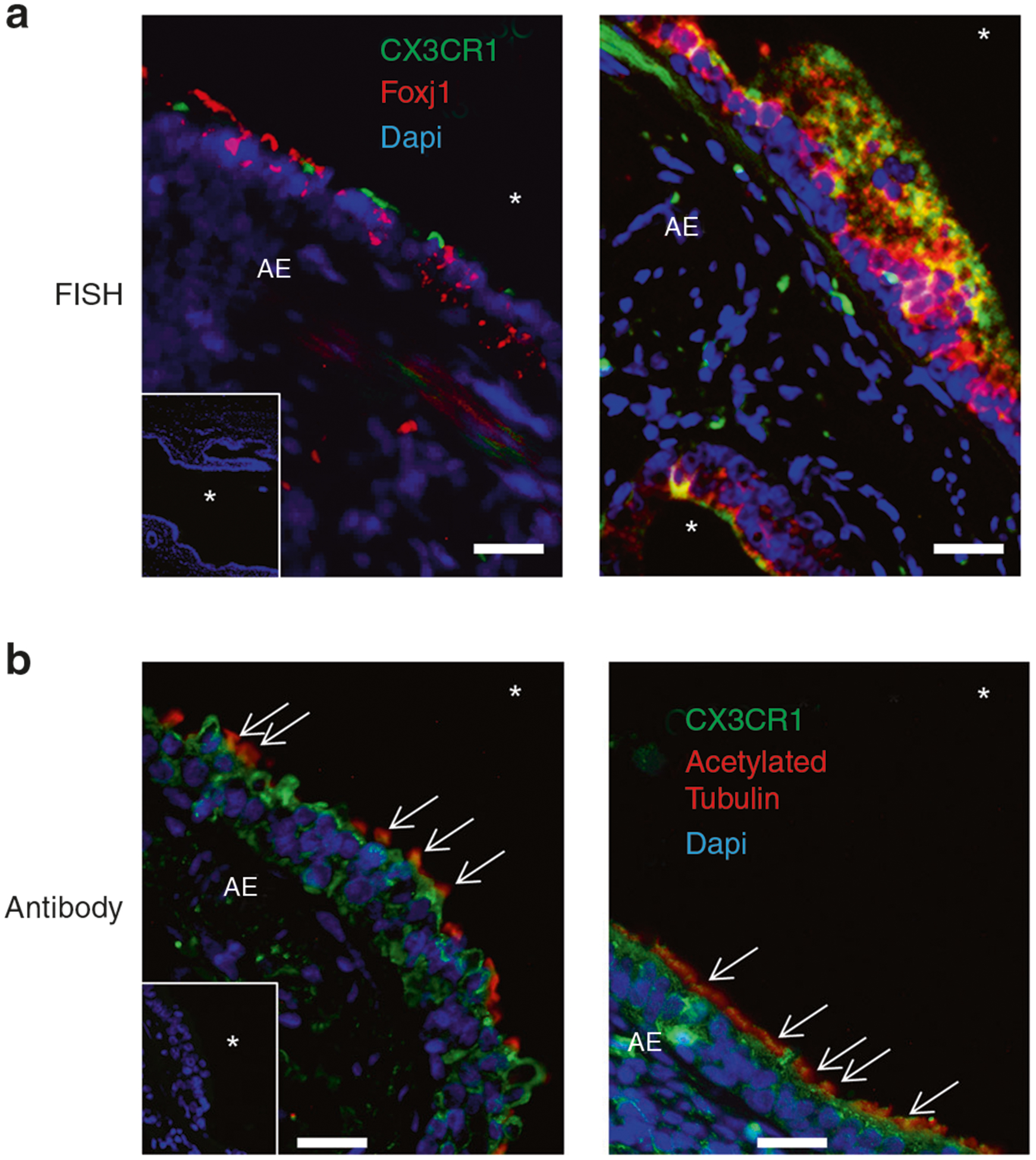
CX3CR1 expression in the pediatric airways. a FISH of pediatric lung samples probed for CX3CR1 (green), Foxj1 (red), and nuclei (blue) for two subjects. In-laid image: negative probe straining for green and red probes and stained for nuclei (blue). Representative images from tissue from two out of three subjects with two slides per subject performed in duplicate. b Lung specimens probed with anti-CX3CR1 antibody (green), anti-acetylated tubulin antibody (red), and counterstained with Dapi nuclear stain (blue). Representative images from tissue from two out four subjects with three slides per subject performed in duplicate. In-laid image: negative control probed with unimmunized rabbit IgG (green) or mouse IgG (red) and counterstained with nuclear stain (blue). Arrows indicate regions of CX3CR1/acetylated tubulin co-localization. White asterisk (*) denotes airway lumen.