


Figure 4. NSDHL deficiency protects from pancreatic adenocarcinoma development in Trp53+/− heterozygotes.
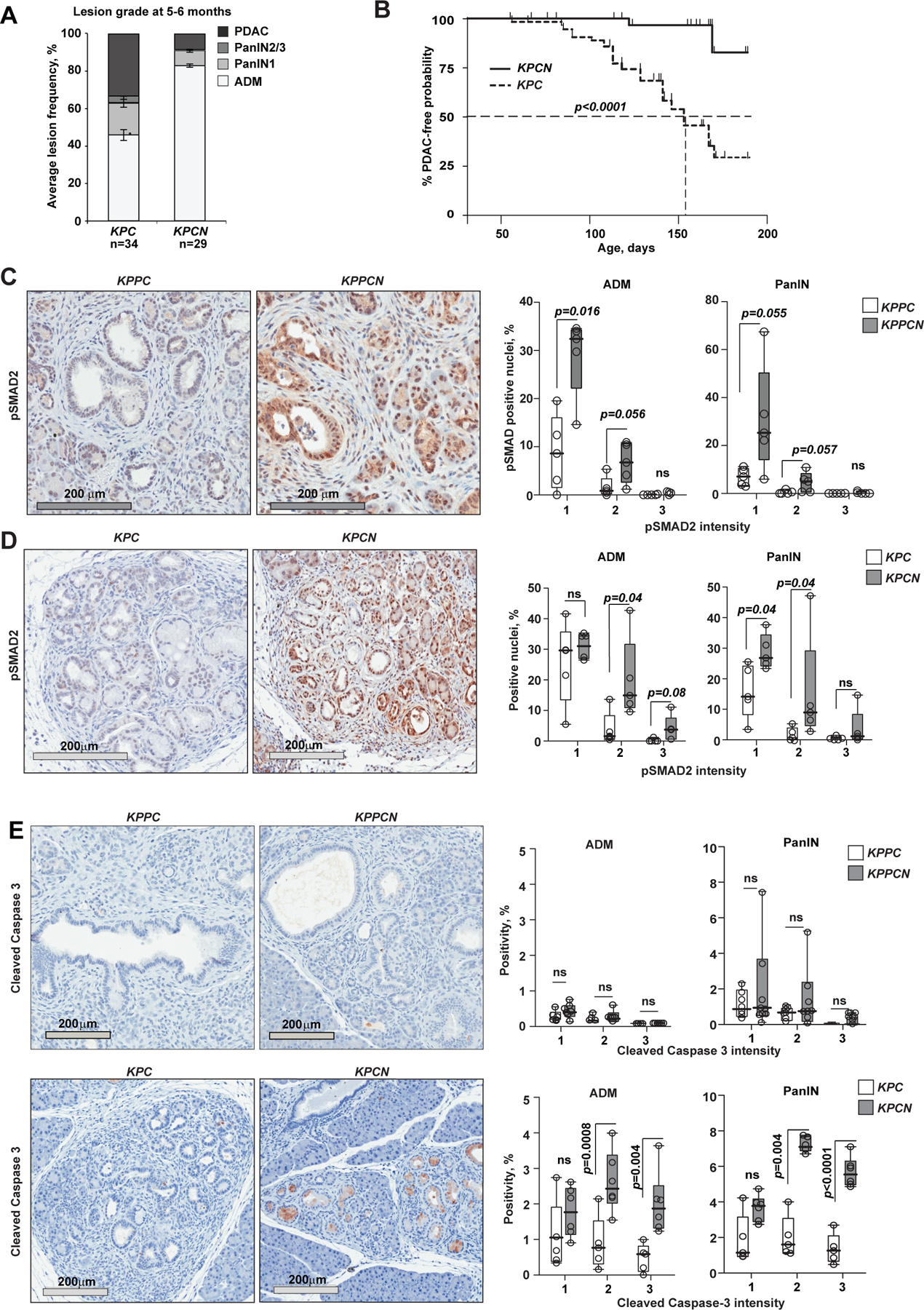
(A) Enumeration of pancreatic epithelial lesion by grade per section in KPC and KPCN mice aged 5–6 months; p=0.035 for PDAC (Fisher’s exact test); p=0.02 for PanIN2/3; p=0.0006 for ADM, Wilcoxon rank-sum test; error bars, SEM. (B) Kaplan-Meier representation of PDAC-free survival of KPC (n=34) and KPCN (n=37) mice. p<0.0001, logrank test. (C, D) Activated TGFβ pathway signaling in NSDHL-deficient pancreatic lesions with homozygous (C) and heterozygous (D) Trp53 knockout as assessed by phosphorylated SMAD2 immunohistochemistry. Top, 5–6 months old KPC and KPCN mice; bottom, 5 week old KPPC and KPPCN pancreatic lesions. Right panel, quantification of pSMAD2-positive nuclei in acinar-to-ductal metaplasia (ADM) and pancreatic intraepithelial neoplasm (PanIN) lesions. (E) Cleaved caspase 3 in pancreatic ADM and PanIN lesions. In C-E, staining intensity was quantified separately in ADM and PanIN lesions; p-values are calculated by Wilcoxon test; ns, not significant; data are represented as boxplots: median (black bar), box (25% to 75% confidence interval), whiskers (full range of measurements). See also Figure S4.