


Figure 1|. Steps in the development of brain metastases in an animal model.
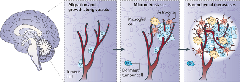
Brain metastatic cancer cells traverse the vascular system and use the outside of vessels as a site of adhesion and migration13,20. Later, the tumour cells use the inflamed brain microenvironment as a niche. Tumour cells interact with activated microglia (macrophage-like cells, shown in yellow) and astrocytes (shown in orange), which provide support for neuronal function. As the metastasis expands, neuronal damage ensues. The brain microenvironment also contains damaged axons, oedema (white halo) and vascular changes (such as the disruption of the blood-brain barrier, indicated by dashed black lines). Both vessel co-option and angiogenesis have been reported in brain metastasis. Dormant solitary tumour cells can also reside in the brain39, constituting a potential source for the development of additional metastases.