


Figure 7.
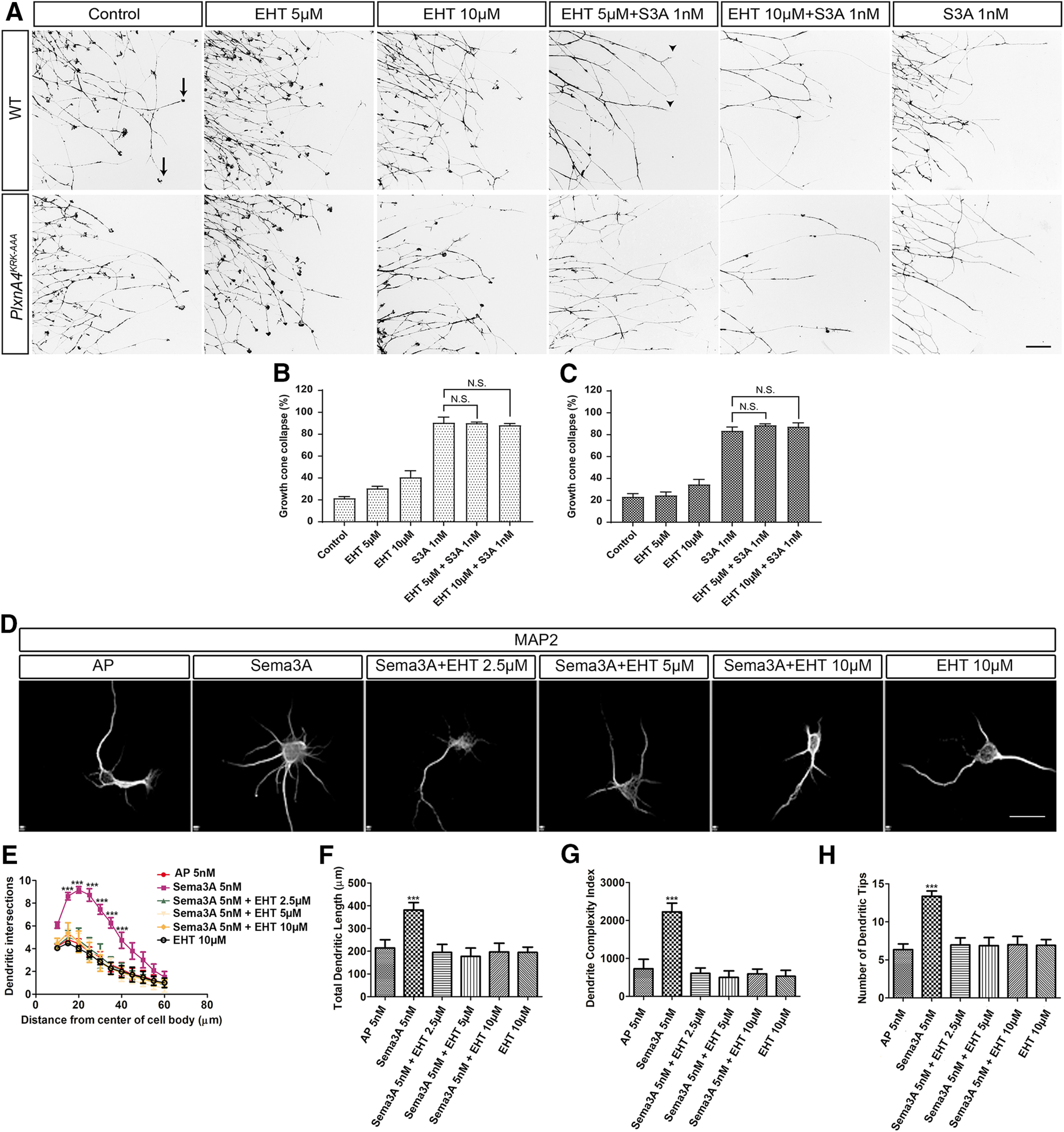
Inhibition of Rac signaling abolishes Sema3A-mediated cortical neuron dendrite elaboration but does not hinder Sema3A-dependent growth cone collapse of WT or PlxnA4KRK-AAA DRG axons in vitro. A, DRG explants from WT and PlxnA4KRK-AAA littermates E13.5 embryos were grown for 48 h, treated for 30 min with the pan-Rac inhibitor EHT 1864 at a concentration of 5 μm, 10 μm, or media alone as control. Then, 1 nm Sema3A or control conditioned media was added for 30 min, followed by fixation and phalloidin-rhodamine staining for assessment of growth cone collapse. Black arrows indicate intact growth cones. Arrowheads indicate collapsed growth cones. B, C, Quantification of collapse response as a mean percentage of collapsed growth cones out of the total ± SEM in WT and PlxnA4KRK-AAA axons, respectively; N.S., Non Significant. Scale bar, 50 µm. D, Representative confocal micrographs of dissociated primary cortical neurons obtained from WT E13.5 embryos. The neurons were treated with 5 nm AP, 5 nm Sema3A, 5 nm Sema3A + 2.5 μm EHT, 5 nm Sema3A + 5 μm EHT, and 5 nm Sema3A + 10 μm EHT. E-H, Sholl analysis of dendritic intersections (E), total dendritic length (F), the DCI (G), and number of dendritic tips (H) for all treatment conditions described above. Data are mean ± SEM from three independent cultures. ***p < 0.001 (two-way ANOVA with post hoc Tukey test). Scale bars: A, D, 25 µm.