


Figure 4.
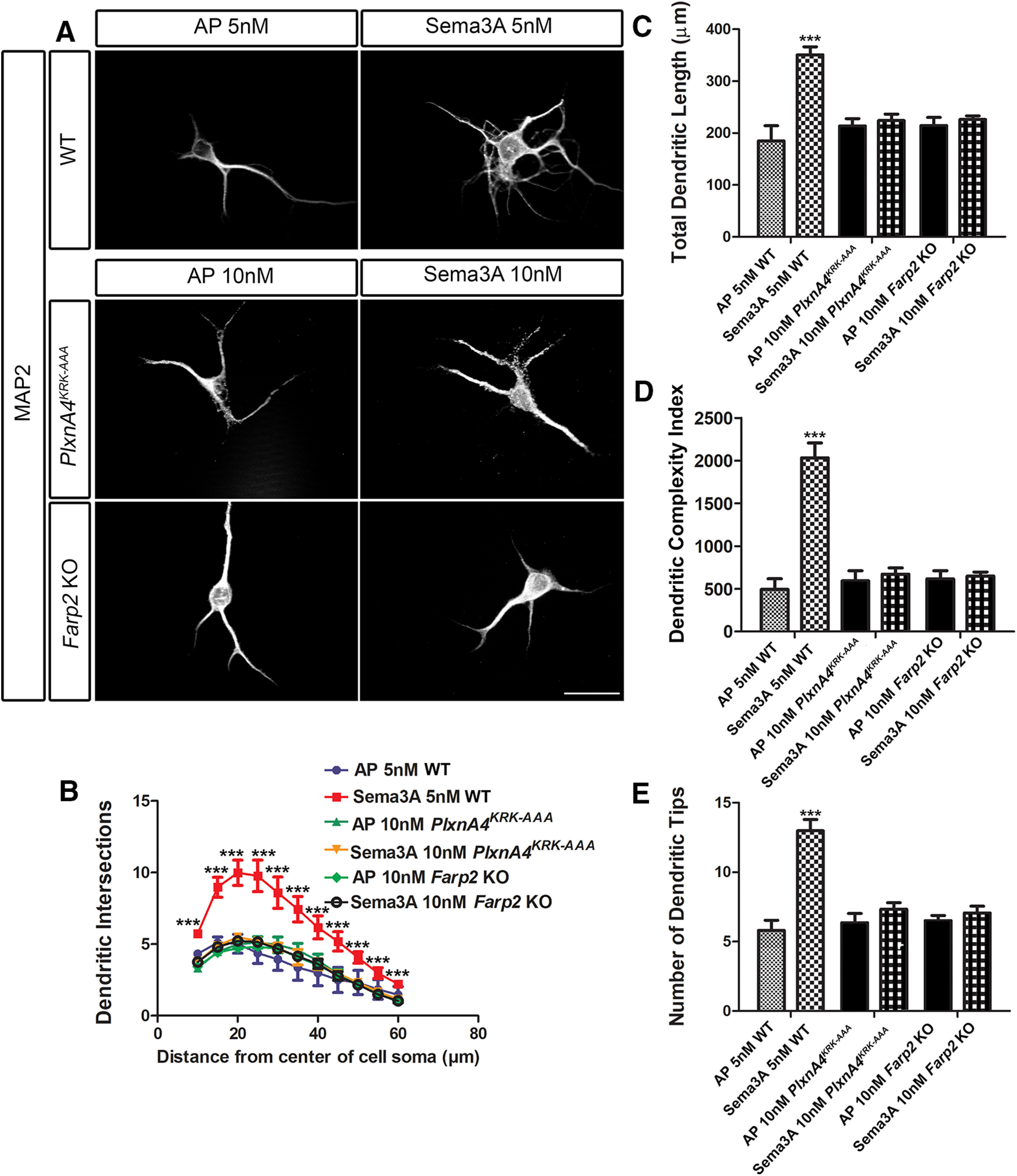
Both PlxnA4KRK-AAA and Farp2 KO cortical neurons are nonresponsive, even to higher concentrations of Sema3A. A, Representative confocal micrographs of dissociated primary cortical neurons obtained from WT, PlxnA4KRK-AAA, and Farp2 KO E13.5 embryos. The WT neurons were treated with AP or Sema3A 5 nm, and the PlxnA4KRK-AAA and Farp2 KO neurons were treated with AP or Sema3A 10 nm for 24 h. B–E, Sholl analysis quantified dendritic growth and branching in WT, PlxnA4KRK-AAA, and Farp2 KO cortical neurons (B), measuring the number of dendritic intersections, the total dendritic length (C), the DCI (D), and the number of dendritic tips (E). Data are mean ± SEM from three independent cultures of each genotype. ***p < 0.001 (two-way ANOVA with post hoc Tukey test). Scale bar, 25 µm.