


Extended Data Figure 6. Structural agreement of hexapeptide prion zipper structures to rPrPRes.
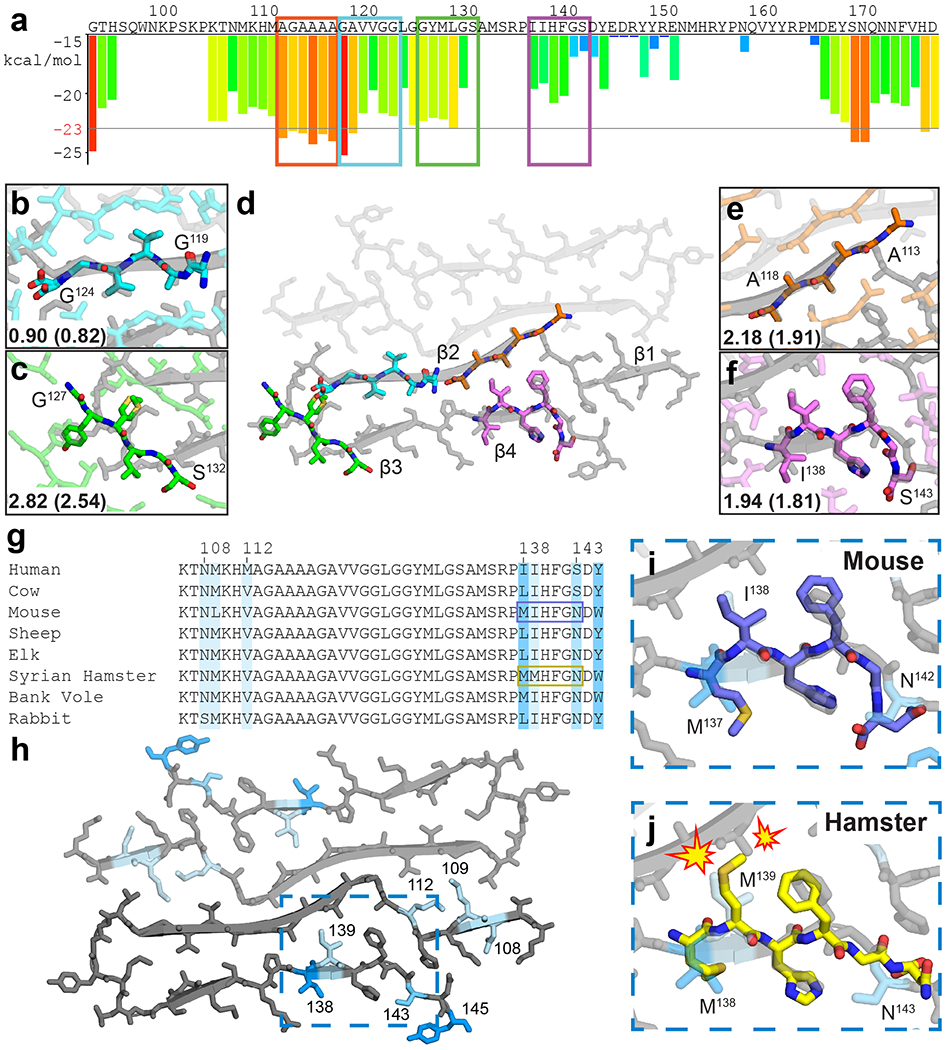
(a) ZipperDB26 profile of the segment encoding for rPrPRes. Bars extending below the line represent hexapeptides predicted to form steric zippers. Peptides with crystal structures aligned to the fibril model are boxed. Two of these were previously published27,28. Alignment of crystal structures 119GAVVGG124 (b), 113AGAAAA118 (c), 127GYMLGS132 (e) and 138IIHFGS143 (f) with rPrPRes (d). Symmetry mates in plane, all atom, and backbone (parentheses) RMSD values against rPrPRes are shown (g) Sequence alignment for species of common interest to prion disease. Darker blues correlates with more variability, loosely defined as more residue mismatches among the species compared, and mapping of these residues onto rPrPRes (h). Hexapeptide structure for muPrP137-142 (mouse numbering) is compatible with rPrPRes (i) while shPrP138-143 is not (j).