


Fig. 4. Mertk knockout disrupts astrocyte lysosome polarization toward dying cells.
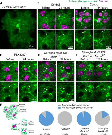
(A) Labeling of astrocyte lysosomes after injection of AAV5.LAMP1-GFP into the subarachnoid space in the adult mouse cortex. (B) Astrocyte response to dying cells in the presence of microglia in adult wild-type mice. Under normal conditions, astrocyte lysosomes exhibited little to no polarization around the dying cell with condensed nuclei (white arrow). (C) After PLX3397-mediated microglia ablation, astrocyte processes polarized lysosomes to gradually digest dying cells (white arrows). (D) Astrocytes failed to polarize lysosomes around dying cells in Mertk−/− mice. KO, knockout. (E) When Mertk is deleted exclusively from microglia (Csf1r-cre:Mertkfl/fl), astrocytes polarized lysosomes around the cell corpses, consistent with their response when microglia were completely absent. (F) Graphical representation and quantification of lysosome-filled thick astrocyte processes polarized around cell corpses in all groups (control, PLX3397-treated, germline Mertk deletion, and microglia-specific Mertk deletion, n = 3 mice per group).