


Fig. 2. Territorial segregation of glial processes during cell corpse engulfment.
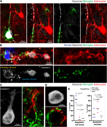
(A) Confocal fluorescence image showing division of labor between microglia (green) and astrocytes (red) during elimination of a neuron (white, yellow arrow) and its apical dendrite, after cell death induction by high-titer adeno-associated virus-9 (AAV9)–green fluorescent protein (GFP) infection. The microglia has converged on the soma and apical dendrite (yellow arrowheads), while the astrocyte surrounds the distal dendrite (white arrowheads), with boundaries observed between processes of these glial cells. (B) Confocal fluorescence image of spontaneous cell death during postnatal development in mouse cortex. The soma (yellow arrow) and neurites (blue arrows), labeled via caspase-3, are engulfed by microglia (green), while the distal dendritic processes are engulfed by astrocytes (red). Intermingling of both glia processes is observed in the middle portion of the dendrite. (C and D) Examples of cells labeled by caspase-3 immunofluorescence showing astrocytes (red) and microglia (green) polarized toward different parts of the dying cell with territorial boundaries (white arrowheads). (E) Quantification of the relative engulfment of cell bodies and dendrites by astrocytes and/or microglia following cell death by high-titer AAV9-GFP infection (n = 3 mice per group, greater than 50 apoptotic neurites and 20 cell bodies per mouse). Statistics with two-way analysis of variance (ANOVA) with Holm Sidak’s multiple comparisons test.