


Figure 2.
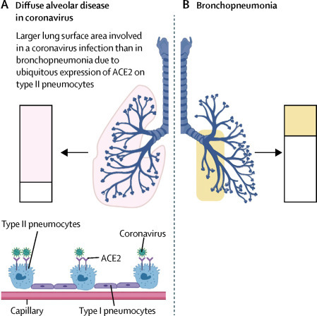
Extent of alveolar lung surface involved in COVID-19 and bronchopneumonia
(A) Some coronavirus family members gain access to the lungs via the ACE2 receptor that is expressed most abundantly on a subpopulation of type II pneumocytes. Shaded boxes indicate the much greater capability for immunothrombosis given the alveolar tropism of SARS-CoV-2. (B) Segmental bronchopneumonia (eg, bacterial and influenza) typically has a different lung distribution, with prominent bronchial tree involvement, including haemorrhagic destruction of trachea and large airways,20 and generally patchy alveolar network disease. There might be large areas with normal perfusion. The slow evolution of COVID-19 with alveolar hypoxia and microthrombosis might result in pulmonary arterial hypertension and the cardiac picture that mostly occurs with hypoxaemia and raised D-dimers. The scale of alveolar and microvascular inflammation, rather than systemic viral infection per se, determines the cardiovascular pattern of disease. Nonetheless, segmental bronchopneumonia could result in a degree of immunothrombosis sufficient to cause a cardiac event, especially in older patients with known or silent cardiac disease. ACE2=angiotensin-converting enzyme 2. COVID-19=coronavirus disease 2019. SARS-CoV-2=severe acute respiratory syndrome coronavirus 2.