


Figure 2. Consciousness level modulated spike rate and timing in deep cortical layers and CL.
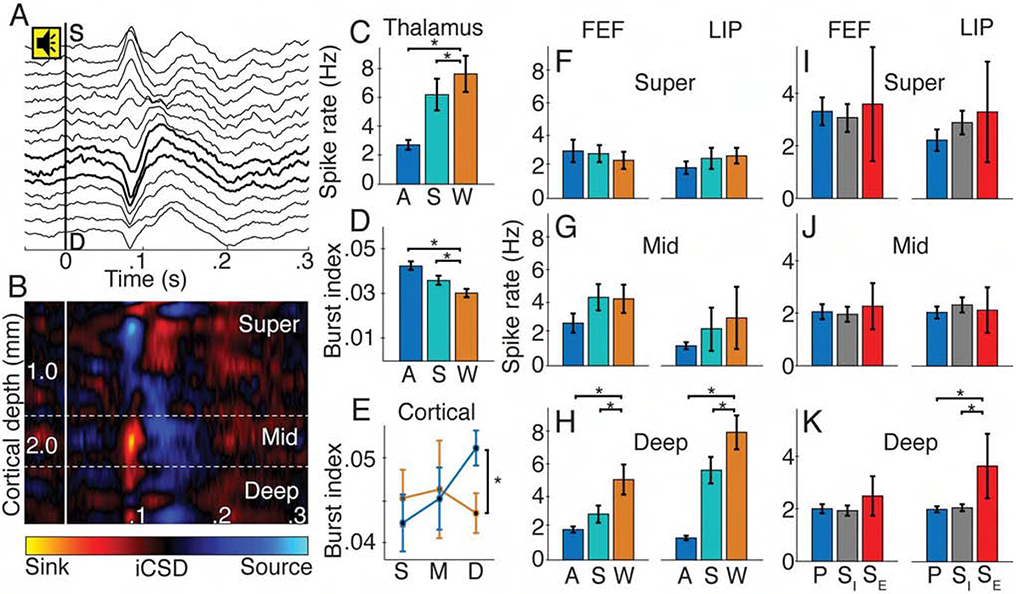
(A) Example sound-aligned evoked potentials from linear multielectrode array in FEF. Tone onset at 0 s. Bold lines show iCSD-defined middle layers. (B) Sound-aligned iCSD corresponding to A. (C) Population CL spike rate (±SE) and (D) CL burst index (±SE) for anesthesia (blue), sleep (teal), and wake (orange) states; * p < 0.05. (E) Cortical (FEF and LIP) burst index (±SE) in superficial (S), middle (M) and deep (D) layers for wakefulness (orange) and anesthesia (blue). (F) Superficial, (G) middle and (H) deep layer spike rates (±SE) in FEF and LIP across states. (I) Superficial, (J) middle and (K) deep spike rates (±SE) in FEF and LIP during effective stimulation (SE, red), ineffective stimulation (SI, gray) and pre-stimulation (P, blue).