


Figure 2.
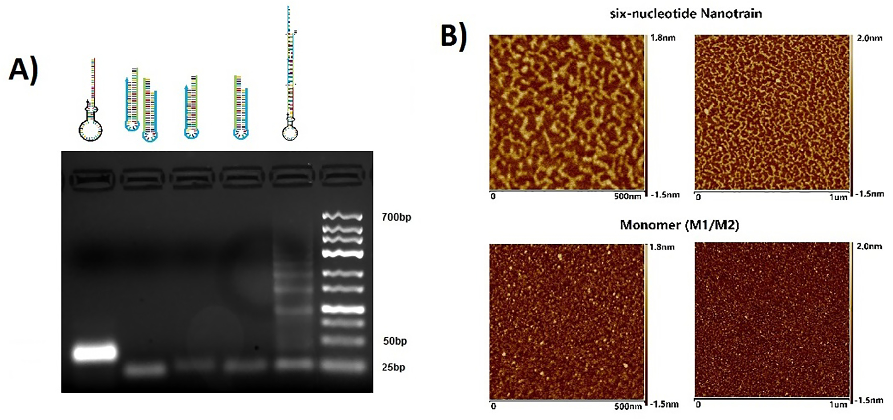
Characterization of nanotrain assembly. (A) Agarose gel image shows migration of (left to right) 6N-Apt-trigger, mixture of 6N-M1 and 6N-M2, 6N-M1, 6N-M2, 6N-Apt-NTrs, DNA ladders. (B) Atomic force microscopy images show the formation of 6-letter nanotrains (top two images) compared to the 6-letter monomers (bottom two images).