


Fig. 6.
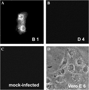
scFv B1 staining SARS pseudovirus in infected Vero E6 cell. (A) scFv B1 but not (B) scFv D4 recognizes SARS pseudovirus in the infected Vero E6 cells. (C) scFv B1 does not stain the mock-infected Vero E6 cells. (D) Pseudovirus-infected Vero E6 cells observed under a phase contrast microscope. Lus/SARS-CoV S protein mammalian expression vector pMT 21-S and pCMVR 8.2 coding for MuLV Gag and Pol, and pHR′-luc coding luciferase cotransfected into 293T cells. After 48 h culture, Vero E6 monolayer cells were fixed and blocked for incubation with purified scFv, and then mouse anti-His-Tag antibody, and finally FITC-labeled anti-mouse IgG. Binding to SARS S pseudovirus in Vero E6 cells was visualized under a fluorescence microscope.