


FIG 2.
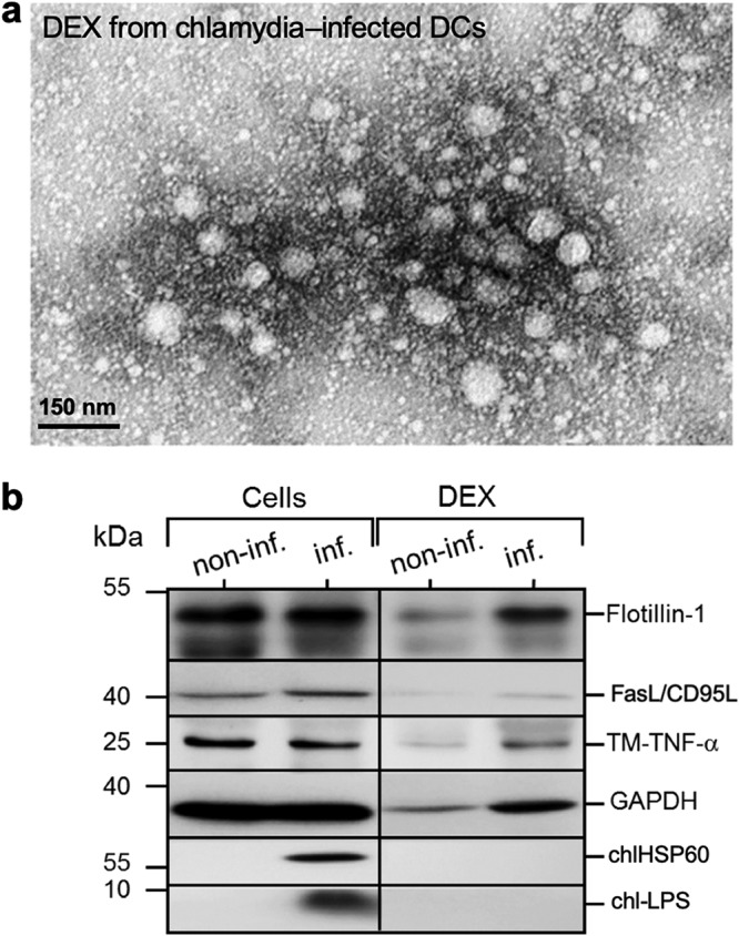
Microscopic and molecular characterization of dexosomes (DEX) released by infected DCs. (a) A TEM image of purified DEX prepared with ExoQuick-TC kit (System Biosciences). (b) Analysis of the detection of distinct DEX proteins. DEX were isolated from the supernatant of Chlamydia-infected DCs (MOI of 10, 48 hpi) using an ExoQuick-TC kit, lysed, and then analyzed by Western blotting with staining for Flotillin-1, FasL/CD95L, TM-TNF-α, GAPDH, Chlamydia HSP60 (chlHSP60), and LPS (chl-LPS).