


Figure 5.
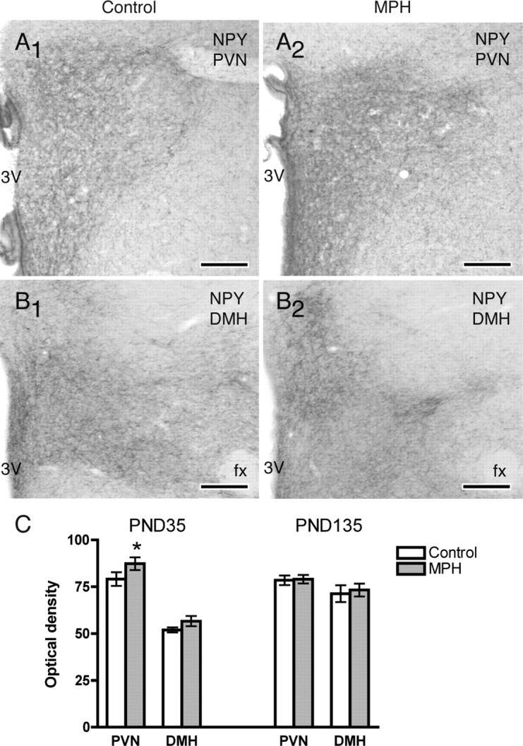
In the hypothalamus, the density of NPY-ir in the PVN and DMH increased with MPH administration (PND35). A, At PND35, increases in the density of NPY-immunoreactive fibers in the PVN were seen in MPH-administered rats (A2) compared with controls (A1). B, In the DMH, MPH-administered rats (B2) showed a more widespread distribution of NPY-labeled fibers than controls (B1). Atlas levels 26 (AP, −1.78 mm from bregma; A1, A2) and 30 (AP, −3.25 mm from bregma; B1, B2) are shown (Swanson, 1992). 3V, Third ventricle; fx, fornix. Scale bars, 500 μm. C, Comparisons of the density of NPY-immunoreactive fibers in the PVN and DMH in control and MPH-exposed rats at PND35 and PND135. A significant (*) increase in NPY-labeled fibers was seen in MPH-administered rats at PND35. *p < 0.05 (Wilcoxon test); n = 6 rats per condition.