


Fig. 14.
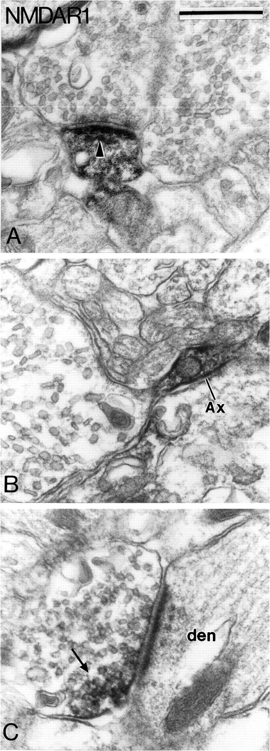
Subcellular localization of NMDAR1 subunit immunoreactivity in the SNc-v. A shows an immunoreactive dendrite that forms an asymmetric synapse (arrowhead) with an unlabeled bouton. B illustrates an immunoreactive preterminal axonal segment that gives rise to a large terminal devoid of immunoreactivity. C depicts an immunoreactive terminal bouton that forms an asymmetric axo-dendritic synapse. Note that the reaction product is confined mainly to a small part of the terminal (arrow). Scale bar (shown inA): 0.5 μm.