


Figure 2.
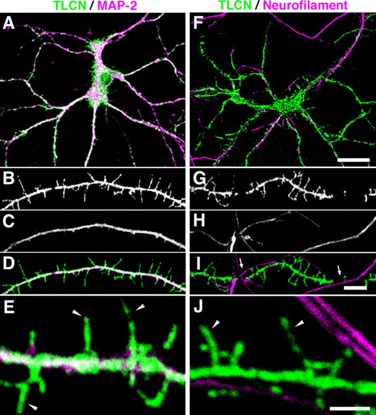
Dendrite-specific localization of TLCN in cultured hippocampal neurons. A–J, Cultured hippocampal neurons at 14 DIV were double-immunostained for TLCN and MAP-2 (dendritic marker) (A–E) or neurofilament (axonal marker) (F–J). In the merged images, TLCN (green) is colocalized with MAP-2 (A, D, E, magenta) but not with neurofilament (F, I, J, magenta). TLCN is frequently excluded from dendritic segments where axons are crossing over (I, arrows). High-power views (E, J) show localization of TLCN on dendritic filopodia (arrowheads). Scale bars: A, F, 30 μm; B–D, G–I, 5 μm; E, J, 2 μm.