


Figure 5.
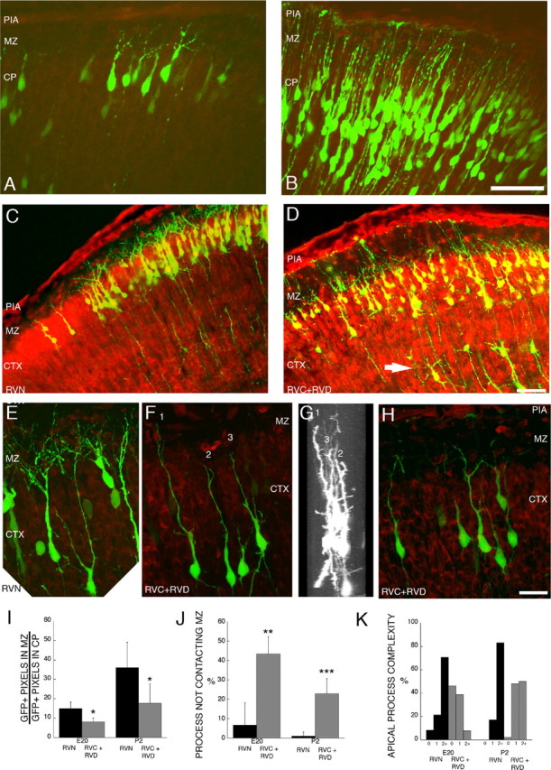
Alterations of leading process and apical dendrite morphology after treatment with RNAi directed against Dab1. Sections stained with the nucleic acid dye propidium iodide (red) from embryos electroporated at E16 and examined at E20 and P2. A, Control (RVN) neurons electroporated at E16 show branched neurites at E20. B, RVC+RVD-treated neurons show less branching in the apical process contacting the MZ. C, D, Low-magnification images of layer 2/3 cortical neurons on P2, 7 d after in utero electroporation on E16. Control RVN electroporated neurons show precise lamination and exuberant dendritic growth in the MZ on P2 (C), whereas Dab1-suppressed cells (RVC+RVD) show disrupted lamination with occasional ectopic deep cells (arrow) and sparse dendrites in the MZ (D). C–F, Flattened z-series of P2 layer 2/3 neurons revealing extensive dendrites in the RVN-treated cells (E) and stunted dendrites that either do not penetrate the MZ (cells 2 and 3) (F–H) or stunted dendrites that do not show extensive secondary and tertiary branching in the MZ (H). G, A 90° rotation of D reveals that cells 1–3 are entirely contained in the optical section and that the stunted dendrites are not attributable to sectioning artifact. Scale bars: B, D, 50 μm; H, 20 μm. I, Quantification of GFP+ pixels in the MZ at E20 and P2, after normalization (see Materials and Methods). Control (RVN) embryos showed approximately twofold more neurites in the MZ compared with RVC+RVD-treated embryos at both E20 (p = 0.014, unpaired t test; df = 6) and P2 (p = 0.025, unpaired t test; df = 7). Single sections from nine RVC+RVD embryos (n = 4 at E20; n = 5 at P2) and eight RVN embryos (n = 4 at E20; n = 4 at P2) were analyzed. J, Quantification of noncontacting apical processes after RVC+RVD treatment shows RNAi-mediated difference at both E20 (p = 0.012, unpaired t test; df = 4) and P2 (p = 0.001, unpaired t test; df = 6). K, Quantification of apical process branching complexity from unbranched (0th order) to second-order and above (2+) branching in RVN- and RVC+RVD-treated cells at E20 and P2. RVC+RVD-treated cells showed less branching than RVN-treated cells at both time points. At E20, morphological analysis was performed on 24 cells in three RVN-treated brains and 26 cells in three RVC+RVD-treated brains. At P2, morphological analysis was performed on 47 cells in five RVN-treated brains and 44 cells in three RVC+RVD-treated brains. Values in I and J are means ± SD. CTX, Cortex.