


Fig. 3.
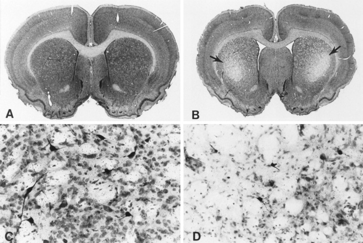
Nissl and NADPH–diaphorase double-stained coronal brain sections at the level of the striatum and anterior commissure from a creatine-fed rat (A, C) and a non-creatine-fed rat (B, D). Rats prefed 1% creatine with subsequent administration of 3-nitropropionic acid resulted in no demonstrable striatal lesions (A) or neuronal loss or alteration (C). In contrast, treatment with 3-nitroproprionic acid alone resulted in bilateral striatal lesions within the caudate–putamen (arrows) (B) with marked neuronal loss and relative preservation of NADPH-diaphorase neurons (dark-stained neurons) (D). Note the ventricular enlargement attributable to striatal atrophy in the 3-nitroproprionic acid-treated rat.