


Fig. 5.
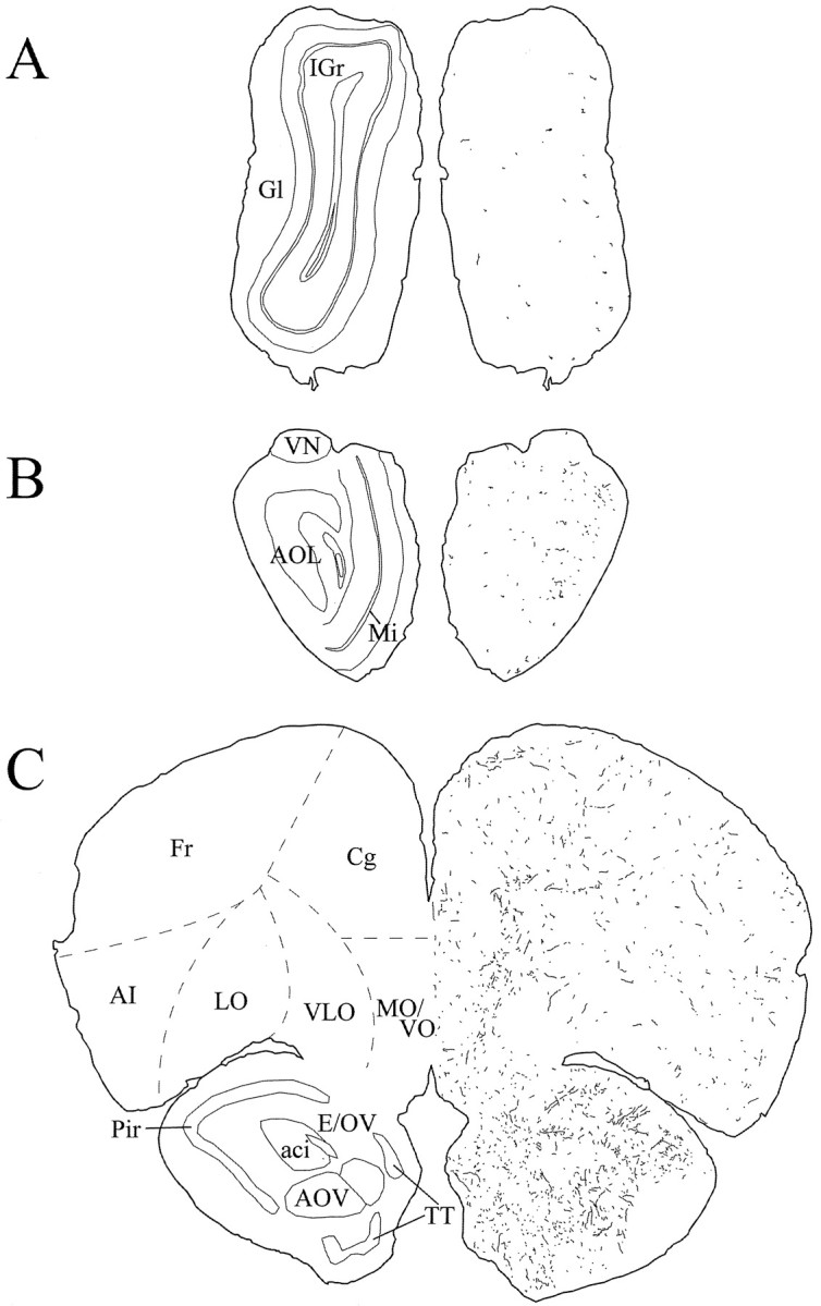
Schematic drawings of 20 μm rostrocaudal coronal sections illustrating the distribution and relative density of hcrt fibers in the prefrontal cortex and the olfactory bulb after immunohistochemistry for hcrt using antibody #2050. aci, Anterior commissure, intrabulbar part;AI, agranular insular cortex; AOL, anterior olfactory nucleus, lateral part; AOV, anterior olfactory nucleus, ventral part; Cg, cingulate cortex;E/OV, ependyma and subependymal layer/olfactory ventricle; Fr, frontal cortex; Gl, glomerular layer of the olfactory bulb; IGr, internal granular layer of the olfactory bulb; LO, lateral orbital cortex; Mi, mitral cell layer of the olfactory bulb; MO/VO, medial/ventral orbital cortex;Pir, piriform cortex; TT, tenia tecta;VLO, ventrolateral orbital cortex; VN, vomeronasal nerve layer.