


Fig. 9.
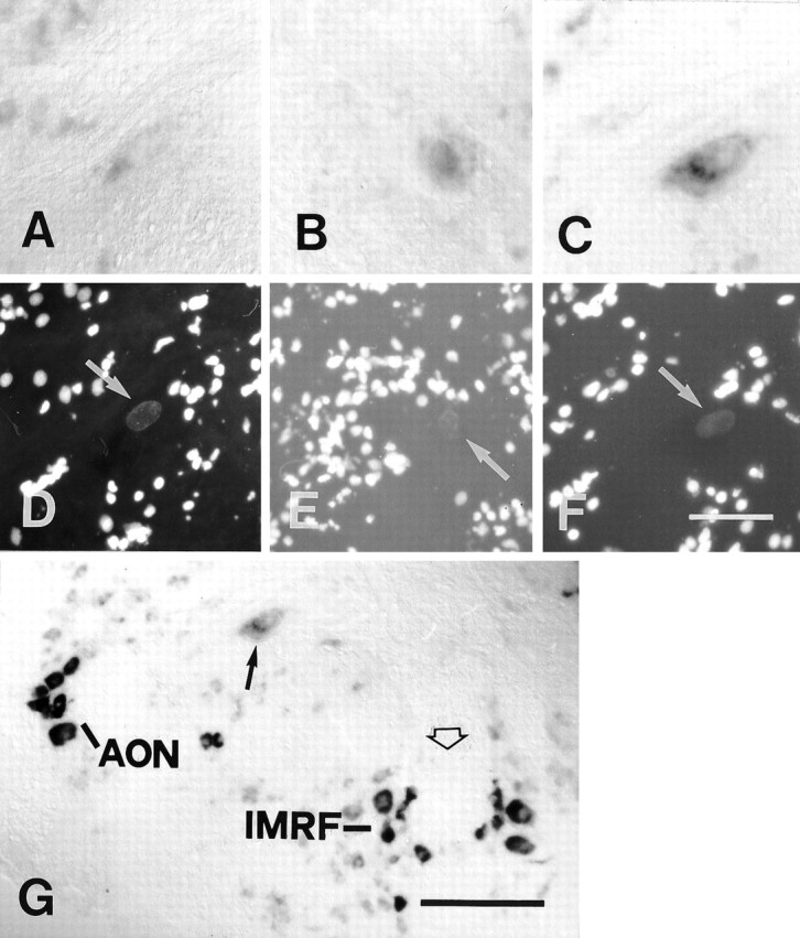
A–G, Labeling for GAP-43 mRNA in the Mauthner cell was not significantly increased after distal and proximal lesion. All images are cross sections. A–C andG show in situ hybridization;D–F show nuclear fluorescence images corresponding toA–C and are included for orientation.Arrows in D–F point to the cell nucleus of the Mauthner cell. A, D, In an unlesioned control animal only weak in situ labeling was detected.B, E, At 14 d after a distal lesion, in situ labeling was faint. C, F, G, At 14 d after a proximal lesion, a slight increase in in situlabeling was seen occasionally (C, F). This increase appeared very weak, however, when compared with that in axotomized neurons that regenerated their axons consistently (seeG, a low-power view of the same section), i.e., the anterior octaval nucleus (AON) or the intermediate reticular formation (IMRF).Arrow in G points out the Mauthner cell, and the open arrow in G depicts the midline of the brain. Scale bar (shown in F forA–F): 50 μm; G, 150 μm.