


Fig. 5.
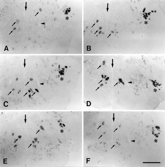
A–F, Expression of L1.1 mRNA was increased between 3 and 56 d after distal lesion. All images are cross sections; dorsal is up. A, Unlesioned control;B, 3 d post-lesion; C, 7 d post-lesion; D, 14 d post-lesion; E, 56 d post-lesion; F, 84 d post-lesion.Large arrows indicate the midline of the brain.Small arrows point to individual cells in the intermediate reticular formation. Asterisks indicate the trigeminal motor nucleus, which constitutively shows intense labeling for L1.1 mRNA as an internal positive control.Arrowheads in A, C, D, andF indicate the ventral dendrite of the Mauthner cell as an additional landmark. A, Labeling was faint in unlesioned controls. B–E, Expression of L1.1 mRNA was increased between 3 and 56 d post-lesion. F, Staining intensity was similar to that in unlesioned controls at 84 d post-lesion (compare with A). Scale bar (shown in F for A–F): 150 μm.