


Fig. 1.
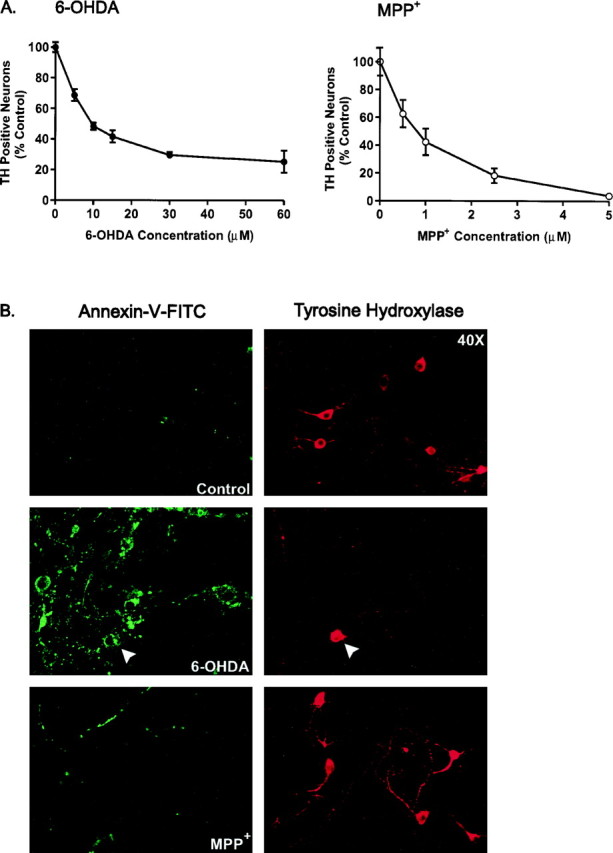
Plasma membrane alterations induced by 6-OHDA and MPP+. A, Cultures were treated with various concentrations of 6-OHDA or MPP+ for 24 and 48 hr, respectively, and processed for TH immunoreactivity, and the number of TH-positive neurons was counted. Data are normalized to control cultures and denote the mean ± SEM of representative determinations made in three separate cultures. Bars with <2% SEM are buried within the symbols. Cells exposed to 6-OHDA or MPP+ exhibited a half-lethal dose of 15 μm and 1 μm, respectively.B, Representative confocal micrographs of mesencephalic cultures stained for TH and annexin-V-FITC visualized at 40× magnification by confocal microscopy. Cultures were treated with 15 μm 6-OHDA or 1 μm MPP+for 6 hr, rinsed, and incubated with annexin-V-FITC for 10 min at room temperature. After imaging, plates were fixed and processed for TH immunoreactivity using a CY3-coupled secondary antibody. Random micrographic fields of annexin-V-stained neurons were relocated by their position on a microwell grid, and the corresponding image of TH immunoreactive neurons was taken.