


Fig. 6.
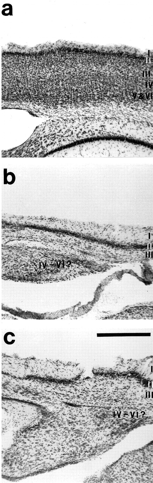
a, Photomicrograph of normal adult neocortex that has been cut coronally and stained for Nissl. The photomicrographs in b and c are from an adult neocortex where a moderate cortical neuroepithelial removal was made at P4. Cortex in this region appears normal in terms of laminar organization (b), although this was not the case for the entire extent of the reduced cortex. In some portions of the cortex, particularly the region toward the caudal end of cortex (c), the laminar organization of the cortex was more disrupted. Dorsal is up; lateral is to theright. Scale bar, 1 mm.