


Fig. 2.
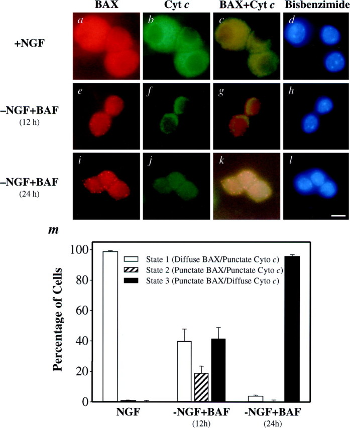
BAX and cytochrome c undergo subcellular redistribution during NGF deprivation in sympathetic neurons. Shown are representative photomicrographs of neurons immunostained for BAX (a, e, i), cytochrome c (b, f, j), or both (c, g, k) and the corresponding bisbenzimide-stained nuclei (d, h, l). Neurons either were maintained in NGF (a–d) or were deprived of NGF in the presence of 50 μm BAF for 12 hr (e–h) or 24 hr (i–l). The cells were fixed and immunostained for BAX and cytochrome c. Then the number of neurons (m) exhibiting diffuse BAX and punctate cytochrome c staining (State 1), punctate BAX and punctate cytochrome c staining (State 2), or punctate BAX and diffuse cytochrome cstaining (State 3) was determined, with 100–150 cells counted for each time point. Mean ± SEM; n ≥ 3. Scale bar, 10 μm.