


Fig. 4.
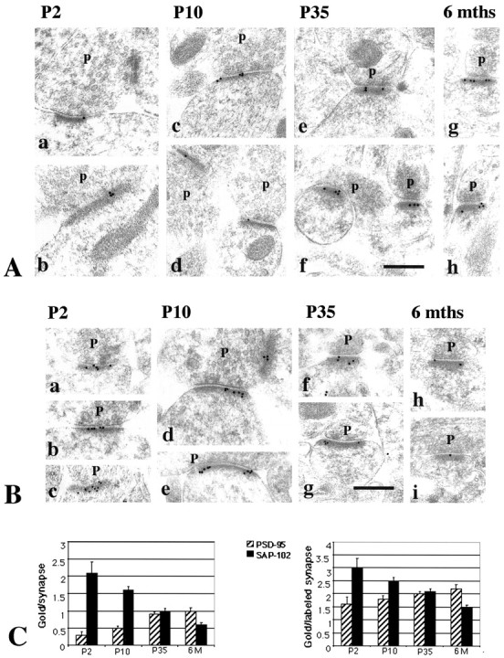
A, Immunogold labeling of PSD-95 in the CA1 stratum radiatum of the hippocampus. Postembedding immunogold labeling (10 nm) was performed using either of two PSD-95 antibodies (b–e, g, h, M, TL; a, f, R, JH), at P2 (a, b), P10 (c, d), P35 (e, f), and 6 months (g, h).p, Presynaptic terminal. Postsynaptic structures include dendrite shafts (b, synapse in a,top right) and spines (c–h); the synapse in the bottom left of a is at the enlarged base of a filopodium extending to the left (data not shown). Scale bar, 0.3 μm. Representative micrographs were chosen to illustrate the increase in PSD-95 labeling in synapses with age.B, Immunogold labeling of SAP-102 in the CA1 stratum radiatum of the hippocampus. Postembedding immunogold labeling (10 nm) was done using either of two SAP-102 antibodies (a, b, d–f, h, i, R, JH; c, g, GP-DB) at P2 (a–c), P10 (d, e), P35 (f, g), and 6 months (h, i). p, Presynaptic terminal. Postsynaptic structures include dendrite shafts (b), protuberances (a, c), and spines (d–i). Scale bar, 0.3 μm. Representative micrographs were chosen to illustrate the decrease in SAP-102 labeling in synapses with age. C, Summary of developmental changes in the number of gold particles per synapse and the number of gold particles per labeled synapse (see Table 2) at P2, P10, P35, and 6 months (6M). Error bars indicate SE.