


Fig. 5.
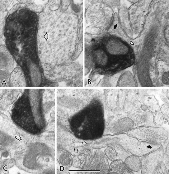
Electron micrographs showing symmetrical synapses of axon terminals used for 3-D reconstructions. A,B, The symmetrical synapses in Figure 4, A andC (FS cell number 1), on the dendritic shaft (open arrow) and on the spine neck (open arrow), respectively. B, Filled arrow, An asymmetrical synapse on the spine head. C, D, The symmetrical synapses in Figure 4, D and F (LTS cell number 1), on the dendritic shaft (open arrow) and on the spine neck (open arrow), respectively. D, Filled arrow, An asymmetrical synapse on the spine head.D, Double arrows, A spine apparatus. Scale bar, 0.5 μm.