


Fig. 3.
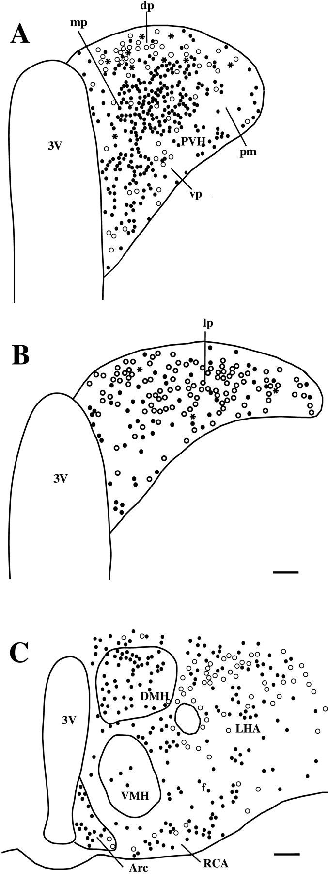
A series of line drawings demonstrating the distribution of Fos-ir (black circles), Fluorogold-ir (white circles), and double-labeled (black stars) neurons in the middle level of the PVH (A), the caudal PVH (B), and the lateral hypothalamic area and arcuate nucleus (Arc) (C), after intravenous injection of LPS, in animals with Fluorogold injected into the T5–T10 levels of the spinal cord. Notice that although retrogradely labeled neurons (whitecircles) are widely distributed in the hypothalamus, most double-labeled neurons were seen in the dorsal parvicellular PVH (dp; A), although a few were seen in the lateroposterior parvicellular PVH (lp;B). DMH, Dorsomedial hypothalamic nucleus; RCA, retrochiasmatic area; VMH, ventromedial hypothalamic nucleus. Other abbreviations are defined in Figure 2 legend. Scale bars: A, B, 75 μm; C, 250 μm.