


Fig. 2.
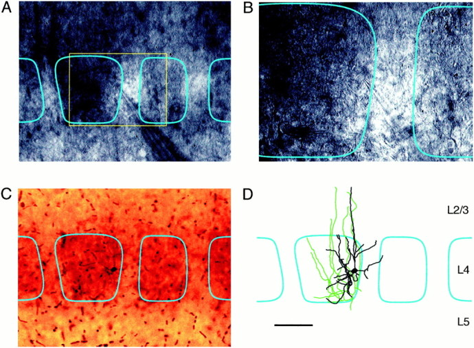
Whole-cell recordings from identified neurons in a specific region of a barrel. Barrels within layer 4 can by visualized at high magnification by enhancing contrast through video microscopy, allowing whole-cell recordings to be made from a given region of a barrel. The neuron in this example is located on the right-hand of the barrel as seen with 10× (A) and 40× (B) water immersion lenses. During the electrophysiological recording the cell is filled with biocytin, allowing the neuron to be visualized later in conjunction with a cytochrome C stain (C). These stains confirm the location of the neuron within the barrel and allow the axonal (green) and dendritic (black) arborizations to be reconstructed (D) in relationship to the barrel boundaries (cyan). Scale bar:A, C, D, 100 μm;B, 50 μm.