


Fig. 1.
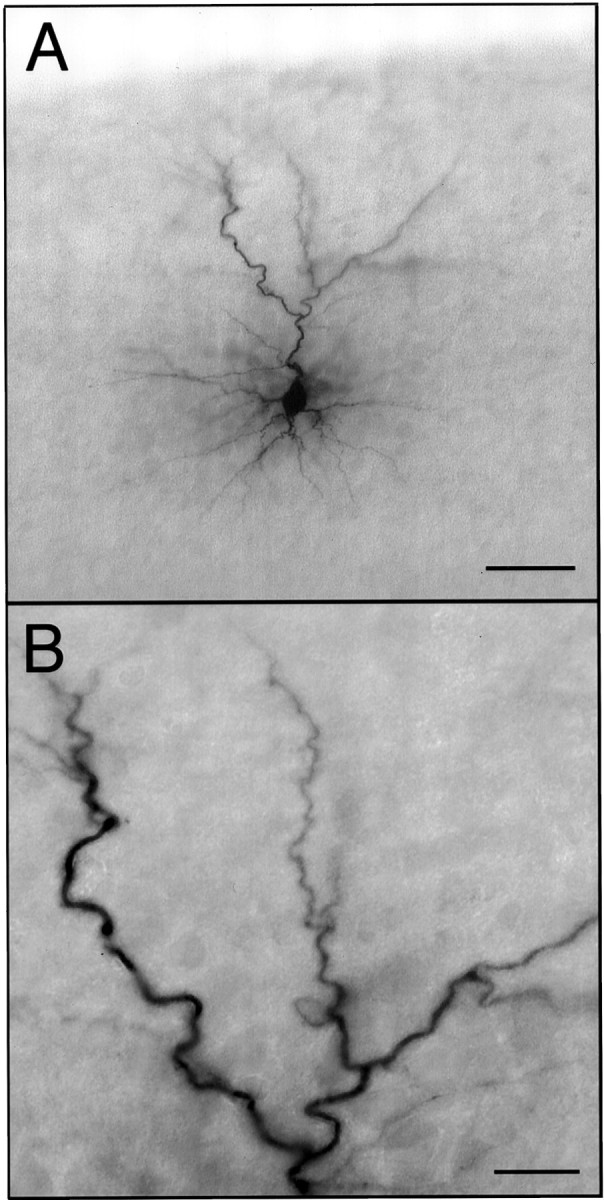
Labeled layer 2 pyramidal neuron.A, The neuron shown is representative of the intracellularly labeled neurons analyzed in this study. The pial surface of the slice is visible at the top of the micrograph.B, A higher-magnification micrograph of a portion of the apical dendritic arbor illustrates the completeness of labeling. Scale bars: A, 50 μm; B, 15 μm.