


Fig. 7.
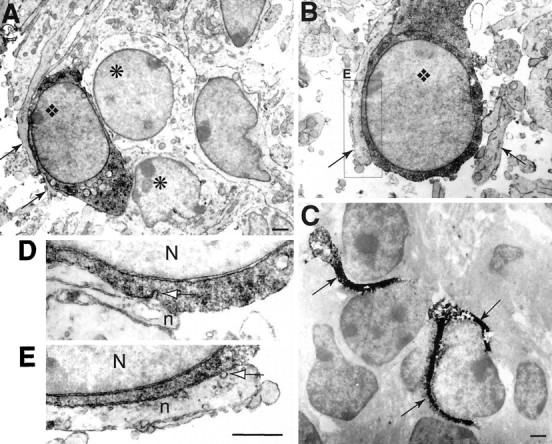
Relationships between neurites and calbindin-positive cells in IZ and CP, as revealed in electron microscopy. A, B, In both CP (A) and IZ (B), calbindin-positive cells (diamond) filled by the dark DAB reaction product are in close contact with neurites (arrows) over long distance. In CP (A), a calbindin-positive cell also contacts unlabeled cells (asterisk). C,In IZ, photoconverted DiI-labeled cortical fibers (DAB reaction product in black) cover cell bodies. D, E, At high magnification (56,000×), no membrane thickening is visible in the region of close apposition between neurites (n) and cell body of the dark-labeled calbindin-positive cells (N, nucleus). In D, the plasmic membrane of the calbindin-positive cell is invaginated in a region apposed to a neurite (white arrow). In E (detail ofB), clear vesicles are visible in the neurite; one (white arrow) appears to fuse to the membrane apposed to the calbindin-positive cell. Scale bars: A, C, 1 μm;D, E, 0.5 μm.